Cleft Lip vs Cleft Palate: 6 Differences Parents Should Understand
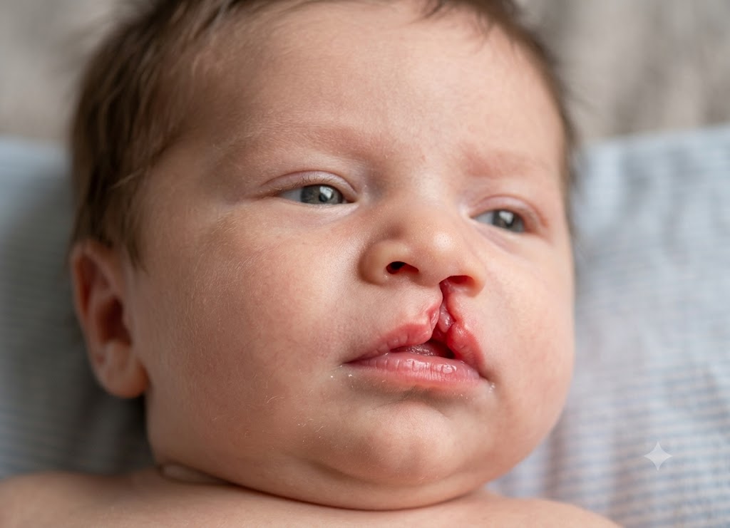
Cleft lip and cleft palate are birth differences that develop when certain facial or oral structures do not completely come together during pregnancy. A cleft lip involves the upper lip and can extend toward the nose, while a cleft palate affects the roof of the mouth. Some babies have only one of these conditions, while others are born with both. Since the two terms are often discussed together, parents may not always understand that they can affect feeding, speech, hearing, dental development, and treatment in different ways.
Learning how cleft lip differs from cleft palate can help parents better understand the diagnosis, upcoming procedures, and long-term care. A cleft lip is generally visible at birth and may sometimes be detected during a prenatal ultrasound, whereas an isolated cleft palate may only become apparent when the inside of the baby’s mouth is examined. Treatment often involves several specialists, including surgeons, pediatricians, feeding experts, dentists, speech-language professionals, and hearing specialists. This article covers six important differences between cleft lip and cleft palate that parents should know.
The fundamental characteristics of a cleft lip and a cleft palate
A cleft lip is a separation involving the upper lip, while a cleft palate is an opening in the roof of the mouth. Both are congenital orofacial conditions and may occur separately or at the same time. They develop when facial structures do not fuse completely during early pregnancy. The severity can vary considerably, from a small notch in the lip to a larger opening that extends through the lip, gum, palate, and into the nasal area. Understanding each condition individually makes their differences easier to recognize.
Defining Orofacial Clefts
Cleft lip and cleft palate are congenital facial conditions, meaning they are present at birth. They develop during the early stages of pregnancy when the tissues responsible for forming the face and mouth do not completely fuse.
These conditions are known collectively as orofacial clefts. They can range from a small, barely noticeable separation to a complete opening involving several facial or oral structures.
                     [Structural Profiles of Orofacial Clefts]
                                         │
         ┌───────────────────────────────┴───────────────────────────────┐
         ▼                                                               ▼
   [Cleft Lip]                                                     [Cleft Palate]
   - Visible opening in the upper lip                              - Opening in the roof of the mouth
   - Develops during early facial formation                         - Develops as the palate forms
   - May affect one or both sides                                  - May involve hard and/or soft palate
A baby may have an isolated cleft lip, an isolated cleft palate, or both together. The size and location of the opening can also vary from one child to another.
Anatomical Features of a Cleft Lip
When discussing what is a cleft lip, it refers to a visible separation in the upper lip. It develops when important facial tissues do not join properly during early fetal development, generally during the fourth through seventh weeks of pregnancy.
The appearance and severity can vary:
- Incomplete Cleft Lip: A smaller opening or notch that remains within the pink portion of the upper lip. This may also be described as a microform cleft.
- Complete Cleft Lip: A deeper opening that extends from the upper lip toward the nostril. In more extensive cases, the underlying lip muscle and upper gum ridge may also be affected.
- Unilateral vs. Bilateral: A unilateral cleft occurs on one side of the upper lip, while a bilateral cleft affects both sides. In bilateral cases, the central section of the upper lip may project forward.
Anatomical Features of a Cleft Palate
A cleft palate is an opening in the roof of the mouth. Unlike a cleft lip, it is primarily an internal condition and can create an abnormal connection between the mouth and nasal cavity.
It develops when the tissues that form the palate do not properly join together during fetal development, generally beginning around the sixth week and continuing through the later stages of palate formation.
The palate consists of two main sections: the hard palate, which forms the bony front portion of the roof of the mouth, and the soft palate, which contains muscle and is located toward the back.
A cleft can affect these structures in different ways:
- Isolated Soft Palate Cleft: The opening is limited mainly to the muscular soft palate toward the back of the mouth.
- Complete Palate Cleft: The separation extends through both the hard and soft portions of the palate.
- Submucous Cleft Palate: The muscles beneath the surface lining of the palate are separated, while the surface itself remains covered. Because the opening is hidden, this type may not be identified until later when feeding or speech concerns develop.
Combined Presentation: Cleft Lip and Palate
Some babies are born with both a cleft lip and a cleft palate. This combined condition involves structures on the outside of the face as well as the inside of the mouth.
[Embryonic Tissue Disruption] ──► [Lip Separation] ──► [Alveolar Ridge Involvement] ──► [Palatal Fusion Failure]
The lip and palate develop during overlapping but different stages of fetal development. The upper lip and front portion of the upper jaw develop from related embryonic structures, while the palate forms later.
When development is disrupted, both areas may remain separated. A more extensive cleft can continue through the upper gum and into the palate.
A combined cleft lip and palate can affect feeding, speech development, hearing, dental alignment, and facial growth. Because several areas may be involved, children usually benefit from coordinated care involving multiple specialists.
6 Core differences between a cleft lip and a cleft palate
Core Difference 1: Anatomical Location and Structural Deficits
The clearest difference between these two conditions is where the opening occurs and which tissues are affected.
                           [Anatomical Cavity Mapping]
                                       │
       ┌───────────────────────────────┴───────────────────────────────┐
       ▼                                                               ▼
 [Cleft Lip Anomalies]                                       [Cleft Palate Anomalies]
 - Mainly affects the upper lip                              - Mainly affects the roof of the mouth
 - Can involve skin, muscle, and gum                         - Can involve bone, muscle, and soft tissue
 - May affect the lip and nostril structure                  - Creates an opening between mouth and nose
The Extent of a Cleft Lip
A cleft lip is an external facial difference affecting the upper lip. The separation can involve the skin, tissue beneath the skin, and the muscles responsible for movement of the lips.
In more extensive cases, the opening may continue upward toward the nostril and affect the shape of the nose. It may also extend through the alveolar ridge, which is the portion of the upper jaw where teeth develop.
The severity ranges from a small notch to a complete separation. It may occur on one side or on both sides of the upper lip.
The Architecture of a Cleft Palate
A cleft palate occurs inside the mouth and involves the structure that normally separates the oral cavity from the nasal cavity.
The roof of the mouth consists of the hard palate at the front and the soft palate toward the back. When these structures do not fuse completely, an opening remains between the mouth and nose.
Because of this opening, food, liquid, and air may pass between the two spaces. An isolated cleft palate may not produce any obvious change in the baby’s external facial appearance.
Core Difference 2: Embryonic Developmental Timelines
The upper lip and palate form during different stages of pregnancy. This helps explain why a baby can have a cleft lip, a cleft palate, or both.
Lip Embryology (Weeks 4 to 7)
The upper lip begins forming very early in pregnancy. During approximately the fourth through seventh weeks, developing facial tissues move together and fuse to create the normal upper lip.
The medial nasal processes and maxillary processes must join correctly for the lip to develop normally.
If this fusion is interrupted during this period, a cleft lip can develop. The causes are usually multifactorial, involving a combination of genetic susceptibility and environmental influences rather than one single factor.
Palate Embryology (Weeks 6 to 12)
The palate forms somewhat later than the upper lip. During early development, palatal shelves grow from the developing upper jaw and initially form on either side of the tongue.
As the jaw and tongue change position, these shelves move upward and toward the center. They then meet and fuse to create the roof of the mouth.
This process continues through the first trimester. If the shelves fail to meet or fuse correctly, a cleft palate may result.
Because the palate forms after much of the lip development has already occurred, a baby can have an isolated cleft palate even when the upper lip has developed normally.
Core Difference 3: Physical Presentation and Clinical Visibility
The location of the cleft affects how easily it can be recognized. A cleft lip is usually visible externally, while an isolated cleft palate may be hidden inside the mouth.
[External Visual Inspection at Birth] ────► Cleft Lip: Usually visible soon after birth.
[Internal Oral Examination]           ────► Cleft Palate: Requires examination inside the mouth.
Visual Characteristics of a Cleft Lip
A cleft lip is generally noticeable when a baby is born. The appearance can range from a small notch in the upper lip to a larger separation that extends toward the nostril.
With a unilateral cleft, only one side of the lip is affected, which can make the nose appear uneven. A bilateral cleft creates openings on both sides and may leave the central portion of the upper lip projecting forward.
Because the difference is visible, parents may become aware of it immediately after delivery.
The Hidden Nature of an Isolated Cleft Palate
An isolated cleft palate may not change the baby’s facial appearance. The lips and outside of the face can look typical even though there is an opening inside the mouth.
Healthcare providers usually identify it by carefully examining the baby’s mouth after birth. The palate is checked to determine whether the hard and soft portions have developed normally.
If an isolated cleft is missed, feeding problems may provide an early clue. Milk may come through the baby’s nose, or the baby may have difficulty maintaining an effective feeding pattern.
A submucous cleft palate can be even harder to identify because the surface tissue remains intact while the underlying muscles are separated. Some children are not diagnosed until they develop feeding or speech difficulties.
Core Difference 4: Primary Functional and Physiological Impacts
Both conditions can affect newborn feeding, but the type and severity of the problem can differ because the lip and palate have different functions.
Challenges unique to a Cleft Lip
One of the main challenges associated with a cleft lip is forming a secure seal around the breast or bottle nipple.
A baby normally uses the lips to create a tight seal during feeding. A separation in the upper lip can make this seal less effective, allowing air to enter and making sucking less efficient.
Some babies may take longer to feed, swallow more air, or become tired during feeding. Feeding techniques, positioning, and specialized nipples can sometimes help.
Challenges unique to a Cleft Palate
A cleft palate can cause more significant feeding and developmental challenges because the mouth and nasal cavity are connected.
Difficulty Creating Suction: Because the roof of the mouth is open, the baby may have difficulty creating the negative pressure needed for normal sucking. Milk can enter the nose, leading to coughing, choking, or nasal leakage. Specialized bottles and feeding systems can help deliver milk more effectively.
Hypernasal Speech Distortions: The soft palate normally moves upward to separate the mouth from the nose during speech. When the palate is affected, air may escape through the nose, which can contribute to a nasal-sounding voice and difficulty producing certain speech sounds.
Middle Ear Fluid Accumulation: The muscles associated with the soft palate help the Eustachian tubes open and ventilate the middle ear. When these muscles do not function normally, fluid can accumulate behind the eardrum. This can contribute to repeated ear infections and temporary hearing difficulties.
Core Difference 5: Surgical Repair Timelines and Approaches
Cleft lip and cleft palate surgeries are usually performed at different stages. The exact timing depends on the child’s health, anatomy, development, and the recommendations of the cleft care team.
                           [Surgical Reconstructive Pathways]
                                           │
         ┌─────────────────────────────────┴─────────────────────────────────┐
         ▼                                                                   ▼
 [Cheiloplasty (Cleft Lip Repair)]                                   [Palatoplasty (Cleft Palate Repair)]
 - Commonly performed during infancy                                 - Commonly performed later in infancy
 - Focuses on restoring lip structure                                - Focuses on closing the oral-nasal opening
 - Reconstructs lip muscles and tissues                              - Reconstructs the palate and muscle function
Cleft Lip Repair (Cheiloplasty)
Cleft lip repair, known as cheiloplasty, is commonly performed during infancy, often around 3 to 6 months of age. The exact timing depends on the baby’s health and the surgical team’s approach.
Some surgeons use general readiness guidelines, including the traditional “Rule of 10s,” which considers the infant’s age, weight, and hemoglobin level before surgery.
During the procedure, the separated lip muscles and tissues are carefully repositioned and joined. The surgeon also works to create a natural shape for the upper lip and improve the appearance and symmetry of the nose and nostril.
The goal is not only cosmetic improvement but also restoration of useful lip function.
Cleft Palate Repair (Palatoplasty)
Repair of a cleft palate is generally performed later than cleft lip repair, often during the first or second year of life. Many teams aim to complete the repair before important speech development occurs.
The timing also considers facial growth, healing, and the child’s overall health.
During palatoplasty, the surgeon brings tissue from both sides of the palate together to close the opening between the mouth and nose. The muscles of the soft palate are also repositioned so they can work together during speech and swallowing.
The exact surgical technique and timing depend on the child’s specific cleft and the recommendations of the craniofacial team.
Core Difference 6: Long-Term Associated Health Complications
Long-term follow-up for cleft conditions can involve several areas of health and development. The specific needs depend on whether the child has a cleft lip, cleft palate, or both.
| Feature | Cleft Lip Complications | Cleft Palate Complications |
|---|---|---|
| Primary Care Focus | Dental development, orthodontic care, and facial symmetry | Hearing monitoring, speech development, and palate function |
| Common Issues | Missing, extra, or misaligned teeth near the cleft; differences in nasal symmetry | Middle ear fluid, recurring ear infections, and speech difficulties |
| Secondary Surgical Needs | Alveolar bone grafting when the gum line is affected; possible facial revisions | Ear tube placement and possible additional palate or speech-related procedures |
Complications linked to a Cleft Lip
Long-term concerns related to a cleft lip often involve the teeth, gums, jaw, and facial symmetry. When the cleft extends through the upper gum line, the teeth developing in that area may be affected.
Children may have missing, extra, unusually shaped, or poorly positioned teeth. This can require long-term dental monitoring and orthodontic treatment.
When the alveolar ridge is involved, an alveolar bone graft may be recommended during childhood to restore the bone in the upper gum and provide support for developing permanent teeth.
Some children may also have additional procedures later to improve the appearance or function of the lip and nose as their face grows.
Complications linked to a Cleft Palate
Long-term concerns associated with cleft palate often focus on hearing, speech, and palate function.
Because the muscles of the palate can affect Eustachian tube function, children with cleft palate are more likely to develop fluid in the middle ear. Regular hearing assessments are therefore an important part of follow-up care. Some children may need ventilation tubes to help manage persistent middle ear fluid.
Speech therapy may also be recommended. Even after the palate has been repaired, some children can develop speech patterns that require professional support.
If the repaired palate does not create an adequate seal between the mouth and nose during speech, a condition known as velopharyngeal insufficiency may occur. In selected cases, another procedure may be considered to improve speech function.
Understanding the Multifactorial Origins of Orofacial Clefts
To understand what is a cleft lip or palate, it is also important to consider why these conditions develop. Orofacial clefts are generally considered multifactorial, meaning several genetic and environmental factors can contribute to their development.
                          [Multifactorial Etiology Web]
                                        │
         ┌──────────────────────────────┴──────────────────────────────┐
         ▼                                                             ▼
 [Genetic Predispositions]                                     [Environmental Factors]
 - Family History                                             - Maternal Smoking
 - Multiple Risk Genes                                        - Nutritional Factors
 - Certain Genetic Syndromes                                  - Maternal Health Conditions
                                                              - Certain Medication Exposures
Is Cleft Lip Genetic?
When parents ask is cleft lip genetic, the answer is that genetics can influence the likelihood of developing an orofacial cleft. However, cleft lip is usually not caused by one single inherited gene.
Orofacial clefts can generally be divided into two broad groups:
- Non-Syndromic Clefts: The child has a cleft without another recognized genetic syndrome or major associated condition. Multiple genes may contribute to susceptibility, and family history can influence risk.
- Syndromic Clefts: The cleft occurs as part of a broader genetic syndrome that may affect several parts of the body. Examples include Van der Woude syndrome, Stickler syndrome, and certain chromosomal conditions.
If a family has a history of cleft lip or palate, a healthcare professional or genetic counselor can help explain the potential recurrence risk.
Environmental Factors and Prevention Strategies
Understanding what causes cleft lip and palate also involves considering environmental factors that may affect fetal development during early pregnancy.
Some recognized risk factors include:
- Maternal Smoking and Alcohol Exposure: Smoking during pregnancy is associated with an increased risk of orofacial clefts. Alcohol exposure may also contribute to developmental risks.
- Nutritional Deficiencies: Adequate folate is important before conception and during early pregnancy. Insufficient folate has been associated with an increased risk of certain birth defects.
- Maternal Health Conditions: Certain maternal health conditions, including poorly controlled diabetes, may increase the risk of birth defects.
- Medication Exposures: Some medications taken during pregnancy, including certain anti-seizure medications and retinoid medications, can increase the risk of developmental abnormalities.
Not every cleft can be prevented, and in many cases the exact cause cannot be identified.
Additional information is vital for understanding cleft conditions
A complete understanding of cleft conditions requires more than simply comparing the lip and palate. Families may also need to understand the different types of clefts, how they are diagnosed, possible causes, and the roles of different healthcare professionals.
Treatment is often a long-term process because cleft conditions can influence feeding, speech, hearing, dental development, facial growth, and appearance at different stages of childhood.
Classifications and Structural Types of Orofacial Clefts
Orofacial clefts can be classified according to their location, size, completeness, and whether one or both sides are affected.
                       [Orofacial Cleft Classification Matrix]
                                          │
         ┌────────────────────────────────┴────────────────────────────────┐
         ▼                                                                 ▼
 [Cleft Lip Categories]                                            [Cleft Palate Categories]
  ├── Unilateral (One Side)                                        ├── Incomplete (Soft Palate)
  ├── Bilateral (Both Sides)                                      ├── Complete (Hard + Soft Palate)
  └── Incomplete vs. Complete                                     └── Submucous (Hidden Muscle Split)
Cleft Lip Classifications
- Unilateral Cleft Lip: The opening affects only one side of the upper lip. It is more frequently seen on the left side.
- Bilateral Cleft Lip: Both sides of the upper lip are separated, leaving a central section of tissue between the two openings.
- Incomplete vs. Complete: An incomplete cleft does not extend completely toward the nostril, while a complete cleft reaches toward the nostril and may also involve the underlying gum ridge.
Cleft Palate Classifications
- Incomplete Cleft Palate: The opening primarily affects the soft palate toward the back of the mouth.
- Complete Cleft Palate: The separation extends through both the hard and soft palate.
- Submucous Cleft Palate: The surface lining appears intact, but the muscles beneath it have not joined properly. It can be difficult to identify and may become apparent through feeding or speech concerns.
Clinical Diagnostic Methods
The method used to diagnose an orofacial cleft depends on whether the condition is visible externally or located inside the mouth.
Prenatal Diagnostic Screening
A cleft lip, especially a more extensive or bilateral cleft, may sometimes be detected during a routine 20-week anatomy ultrasound. Prenatal imaging can identify an opening in the upper lip and may show changes in the shape of the nose.
Early identification allows parents and healthcare professionals time to discuss the condition and arrange appropriate specialist care.
However, small cleft lips and isolated cleft palates may be difficult to detect before birth. The baby’s position and the tongue can make the roof of the mouth difficult to visualize during a routine ultrasound.
Postnatal Diagnostic Confirmation
An isolated cleft palate is usually identified through a newborn physical examination. The healthcare provider examines the inside of the baby’s mouth to assess the hard and soft palate.
Submucous cleft palate can be more challenging to diagnose because the surface appears intact. It may not be discovered until a child develops symptoms such as feeding difficulties, nasal leakage, or speech changes.
Underlying Causes and Genetic Risk Factors
Orofacial clefts are generally multifactorial conditions. This means that a combination of genetic susceptibility and environmental influences may contribute to their development.
The facial structures develop and fuse during several important weeks of early pregnancy. A disruption during these stages can interfere with normal formation.
[Genetic Susceptibility (Risk Genes / Family History)] + [Environmental Factors]
                                           │
                                           ▼
                     [Interrupted Facial Fusion] ──► Orofacial Cleft
Genetic and Syndromic Influences
Genetic factors can increase susceptibility to cleft lip and palate. A child may have a higher chance of developing an orofacial cleft when a close family member has previously been affected.
Clefts can occur by themselves or as part of a genetic syndrome. Van der Woude syndrome, for example, can include cleft lip or palate along with characteristic pits in the lower lip.
Pierre Robin sequence can involve a small lower jaw that changes the position of the tongue and can interfere with normal palate development.
Environmental Modifiers and Prevention
- Nutritional Deficiencies: Adequate folic acid (vitamin B9) before conception and during early pregnancy supports normal fetal development. Low folate levels have been associated with an increased risk of certain birth defects.
- Maternal Health Conditions: Certain maternal conditions, including poorly controlled diabetes, may increase the likelihood of orofacial clefts.
- Teratogenic Exposures: Tobacco exposure and alcohol use during pregnancy can increase developmental risks. Some medications, including certain anti-seizure drugs and retinoids, are also associated with a higher risk of birth defects.
The Multidisciplinary Cleft Care Team Model
Because cleft conditions can affect several aspects of a child’s health and development, treatment commonly involves a coordinated team of specialists. Care may continue from infancy through childhood and adolescence.
The Core Craniofacial Team
- Craniofacial/Plastic Surgeon: Plans and performs procedures to repair the cleft and may provide additional surgeries as the child grows. Lip repair is commonly performed during infancy, while palate repair usually follows later.
- Speech-Language Pathologist (SLP): Helps with feeding during infancy and later monitors speech development. The SLP can provide therapy if speech sounds or resonance are affected.
- Pediatrician: Provides routine medical care, monitors growth and development, and helps ensure the child is healthy enough for procedures.
Dental, Otologic, and Psychosocial Support
Orthodontist and Oral Surgeon: Monitor tooth development, jaw growth, and the alignment of the upper jaw. When the cleft affects the gum ridge, an alveolar bone graft may be recommended during childhood.
Otolaryngologist (ENT) and Audiologist: Monitor ear health and hearing. Children with cleft palate are more likely to experience middle ear fluid and may require ear tubes. Regular hearing tests help identify problems early.
Craniofacial Psychologist or Social Worker: Can provide emotional and social support to children and families as they navigate treatment, surgeries, facial differences, and other challenges that may arise during development.
Conclusion
Cleft lip and cleft palate are closely related birth conditions, but they affect different parts of the face and mouth. A cleft lip mainly involves the upper lip and may also affect the nose and gum line, while a cleft palate affects the roof of the mouth and can have a stronger impact on feeding, speech, and middle ear function.
Both conditions can be treated, although the timing and type of treatment depend on the child’s specific anatomy and needs. Some children may require several stages of care involving surgery, feeding support, speech therapy, hearing evaluations, dental treatment, and orthodontic care.
For babies diagnosed with cleft lip, cleft palate, or both, early evaluation by a specialized cleft or craniofacial team can help families understand the condition and plan appropriate care throughout childhood.
Frequently Asked Questions
1. What is a cleft lip?
A cleft lip is an opening or separation in the upper lip that develops before birth. It occurs when the tissues forming the lip do not completely fuse during fetal development. The opening may be very small or may extend upward toward the nose. It can affect one side or both sides of the upper lip.
2. What is the difference between cleft lip and cleft palate?
A cleft lip affects the upper lip, whereas a cleft palate affects the roof of the mouth. A baby can have either condition separately or have both at the same time. Cleft lip is usually visible externally, while an isolated cleft palate may only be identified by examining the inside of the mouth. Both conditions can affect feeding, but cleft palate is particularly associated with speech and ear-related concerns.
3. Can cleft lip and cleft palate happen together?
Yes. Some babies are born with both cleft lip and cleft palate. In these cases, the opening affects both the upper lip and structures inside the mouth. Depending on the severity, the condition can influence feeding, speech, hearing, dental development, and facial growth. A multidisciplinary cleft team can coordinate treatment as the child develops.
4. How are cleft lip and cleft palate treated?
Treatment generally involves surgery, although the timing varies according to the type and severity of the cleft. Cleft lip repair is usually performed during infancy, while cleft palate repair often occurs later during the first or second year of life. Some children may also need feeding support, speech therapy, hearing care, dental treatment, orthodontics, or additional procedures. The care plan is tailored to each child.
5. What causes cleft lip?
Cleft lip usually develops because of a combination of genetic and environmental factors, although the exact cause may not always be known. Factors associated with increased risk can include family history, smoking during pregnancy, certain maternal health conditions, and exposure to particular medications during pregnancy. Having a baby with a cleft is not necessarily the result of anything a parent did or failed to do. A healthcare provider or genetic counselor can help families understand possible causes and recurrence risk.