7 Things to Know About Clotting Factors and How Blood Clots Form
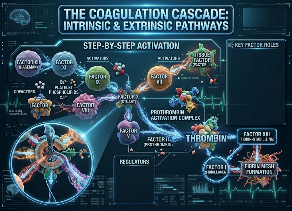
Clotting factors are a group of blood proteins that step in the moment a vessel gets damaged, working together to stop the bleeding. The instant an injury happens, platelets show up first and form a rough temporary patch over the wound.
From there, clotting factors take over, running through an orderly chain reaction, the coagulation cascade, that produces fibrin, a mesh-like protein that reinforces the patch and locks the seal in place.
Getting a handle on how clotting factors work makes it much easier to understand how clots actually form, and why the whole system depends on staying in balance. When clotting activity runs too low, the result can be easy bruising, frequent nosebleeds, or bleeding that just won’t quit.
When it runs too high, the danger shifts toward unwanted clots forming inside the vessels themselves, raising the risk of deep vein thrombosis, pulmonary embolism, heart attack, or stroke. Below are seven essential facts about clotting factors, how they protect the body, and why abnormal clotting is always worth a doctor’s attention.
What Are Clotting Factors?
Clotting factors are specialized blood proteins whose job is to stop bleeding once a vessel has been injured. The liver manufactures most of them, and they sit dormant in the bloodstream until they’re actually called into action. The moment a cut or internal injury occurs, these proteins switch on in a tightly sequenced chain reaction known as the coagulation cascade.
This whole process runs in parallel with platelets, small blood cells that arrive at the wound first. Platelets build the initial plug, and clotting factors follow up by generating fibrin, a tough, thread-like protein that binds the clot into something solid. A useful way to picture it: platelets are the first bandage, and fibrin is the net that keeps that bandage from falling apart. Between the two, the vessel gets sealed and blood loss stays under control.
Balance is everything here. Not enough of certain clotting factors, and bleeding drags on longer than it should, or bruises show up too easily. Too much clotting activity, and clots start forming inside vessels where they don’t belong, which can turn dangerous fast. That’s exactly why unexplained bruising, repeated nosebleeds, leg swelling, chest pain, or sudden shortness of breath should never be brushed off. They’re worth a trip to a healthcare provider.
7 Key Facts About Coagulation Factors and the Clotting Cascade
These seven facts cover the essentials: clotting factors start out as inactive protein precursors, they operate inside a sequential amplification system, three factors in particular, X, II, and I, carry the process to its conclusion, Vitamin K is non-negotiable for building them correctly, the whole cascade exists to produce a stable fibrin clot, two separate pathways can kick the process off, and the resulting clot is only ever meant to be temporary.
Together, these facts paint a full picture of hemostasis, from the very first molecular trigger all the way through to the clot’s eventual breakdown. We’ll walk through each one in detail, starting with what a coagulation factor actually is.
Cellular Nature and Activation of Clotting Factors
Coagulation factors, the technical name for clotting factors, are a family of specialized proteins that drift through blood plasma, coordinating the biochemical work of building a clot. Most of them exist in plasma as zymogens: inactive precursor forms waiting to be switched on. Out of the 13 principal factors, the liver produces the majority.
They’re labeled with Roman numerals (Factor I, Factor II, and so on), but that numbering simply reflects the order in which scientists discovered them historically, not the order in which they actually act during the cascade.
There’s a good reason these factors stay dormant while circulating: it’s a built-in safety mechanism. If they were constantly switched on, the body would be flooded with unprovoked clots throughout the vascular system. So when a vessel wall gets breached, these zymogens undergo a targeted cleaving process, an already-active enzyme snips off a small piece of the precursor, exposing the active site underneath.
Once a factor is activated (marked with a lowercase “a,” as in Factor Xa), it becomes a precise enzyme capable of cleaving the next zymogen down the line. Most factors function this way, as enzymes, though a handful, including Factor V and Factor VIII, work differently, acting as cofactors that latch onto the reaction and dramatically speed it up rather than triggering it directly.
The Amplification Dynamics of the Coagulation Cascade
The coagulation cascade functions as a tightly coordinated, step-by-step amplification system built to seal a vascular leak fast. Because it runs as an enzymatic chain reaction, even a small injury can set off a disproportionately large response.
One activated factor early in the sequence is enough to switch on thousands more further down the line. That domino effect produces a sudden surge of thrombin, more than enough material to rapidly build a stable barrier right where the injury occurred.
                    Structural Triggers of the Coagulation Cascade
                                           │
         ┌─────────────────────────────────┴─────────────────────────────────┐
         ▼                                                                   ▼
                 Extrinsic Pathway                                    Intrinsic Pathway
        - Main physiological trigger                          - Secondary amplification loop
        - Set off by external tissue trauma                   - Set off by internal vessel damage
        - Tissue Factor (III) binds Factor VII                - Factor XII binds exposed collagen
         │                                                                   │
         └─────────────────────────────────┬─────────────────────────────────┘
                                           ▼
                                     Common Pathway
                              - Both pathways converge at Factor X
                              - Prothrombin (II) becomes Thrombin (IIa)
                              - Fibrinogen (I) becomes Fibrin mesh
To make sense of all this, the cascade is generally broken into three connected stages: the extrinsic pathway, the intrinsic pathway, and the common pathway where they meet.
Initiation Pathways: Extrinsic vs. Intrinsic Triggers
The intrinsic and extrinsic pathways don’t respond to the same kind of damage. Think of them as two separate detection systems, each tuned to a different type of injury.
The Extrinsic Pathway (Primary Initiator): This one fires in response to trauma affecting the vessel wall from the outside, a deep cut, a crush injury, anything that breaches the wall directly. Once the protective endothelial lining is broken, the tissue layer beneath it is exposed to passing blood. Cells in that exposed layer carry a protein called Tissue Factor (also known as Factor III). The moment Tissue Factor makes contact, it binds tightly to Factor VII, converting it into VIIa. Together, this TF-VIIa complex acts fast, immediately jumping into the common pathway by activating Factor X. It’s essentially the spark that gets the whole process moving.
The Intrinsic Pathway (Amplification Loop): This one responds to damage occurring inside the vessel itself, and everything it needs is already circulating in the blood. It kicks off when blood makes contact with a negatively charged surface, typically the collagen fibers exposed when the vessel’s inner lining is compromised. Factor XII binds to that exposed collagen first, setting off a chain reaction that runs through Factors XI, IX, and VIII in sequence. This pathway is slower to get started, but it functions as a critical backup, feeding a steady stream of thrombin into the system to keep the clot durable.
The Core Mechanics of the Common Pathway
The common pathway is where both branches converge, the final stretch where a chemical signal turns into an actual physical seal. Three factors drive this last stage.
Factor X activated to Xa ──► Prothrombin (II) cleaved to Thrombin (IIa) ──► Fibrinogen (I) converted to FibrinFactor X (Stuart-Prower Factor): This is the entry point into the common pathway, activated by signals from both upstream branches. Once converted to Factor Xa, it pairs up with its cofactor, Factor Va, on a platform built from calcium ions and platelet membranes, together forming what’s called the prothrombinase complex.
Prothrombin (Factor II): This is the direct precursor to thrombin (Factor IIa), arguably the single most important enzyme in the whole cascade. Thrombin doesn’t just build the clot’s final structure, it also loops back and reactivates Factors V, VIII, and XI, creating a feedback loop that accelerates its own production.
Fibrinogen (Factor I): A soluble protein that circulates in large quantities in the blood. Thrombin trims small fragments off fibrinogen, converting it into fibrin monomers that spontaneously link together into a strand-like web.
The Critical Role of Vitamin K in Synthesis and Anchoring
The liver can’t build fully functional clotting factors without Vitamin K. It serves as a required cofactor for an enzyme called gamma-glutamyl carboxylase, which chemically modifies four specific factors: II, VII, IX, and X.
     Liver builds inactive precursors of Factors II, VII, IX, and X
                                │
                                ▼
        Vitamin K activates gamma-glutamyl carboxylase enzyme
                                │
                                ▼
     Carboxylation attaches negative Gla residues onto the factors
                                │
                                ▼
     Gla residues bind securely to positive calcium ions on platelets
                                │
                                ▼
       Clotting factors anchor precisely at the site of injury
Without Vitamin K, the liver still churns out these four proteins, but they come out structurally incomplete, missing the chemical tags needed to bind calcium.
That calcium-binding step works like a molecular anchor, letting the factors attach firmly to activated platelets right where the injury is. Skip that step, and the factors just drift off into general circulation, too diluted and too slow to actually stop active bleeding.
Achieving a Stable Fibrin Mesh
The whole point of the coagulation cascade is to turn liquid blood into a firm, cross-linked fibrin structure exactly where the vessel has torn, stopping serious blood loss in its tracks.
When thrombin converts fibrinogen into fibrin monomers, those strands link up into a loose web on their own. This soft, gel-like structure is enough to trap red blood cells and platelets, forming a basic plug.
But that first-pass mesh is only held together by weak hydrogen bonds, and it can’t hold up against the ongoing pressure of blood flow for long. To lock things down, thrombin activates Factor XIII, the fibrin-stabilizing factor, turning it into Factor XIIIa.
This enzyme forges strong, permanent chemical bonds between neighboring fibrin strands, transforming the soft gel into a tough, stable, insoluble barrier that stays anchored to the vessel wall, stopping the bleeding and buying the tissue time to heal.
Fibrinolysis: The Systematic Dissolution of Clots
A fibrin clot was never meant to be permanent, it’s a temporary scaffold. The body relies on a careful balance between clot-building forces and clot-dissolving ones, a process called fibrinolysis. Once the vessel wall has healed, the clot has to be broken down methodically, so it doesn’t dislodge or permanently block blood flow.
     Inactive plasminogen sits trapped inside the fibrin mesh
                          │
                          ▼  ◄── Healed endothelial cells release t-PA
     Active plasmin is generated
                          │
                          ▼  ◄── Plasmin cuts through the fibrin strands
     Fibrin fragments (FDPs) and D-dimer released into the bloodstream
The key player here is plasmin, which circulates in an inactive form called plasminogen. As the clot initially forms, plasminogen molecules get trapped inside the fibrin web. Once the tissue underneath starts healing, nearby endothelial cells slowly release tissue plasminogen activator (t-PA), converting the trapped plasminogen into active plasmin, which then slices through the cross-linked fibrin like a pair of molecular scissors.
The end result is a set of small, soluble fragments called Fibrin Degradation Products (FDPs), including a well-known marker called D-dimer. Immune cells clear these fragments from the blood, and normal flow through the repaired vessel resumes.
Classification of Genetic Bleeding Disorders
Inherited bleeding disorders happen when specific clotting factors, or the proteins that carry them, are missing or structurally faulty. Without the right protein in place, the coagulation cascade can’t build a stable fibrin mesh, and the body loses its ability to seal damaged vessels properly.
                      Genetic Bleeding Disorder Pathways
                                       │
        ┌──────────────────────────────┼──────────────────────────────┐
        ▼                              ▼                              ▼
  Von Willebrand Disease         Hemophilia A (Classic)         Hemophilia B (Christmas)
  - Most common inherited form   - Deficient Factor VIII        - Deficient Factor IX
  - Autosomal dominant trait     - X-linked recessive trait     - X-linked recessive trait
  - Disrupts platelet anchoring  - Impairs intrinsic pathway    - Nearly identical symptoms
How severe these conditions get depends directly on how much active factor is still circulating in the plasma. Pinpointing the exact defect matters, since modern treatment relies on replacing the specific missing protein.
Von Willebrand Disease (VWD)
Von Willebrand disease is the most common inherited bleeding disorder there is. Unlike hemophilia, it follows an autosomal dominant inheritance pattern, meaning it shows up in men and women at roughly equal rates.
It’s caused by a defect or shortage of von Willebrand factor (vWF), a protein that handles two separate jobs in hemostasis:
Platelet Anchoring: vWF acts as a bridge, binding to exposed collagen at the injury site and holding passing platelets in place to form the initial plug.
Factor VIII Stabilization: vWF also binds to circulating Factor VIII, shielding it from being broken down too quickly before it gets its chance to act in the common pathway.
Because vWF is tied up in both of these roles, people with VWD can experience impairment on two fronts at once. That said, symptoms are usually milder than hemophilia, typically showing up as frequent nosebleeds, easy bruising, or bleeding that runs long after dental work.
Hemophilia A (Classic Hemophilia)
Hemophilia A is the most common severe inherited bleeding disorder. It follows an X-linked recessive inheritance pattern, meaning the affected gene sits on the X chromosome, so mothers are usually symptom-free carriers, while the disease itself shows up almost exclusively in males.
The root cause is a shortage, or complete absence, of functional Factor VIII. Within the intrinsic pathway, Factor VIII acts as a required cofactor, pairing with Factor IXa to form the “tenase” complex responsible for activating Factor X.
Without enough Factor VIII, that amplification step stalls, and the thrombin surge needed for a stable clot never happens. People with severe Hemophilia A (under 1% of normal factor activity) tend to experience deep, prolonged bleeding after even minor injuries, along with spontaneous bleeding into joints and muscles.
Hemophilia B (Christmas Disease)
Hemophilia B, once known as Christmas disease, follows the same X-linked recessive pattern and produces symptoms that are nearly impossible to tell apart from Hemophilia A just by looking.
The real difference comes down to which protein is missing: Hemophilia B stems from a Factor IX deficiency. Once activated to IXa, Factor IX partners directly with Factor VIIIa to push the common pathway forward.
Because both disorders knock out the exact same step in the intrinsic pathway, distinguishing between them requires a specific lab test measuring Factor VIII and Factor IX activity levels separately.
Clinical Severity and Modern Treatment Strategies
Treatment for these disorders comes down to replacing whatever protein is deficient, based on the patient’s baseline factor activity.
| Bleeding Disorder | Deficient Protein | Inheritance Pattern | Main Treatment |
|---|---|---|---|
| Von Willebrand Disease | von Willebrand Factor (vWF) | Autosomal Dominant | Desmopressin (DDAVP), or vWF-containing concentrates |
| Hemophilia A | Factor VIII | X-Linked Recessive | Recombinant Factor VIII concentrates |
| Hemophilia B | Factor IX | X-Linked Recessive | Recombinant Factor IX concentrates |
Desmopressin (DDAVP): For milder cases of Von Willebrand disease or mild Hemophilia A, doctors often reach for desmopressin. It prompts endothelial cells to release their stored vWF and Factor VIII directly into circulation, temporarily raising factor levels without needing a transfusion.
Targeted Factor Concentrates: For severe deficiencies, or ahead of surgery, patients get intravenous infusions of purified, concentrated clotting factors. This keeps the cascade stabilized, prevents spontaneous bleeding episodes, and lets people maintain a normal quality of life.
Clinical Screening Panel for Coagulation Function
Doctors assess how well the coagulation cascade is functioning using a specific panel of blood tests, drawn into a light-blue-top tube containing sodium citrate. Citrate works as a reversible anticoagulant, binding up calcium ions and pausing all clotting activity until the sample reaches the lab.
                    Initial Coagulation Screening Strategy
                                     │
         ┌───────────────────────────┴───────────────────────────┐
         ▼                                                       ▼
   Prothrombin Time (PT / INR)                Activated Partial Thromboplastin Time (aPTT)
   - Tests Extrinsic & Common Pathways        - Tests Intrinsic & Common Pathways
   - Measures Factor VII function              - Measures XII, XI, IX, VIII function
   - Tracks Warfarin therapy                   - Tracks Heparin therapy
By reintroducing calcium and specific triggers into the citrated plasma under controlled lab conditions, technicians can time exactly how long it takes a stable fibrin mesh to form.
Prothrombin Time (PT) and International Normalized Ratio (INR)
The PT test checks how well the extrinsic and common pathways are functioning.
The Lab Process: The lab adds calcium and thromboplastin (a mix of Tissue Factor and phospholipids) to the plasma sample, simulating outside tissue injury and immediately activating Factor VII. The test times how long it takes fibrinogen to convert into fibrin. A normal PT typically falls between 11 and 13.5 seconds.
What Results Mean: An isolated prolonged PT, with everything else normal, usually points to a Factor VII problem specifically. A prolonged PT can also signal advanced liver disease, since the liver builds these proteins, or a significant Vitamin K deficiency.
Because thromboplastin reagents differ in sensitivity from lab to lab, the World Health Organization standardized PT results into the International Normalized Ratio, or INR. In a healthy person on no medication, a normal INR sits at 1.1 or below.
INR is the main tool doctors use to monitor patients on Warfarin (Coumadin). Warfarin deliberately suppresses Factors II, VII, IX, and X to keep the INR within a therapeutic range of 2.0 to 3.0.
Activated Partial Thromboplastin Time (aPTT)
The aPTT test evaluates how well the intrinsic and common pathways are working.
The Lab Process: A chemical activator (silica or celite, typically) along with phospholipids gets mixed into the plasma to mimic the exposed collagen of a damaged vessel wall. This triggers Factor XII and sets off a chain through Factors XI, IX, and VIII. A normal aPTT usually falls between 30 and 40 seconds.
What Results Mean: An isolated prolonged aPTT points to a defect somewhere in the intrinsic pathway, making it the go-to screening test for Hemophilia A or B. It’s also used to monitor patients on intravenous unfractionated heparin, with a therapeutic target usually set at 1.5 to 2.5 times the patient’s baseline. A prolonged aPTT can also flag an autoimmune inhibitor like the lupus anticoagulant, which interferes with the phospholipids used in the test itself.
Advanced Diagnostic Pathways for Abnormal Screening Results
When a PT or aPTT result comes back abnormal, doctors follow a structured process to pin down exactly what’s wrong.
        Abnormal / Prolonged Screening Result (PT or aPTT)
                            │
                            ▼
                 Perform a Plasma Mixing Study
                            │
              ┌─────────────┴─────────────┐
              ▼                           ▼
     Clotting Time Corrects       Clotting Time Stays Prolonged
     → Factor Deficiency          → Factor Inhibitor Present
     → Next: Factor Assay         → Next: Inhibitor Screen
Plasma Mixing Studies: This test figures out whether a prolonged clotting time comes from a missing protein or an antibody actively blocking the process. The patient’s plasma gets mixed 50:50 with normal donor plasma. If the clotting time corrects, the diagnosis is a factor deficiency, the donor plasma supplied what was missing. If it stays prolonged, that points to an inhibitor: an antibody in the patient’s blood interfering with the donor’s proteins.
Specific Clotting Factor Assays: Once a deficiency is confirmed, individual factor assays measure exactly how much active protein is present, for example, testing Factor VIII activity to confirm Hemophilia A.
Specialized Coagulation Panels: When doctors need to look beyond the standard pathways, say, investigating unexplained clots inside a deep vein, they’ll order specialized tests. That can include D-dimer levels to gauge clot breakdown, or screening for inherited clotting tendencies like the Factor V Leiden mutation, or deficiencies in natural anti-clotting proteins like Protein C and Protein S.
How Warfarin Interferes with the Coagulation Cascade
Unlike fast-acting anticoagulants such as heparin, Warfarin (Coumadin) doesn’t directly block clotting factors already circulating in the blood. Instead, it works as a Vitamin K antagonist, disrupting the liver’s ability to manufacture functional factors in the first place.
        Warfarin enters liver cells and binds to the VKORC1 enzyme
                            │
                            ▼
        VKORC1 inhibition blocks Vitamin K recycling
                            │
                            ▼
        Functional Vitamin K deficiency prevents carboxylation
                            │
                            ▼
        Liver secretes non-functional PIVKAs lacking Gla anchors
                            │
                            ▼
        Precursors float freely, unable to bind calcium or clot
The Biochemical Blockade: VKORC1 Inhibition
To build functional versions of Factors II, VII, IX, and X, the liver enzyme gamma-glutamyl carboxylase needs a steady supply of active Vitamin K to add carboxyl groups onto these proteins. Once it’s done its job, that Vitamin K becomes oxidized and inactive.
Warfarin targets an enzyme called VKORC1, whose job is recycling that inactive Vitamin K back into a usable form. By shutting that recycling process down, Warfarin effectively creates a functional Vitamin K shortage.
The Production of PIVKAs
With recycling blocked, the liver keeps assembling Factors II, VII, IX, and X, but can’t finish the chemical modifications they need. These incomplete proteins, called PIVKAs (Proteins Induced by Vitamin K Absence), get released into the blood anyway.
Since PIVKAs can’t bind calcium or attach to activated platelets, they just drift harmlessly through the plasma, slowing the cascade down and delaying clot formation.
Clinical Dynamics of Warfarin Therapy
Why the Effect Is Delayed: Warfarin only prevents new factors from being built correctly, it does nothing to factors already circulating. So its full effect depends on how long those existing factors stick around:
- Factor VII clears fastest, with a half-life of only about 6 hours, which is why PT results shift quickly after starting Warfarin.
- Factor II (Prothrombin) has a much longer half-life, 60 to 72 hours, which is why the medication’s full effect takes 3 to 5 days to fully settle in.
Monitoring and Reversal: Because Warfarin’s effect shifts based on diet and other medications, regular PT/INR monitoring is required. For most conditions, atrial fibrillation, deep vein thrombosis, doctors aim to keep INR stable between 2.0 and 3.0.
If INR climbs too high, or bleeding occurs, Vitamin K can restore normal factor production within several hours for non-emergencies. In true emergencies, Fresh Frozen Plasma or Prothrombin Complex Concentrates supply active factors immediately.
Coagulation vs. Thrombosis: Physiological vs. Pathological
Both terms describe blood shifting from liquid to solid, but the similarities end there.
Coagulation (Physiological Hemostasis): This is the normal, protective response to an actual injury. When a vessel is torn, the cascade builds a localized fibrin mesh to seal the breach, and once healing is done, the body safely dissolves that temporary scaffold on its own.
Thrombosis (Pathological Occlusion): This is when a clot, a thrombus, forms inside a vessel that wasn’t meaningfully injured. Rather than stopping bleeding, it blocks blood flow, cutting off oxygen and nutrients to whatever tissue lies downstream. A thrombus in an artery can trigger a heart attack or stroke; one in a deep vein causes DVT. If a fragment breaks off and travels to the lungs, it becomes a life-threatening pulmonary embolism.
Virchow’s Triad: The Triggers of Thrombosis
To explain what pushes clotting into pathological territory, clinicians use a framework called Virchow’s Triad, three factors that tip the balance toward dangerous clotting.
                    Virchow's Triad of Pathological Thrombosis
                                       │
        ┌───────────────────────────────┼───────────────────────────────┐
        ▼                               ▼                               ▼
  Endothelial Injury              Abnormal Blood Flow              Hypercoagulability
  - Damage to vessel lining       - Pooling or turbulence           - Systemic shift toward clotting
  - From hypertension or          - From immobility, atrial         - From genetic mutations, cancer,
    atherosclerosis                 fibrillation, varicose veins       or pregnancy
Endothelial Injury: Direct damage to the smooth inner lining of a vessel, often caused by high blood pressure, smoking, or cholesterol buildup. This exposes tissue factor and collagen to blood flow, triggering both pathways inside an otherwise intact vessel.
Abnormal Blood Flow: Sluggish pooling or chaotic turbulence, as seen during long periods of immobility or in atrial fibrillation, lets activated clotting factors build up in one spot instead of being diluted through normal circulation, sparking a clot.
Hypercoagulability: A systemic tilt toward clotting, whether inherited (like the Factor V Leiden mutation) or acquired (from cancer, pregnancy, birth control, or severe inflammation). In these states, the body produces more pro-clotting factors and fewer natural inhibitors, making the whole cascade hypersensitive.
Conclusion
Clotting factors are essential to sealing an injured vessel, working alongside platelets, calcium, Vitamin K, and fibrin to keep bleeding under control. When these factors are missing, reduced, overactive, or thrown off by medication or disease, the body can either bleed too freely or clot when it shouldn’t. Unusual bruising, prolonged bleeding, repeated nosebleeds, heavy periods, one-sided leg swelling, chest pain, sudden shortness of breath, or stroke-like symptoms all warrant medical evaluation.
Frequently Asked Questions
1. What are clotting factors?
Proteins in the blood, mostly made by the liver, that stay inactive until an injury triggers a sequence that produces fibrin, the substance that reinforces the platelet plug into a stable clot.
2. How do clotting factors help blood clots form?
Through the coagulation cascade: each activated factor sets off the next, eventually producing thrombin, which converts fibrinogen into fibrin strands that hold the clot together.
3. What happens if clotting factors are low?
Easier bruising, frequent nosebleeds, heavy periods, or bleeding that lasts too long after injury, dental work, or surgery. Hemophilia is one example; liver disease, Vitamin K deficiency, and certain medications can also lower clotting factor activity.
4. Can clotting factors cause harmful blood clots?
Yes, abnormal clotting activity raises the risk of DVT, pulmonary embolism, heart attack, or stroke. Some clotting problems are inherited, while others stem from illness, surgery, immobility, cancer, pregnancy, smoking, or medication.
5. How are clotting factor problems diagnosed?
Through blood tests measuring clotting speed and specific factor function, including PT, aPTT, fibrinogen testing, and individual factor assays, alongside a review of bleeding history, family history, medications, and liver function.