What Is Mucosa? A Complete Guide to the Body’s Inner Lining
Every day, the human body relies on specialized tissue layers that quietly protect organs and keep them running smoothly. One of the most important of these layers is the mucosa, commonly called a mucous membrane. While most people have heard the word, far fewer understand just how much work this thin lining actually does.
Mucosa is a moist tissue that lines the parts of the body exposed to the outside world the digestive tract, airways, urinary system, and reproductive organs. It works as a protective shield, keeping out bacteria and irritants, while also helping the body digest food, breathe, and mount immune defenses.
Unlike regular skin, mucosa is built to stay soft, moist, and flexible. It’s packed with cells that produce mucus a slippery substance that lubricates tissue, traps particles, and shields delicate surfaces underneath. In the lungs, this mucus catches dust and germs before they go further; in the gut, it shields the stomach and intestines from irritation.
When the mucosa gets damaged or inflamed, it can throw off the body’s balance leading to dryness, discomfort, or disrupted organ function. Because mucosal tissue shows up in so many different places, problems with it can look very different depending on where they occur.
This guide breaks down what mucosa is, where you’ll find it, how it works, its different types, common conditions that affect it, and simple ways to keep it healthy.
What Are the Primary Layers of Mucosa?
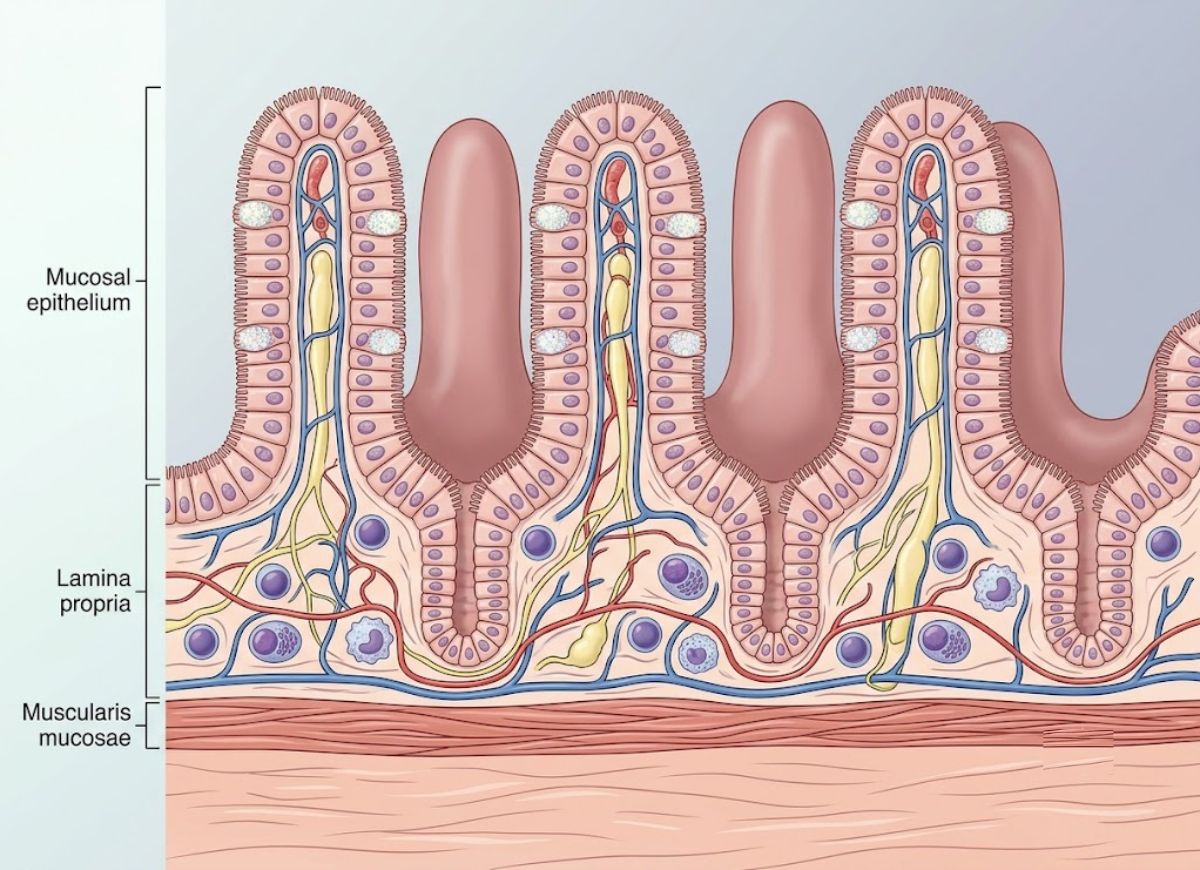
Mucosa is built from three core layers: the epithelium, the lamina propria, and the muscularis mucosae each with its own cell types and job to do.
These layers work together as a flexible, responsive barrier lining the body’s internal passageways. The exact makeup of each layer shifts depending on the organ, but this three-part structure is the blueprint behind every mucous membrane in the body.
The Epithelial Layer of the Mucosa
The epithelium is the outer layer of the mucosa the part in direct contact with whatever passes through an organ. Its job is to act as a gatekeeper, controlling protection, secretion, and absorption. This is also the layer that varies the most, shaped entirely by what its specific location needs.
Epithelial cells sit tightly packed together, held in place by junctions like tight junctions and desmosomes, forming a barrier that regulates what gets in and out. Depending on location, this tissue can be a single cell layer or several layers stacked up, and the cells themselves may be flat, cube-shaped, or tall and column-like.
Where an organ faces heavy wear the mouth, throat, esophagus, or vagina the mucosa is covered in tough, stratified squamous epithelium. Multiple cell layers give it durability, and worn-out surface cells are constantly replaced by fresh ones pushing up from below.
The stomach and intestines take a different approach: a single layer of tall columnar cells built for secretion and nutrient uptake. In the stomach, these cells dip down into pits that house glands producing acid, enzymes, and mucus.
In the small intestine, those columnar cells are covered in microvilli tiny finger-like extensions that massively expand the surface available for absorbing nutrients. Scattered among them are goblet cells, which manufacture mucin, the main building block of mucus.
The respiratory tract uses yet another design: pseudostratified ciliated columnar epithelium. Tiny hair-like cilia beat in sync, sweeping mucus (along with trapped dust and germs) up and out of the lungs a defense system often nicknamed the “mucociliary escalator.”
The Function of the Lamina Propria
Sitting just beneath the epithelium, the lamina propria is a layer of loose connective tissue that supports the lining above it while housing blood vessels, lymphatic channels, nerve endings, and a large concentration of immune cells. It anchors the epithelium and serves as a highway for nutrients, signals, and defense.
Unlike the densely packed epithelium, the lamina propria is looser and springier, made of collagen and elastin fibers suspended in a gel-like matrix. That combination gives the mucosa both strength and the ability to stretch without tearing.
A dense network of capillaries runs through this layer, delivering oxygen and nutrients to the epithelium above (which has no blood supply of its own) and carrying away waste. In absorptive tissue like the small intestine’s lining, these vessels are essential for moving digested nutrients into the bloodstream.
Lymphatic vessels here drain excess fluid and shuttle absorbed fat into circulation. This lymphatic network also plays a central role in immune monitoring.
One defining feature of the lamina propria is its dense population of immune cells lymphocytes, plasma cells, macrophages, and mast cells collectively known as Mucosa-Associated Lymphoid Tissue (MALT). MALT acts as the body’s frontline defense at mucosal surfaces, constantly screening for threats and reacting to them.
Plasma cells in this layer manufacture antibodies, especially Immunoglobulin A (IgA), which gets pushed through the epithelium into the mucus layer to neutralize toxins and block invading microbes.
The lamina propria also contains nerve endings that sense chemical and mechanical changes in the environment, feeding that information back to the nervous system to help regulate secretion and movement.
The Purpose of the Muscularis Mucosae
The muscularis mucosae is a thin sheet of smooth muscle that creates small folds and ridges in the mucosal surface, boosting the area available for absorption and secretion and improving contact between the lining and whatever passes over it.
This layer marks the deepest edge of the mucosa, separating it from the submucosa below. It’s typically made of two thin muscle sheets one circular, one longitudinal that contract involuntarily under the control of local nerve networks.
Its movements are subtle but important, especially in the digestive tract. Contractions fold the mucosa into ridges rugae in the stomach, plicae circulares in the small intestine.
These macro-level folds, paired with microscopic villi and microvilli, multiply the surface area available for digestion and absorption many times over. Without this muscle layer, the mucosal surface would be much flatter and far less effective.
The gentle movements it generates also stir the contents at the epithelial surface, clearing away stagnant fluid and mucus so nutrients stay in contact with absorptive cells. It also helps push out gland secretions, preventing blockages.
This muscle layer lets the mucosa move on its own, independent of the much larger muscularis externa responsible for major peristaltic waves. That independence allows the mucosa to fine-tune its surface without disrupting the broader movement of material through the tract. Not every mucous membrane has this layer the oral cavity lacks it but where it exists, it’s key to efficient function.
Function of the Mucosa
The mucosa’s core jobs protection, secretion, and absorption work together to keep organs functioning, fend off external threats, and regulate what enters the body.
Sitting at the boundary between the body’s interior and the outside world, mucosa has to strike a careful balance: sturdy enough to stop pathogens and irritants, yet permeable enough to let through what the body actually needs, like nutrients, water, and oxygen.
How Does the Mucosa Act as a Protective Barrier?
The mucosa protects the body mainly through its continuous epithelial layer reinforced by tight junctions and through a coating of mucus that traps invaders and keeps surfaces lubricated. Together, these form the body’s first line of defense against a nonstop stream of potential threats.
The physical barrier comes from epithelial cells bound tightly together by junction proteins, sealing off gaps that could otherwise let water, microbes, or dissolved substances leak through unchecked.
Cells here also turn over quickly in areas like the gut, they’re replaced every few days which helps sweep away any pathogens that managed to attach to the surface.
Covering it all is a layer of mucus from goblet cells: a hydrogel made mostly of water, salts, mucin proteins, and fats. It cuts down friction, keeps the epithelium hydrated, and traps debris, particles, and microbes.
That mucus layer isn’t passive, either it’s chemically active. It contains lysozyme (which breaks down bacterial walls), lactoferrin (which starves bacteria of iron), and antimicrobial peptides that disrupt microbial membranes.
The strongest weapon in this system is secretory Immunoglobulin A (sIgA), made by plasma cells in the lamina propria and pushed into the mucus layer. sIgA binds to bacteria and viruses, physically blocking them from attaching to epithelial cells a strategy called immune exclusion. Combined, these physical, chemical, and immune defenses make mucosa an unusually effective protective system.
Substances Secreted by the Mucosa
The mucosa secretes a range of substances mucus from goblet cells, digestive enzymes, hormones from enteroendocrine cells, and ions like acid and bicarbonate all essential for digestion, lubrication, protection, and regulating the body’s internal chemistry.
Secretion is one of the mucosa’s main jobs, letting it actively shape the environment inside the organs it lines. These secretions come from specialized cells built into the epithelium or glands that dip down into the lamina propria.
Mucus, as mentioned, is the most widespread secretion, coming from goblet cells throughout the respiratory, digestive, and urogenital tracts. In the stomach, a thick bicarbonate-rich mucus layer is critical for shielding cells from corrosive stomach acid and pepsin.
The GI tract is also a major producer of enzymes and acid. Chief cells release pepsinogen, which becomes active pepsin once it meets the hydrochloric acid secreted by parietal cells. In the small intestine, enterocytes produce brush-border enzymes like lactase and sucrase that finish breaking down carbohydrates.
The gut mucosa also doubles as the body’s largest endocrine organ, dotted with enteroendocrine cells that release hormones in response to what’s passing through. G cells release gastrin to stimulate acid production; I cells release cholecystokinin (CCK) in response to fat and protein, triggering gallbladder contraction and pancreatic enzyme release. These hormones coordinate digestion either locally or through the bloodstream.
The mucosa also secretes water and electrolytes like chloride and bicarbonate, creating the right fluid conditions for enzymes to work and for neutralizing acid.
How Does the Mucosa Facilitate Absorption?
The mucosa boosts absorption by dramatically expanding its surface area through folds, villi, and microvilli, and by using specialized transport proteins in epithelial cell membranes to move nutrients, water, and electrolytes from the gut into the bloodstream.
While protection and secretion happen throughout the body, absorption is the small intestine’s specialty and its structure reflects that. The intestinal lining folds at three different scales to maximize contact with digested food.
At the largest scale are plicae circulares permanent circular folds visible to the naked eye. Covering those folds are millions of finger-like villi. And covering each cell on those villi are microvilli, forming what’s called the brush border.
Together, these three levels of folding expand the small intestine’s absorptive surface roughly 600-fold reaching an area comparable to a tennis court.
Nutrients cross the epithelial membrane through several different mechanisms. Small, fat-soluble molecules like fatty acids diffuse directly through cell membranes. Carrier proteins move molecules like fructose via facilitated diffusion. For glucose and amino acids, the key mechanism is secondary active transport, where absorption is coupled to sodium moving down its gradient powered by the Na+/K+ pump.
Water follows passively by osmosis, drawn along the gradient created as solutes move from the gut into tissue. Tight junctions, while blocking larger molecules, still allow water and small ions through efficiently.
The Different Classifications of Mucosa Found Throughout the Body
There are four main types of mucosa, categorized by where they’re located and what they do: oral mucosa, respiratory mucosa, gastrointestinal mucosa, and urogenital mucosa.
Each type combines a distinct epithelial makeup, gland structure, and connective tissue suited to its specific challenges whether that’s resisting abrasion, filtering air, digesting food, or supporting reproduction.
Looking closer at each category shows how the same basic three-layer mucosal structure gets reshaped for very different jobs and nowhere is that clearer than in the epithelium.
What Characterizes the Oral and Respiratory Mucosa?
Oral and respiratory mucosa share a focus on protection and filtration but they get there differently. Oral mucosa uses a tough, stratified squamous epithelium built to resist wear, while respiratory mucosa relies on ciliated pseudostratified columnar epithelium designed to trap and sweep away inhaled particles.
Both sit at entry points to major body systems, but they’re shaped by very different types of exposure. The mouth deals with constant mechanical stress from chewing and talking, while the airways have to manage a nonstop flow of air carrying dust and pathogens.
Oral mucosa is mostly non-keratinized stratified squamous epithelium, offering a tough, layered surface. It splits into subtypes by function: masticatory mucosa on the gums and hard palate is keratinized for maximum chewing resistance, lining mucosa on the cheeks, lips, and mouth floor stays flexible for movement, and specialized mucosa on the tongue houses taste buds. The lamina propria in chewing surfaces is dense and firmly anchored to resist shearing forces.
Respiratory mucosa lines most of the nasal cavity and airways down into the lungs. Its defining feature is pseudostratified ciliated columnar epithelium packed with mucus-producing goblet cells together forming the mucociliary escalator. Inhaled particles, pollen, and microbes get trapped in mucus, and coordinated cilia sweep that mucus upward toward the throat, where it’s swallowed or coughed out cleaning the air before it reaches the lungs. The nasal lining’s rich blood supply also helps warm and humidify incoming air.
The Composition of the Gastrointestinal Mucosa
GI mucosa changes dramatically along the length of the digestive tract protective stratified squamous epithelium in the esophagus, secretory columnar epithelium with gastric pits in the stomach, and absorptive columnar epithelium with villi and microvilli in the intestines.
This variation reflects how many different jobs the GI tract handles, from simply moving food along to complex chemical digestion, hormone release, and nutrient absorption all while defending against a harsh internal environment.
The esophagus is essentially a passageway, and its thick, non-keratinized stratified squamous lining withstands friction from swallowed food. Scattered mucus glands in the lamina propria help keep things lubricated.
The stomach’s lining is built for secretion in an acidic environment. Its columnar epithelium folds into gastric pits leading to glands containing mucous neck cells (mucus), parietal cells (acid and intrinsic factor), chief cells (pepsinogen), and G cells (gastrin). A thick, alkaline mucus coating protects the stomach from digesting itself.
The small intestine’s mucosa is optimized for absorption plicae circulares, villi, and microvilli maximize surface area, while goblet cells scattered among enterocytes add mucus production. At the base of the villi, intestinal crypts house stem cells for renewal and Paneth cells that secrete antimicrobial substances. The large intestine, by contrast, has no villi and a flatter surface, since its job is mainly reabsorbing water and lubricating waste its epithelium is dense with goblet cells.
What Defines the Urogenital Mucosa?
Urogenital mucosa covers the urinary and reproductive tracts, handling protection, secretion, and reproductive functions including a unique stretchy transitional epithelium in the bladder and varying stratified squamous or columnar linings in the reproductive organs.
This system needs to handle major volume changes (the bladder), resist infection, and support complex reproductive processes like gamete transport and implantation and its epithelium adapts accordingly.
The renal pelvis, ureters, and bladder are lined with transitional epithelium (urothelium), built to resist urine’s toxicity and stretch dramatically as the bladder fills. Empty, it’s several cell layers thick with rounded surface cells; as the bladder fills, those cells flatten and the layers thin out, allowing expansion without losing its protective seal.
Female reproductive mucosa varies by location. The vagina uses non-keratinized stratified squamous epithelium for protection during intercourse and childbirth; its cells store glycogen that resident bacteria convert into lactic acid, creating an acidic, pathogen-suppressing environment. The uterine lining (endometrium) is simple columnar epithelium with glands that undergo dramatic cyclical changes tied to hormone levels thickening for potential implantation, then shedding during menstruation. The fallopian tubes are lined with ciliated columnar epithelium that helps move the egg toward the uterus.
Male reproductive mucosa varies too the epididymis and ductus deferens use pseudostratified columnar epithelium with long stereocilia to aid sperm maturation. The urethra shifts from transitional epithelium near the bladder to pseudostratified columnar and finally stratified squamous epithelium near the exit. Mucous glands along the way contribute to semen composition.
Common Conditions Affecting the Mucosa
The mucosa faces a range of conditions, from inflammatory diseases like IBD and mucositis to general wear from poor diet or stress with immune health, nutrition, and lifestyle all playing a role in keeping it resilient.
Its intricate structure lets the mucosa handle everything from immune defense to nutrient absorption but that same complexity makes it vulnerable to disruption. Understanding what supports mucosal health helps in preventing and managing these problems.
Mucosa Contributing to the Immune System
The mucosa is a cornerstone of the body’s defenses, largely thanks to Mucosa-Associated Lymphoid Tissue (MALT) actually the largest component of the entire immune system, spread across the digestive, respiratory, and urogenital linings.
MALT constantly screens for antigens pathogens, food particles, airborne allergens that contact these surfaces, backed by lymphocytes, plasma cells, and macrophages ready to respond quickly.
A key output of this system is secretory IgA, the most abundant antibody in mucosal secretions. Rather than circulating in blood, it’s pushed directly into the mucus layer, where it neutralizes toxins and blocks pathogens from attaching to mucosal surfaces immune exclusion in action.
MALT includes specialized structures like Peyer’s patches in the small intestine and the tonsils, both featuring M cells that shuttle antigens from the gut lumen to immune cells below, triggering a response without damaging tissue.
Gut-associated lymphoid tissue (GALT), part of MALT, also has to tell the difference between real threats and harmless material like food proteins or normal gut bacteria. It suppresses reactions to these benign substances a process called oral tolerance preventing unnecessary inflammation and allergies.
Though immune responses often start locally, they can spread system-wide: activated immune cells can travel between mucosal sites, extending protection across the body after a single exposure.
Common Diseases Affecting the Mucous Membranes
Mucous membranes are prone to a range of diseases, generally involving inflammation, ulceration, or barrier breakdown. Mucositis is one of the more severe a painful inflammation and ulceration of the digestive tract’s lining, often triggered by chemotherapy or radiation, which damage rapidly dividing epithelial cells. It can cause intense pain, trouble swallowing, and higher infection risk.
Inflammatory Bowel Disease (IBD) including Crohn’s disease and ulcerative colitis involves the immune system mistakenly attacking the GI lining, causing chronic inflammation, deep ulcers, and lasting damage that impairs nutrient absorption and causes pain and diarrhea.
Peptic ulcers are open sores in the stomach or upper small intestine lining, usually caused by H. pylori infection or long-term NSAID use both of which erode the protective mucus layer and let stomach acid damage the tissue beneath.
Celiac disease is an autoimmune reaction to gluten that damages the villi lining the small intestine, severely limiting nutrient absorption and leading to malnutrition and other symptoms.
Allergic rhinitis (hay fever) is inflammation of the nasal mucosa triggered by an overreaction to allergens like pollen or dust, causing sneezing, congestion, and a runny nose.
Sjögren’s syndrome is an autoimmune condition that attacks the glands producing tears and saliva, causing severe dryness of the eyes and mouth, raising infection risk, and damaging mucosal surfaces over time.
Diet and Lifestyle Choices to Support Mucosal Integrity
Diet and daily habits play a major role in keeping the mucosal barrier strong especially in the gut, where mucosal health is closely tied to overall wellbeing.
Dietary fiber from fruits, vegetables, and whole grains isn’t digested directly but gets fermented by gut bacteria, producing short-chain fatty acids like butyrate. Butyrate fuels colon cells, strengthens the mucosal barrier, reduces inflammation, and boosts mucus production.
Probiotic foods (like yogurt and kefir) and prebiotic fiber (like garlic and onions) support a balanced gut microbiome, which crowds out harmful bacteria, reinforces the mucosal barrier, and helps regulate local immune activity.
Certain nutrients matter too: Vitamin A supports epithelial cell renewal, Vitamin D helps regulate immune responses, and zinc is important for tissue repair.
Lifestyle factors matter just as much. Chronic stress can disrupt the gut microbiome, increase intestinal permeability (“leaky gut”), and drive inflammation weakening this defensive layer. Meditation, exercise, and good sleep can help counteract these effects.
Since mucus is mostly water, staying hydrated is essential for producing enough high-quality mucus to lubricate and protect tissues properly.
Heavy alcohol use, processed foods high in sugar and unhealthy fats, and medications like NSAIDs can directly irritate and damage the mucosal lining. Cutting back on these helps prevent chronic inflammation and keeps the mucosa intact.
The Difference Between Mucosa and Serosa
Mucosa and serosa differ in where they’re located, how they’re built, and what they’re for.
Mucosa lines passages that open to the outside world the digestive, respiratory, urinary, and reproductive tracts. It’s built from three layers: an epithelium on the surface, a lamina propria of connective tissue underneath (carrying blood vessels, nerves, and MALT), and a thin muscularis mucosae at the base. This structure supports its varied roles: protection, mucus secretion, and nutrient absorption.
Serosa, by contrast, lines the body’s sealed internal cavities pleural, pericardial, and peritoneal and wraps around the organs inside them. It’s much simpler: a thin layer of flat epithelial cells (mesothelium) sitting on a light layer of connective tissue.
In terms of location, mucosa lines tracts open to the outside world (stomach, lungs, bladder), acting as a barrier between inside and outside. Serosa lines sealed internal cavities (like the space around the lungs and heart) and forms the outer covering of the organs within.
In terms of secretion, mucosa produces mucus thick, glycoprotein-rich fluid that traps debris and protects tissue from pathogens and harsh substances like stomach acid. Serosa produces a thin, watery serous fluid, similar to blood plasma but with less protein, purely for reducing friction.
And in terms of role, mucosa’s functions vary widely by location absorption in the intestine, protection in the stomach, gas exchange in the lungs, immune defense everywhere. Serosa has one job: letting organs like the heart, lungs, and intestines move freely against each other without friction damage.
FAQs
1. What is the functional mucosa?
It’s the mucous membrane tissue carrying out specific jobs depending on where it’s located protecting tissue, producing mucus, supporting immunity, and aiding digestion or breathing. In the gut, it shields against stomach acid and helps absorb nutrients; in the lungs, it traps and clears dust and bacteria.
2. How to keep mucosa healthy?
Stay hydrated, eat a balanced diet rich in fruits and vegetables, avoid smoking, manage stress, and practice good hygiene. Minimizing exposure to irritants and treating infections early also helps.
3. Can damaged mucosa heal?
Yes mucosal tissue regenerates readily since cells here are constantly replaced. Healing speed depends on the cause and severity of damage; ongoing irritation or chronic conditions can slow it down.
4. What helps repair mucosal lining?
Hydration, good nutrition, and a healthy immune system all support repair. Protein, vitamins, and minerals aid tissue rebuilding, while avoiding tobacco and excess alcohol protects the lining as it heals.
5. Do probiotics help with mucus?
Probiotics may support the gut’s microbial balance, which can indirectly benefit mucosal health and immune function. They don’t directly boost mucus production everywhere, though, and effects vary by strain and individual.
6. Can mucosa repair itself?
Yes through continuous cell renewal, mucosal tissue replaces damaged or aging cells on its own, maintaining protective barriers in areas constantly exposed to food, air, and bacteria.
7. Where is mucosa found in the body?
Throughout areas connecting to the outside world: the mouth, stomach, and intestines; the nose and airways; and parts of the urinary and reproductive systems each with functions suited to its location.
8. What happens when mucosa is damaged?
The protective barrier weakens, making irritation, inflammation, or infection more likely. Symptoms vary by location and severity dryness, pain, sensitivity, or digestive discomfort are common. Persistent damage may need medical evaluation.
Conclusion
Mucosa is a vital protective lining that keeps many body systems running smoothly supporting digestion, breathing, and defense against harmful substances and infection.
Because mucosal tissue is constantly exposed to the outside world, it’s prone to irritation and damage. Fortunately, the body’s natural repair systems, supported by good habits, can restore it.
Understanding where mucosa is, how it works, and what affects it helps you appreciate just how much this thin but powerful tissue does. Staying hydrated, eating well, and maintaining a healthy lifestyle all support long-term mucosal function and overall wellbeing.