7 Causes of Bedsores and How to Prevent Them from Developing
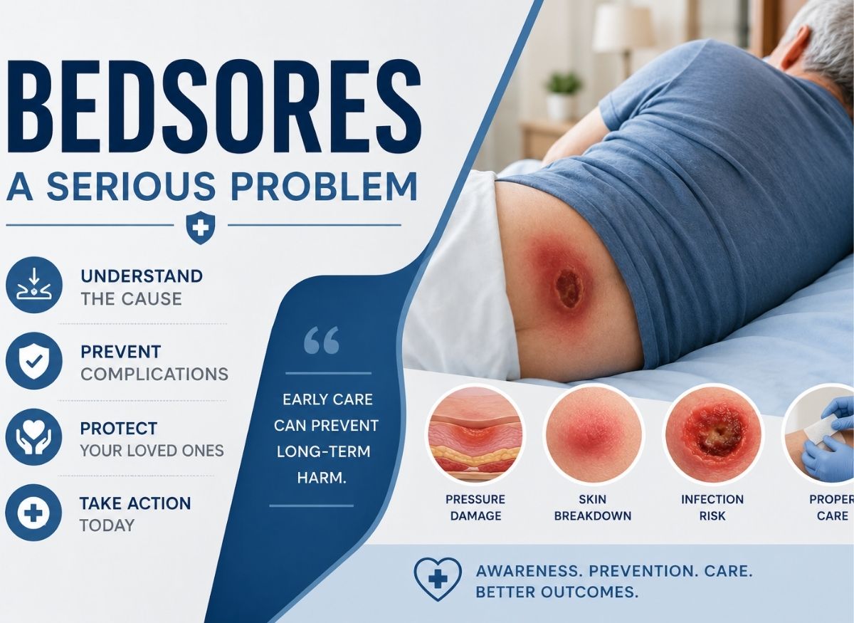
Bedsores, also called pressure ulcers or pressure injuries, can develop surprisingly fast when constant pressure cuts off blood flow to the skin. They often affect people who spend long periods in bed, use wheelchairs, or have limited mobility due to illness, injury, or aging. They may start as mild redness or tenderness, but they can quickly become painful open wounds that are hard to heal if ignored.
The good news is that most bedsores are preventable with early awareness, consistent skin care, and simple daily habits that take pressure off vulnerable areas. Knowing what causes them is one of the most effective ways to stop them before they start. Bedsores commonly form when pressure, friction, moisture, or poor circulation damage the skin over bony areas such as the hips, tailbone, heels, and elbows.
What Are Bedsores?
Bedsores, also known as pressure ulcers, pressure sores, or pressure injuries, are areas of damaged skin and underlying tissue caused by prolonged pressure on the body. They usually develop when someone stays in one position for too long, which reduces blood flow to the skin and keeps tissues from getting enough oxygen and nutrients. Over time, this lack of circulation can damage the skin and create painful wounds that range from mild redness to deep open sores.
Bedsores most often form over bony areas with little natural cushioning, such as the tailbone, hips, heels, ankles, elbows, and shoulder blades. People who spend long periods in bed, use wheelchairs, or have limited mobility due to illness, surgery, or aging are at higher risk. Although bedsores can become serious if left untreated, they are often preventable with proper repositioning, skin care, and early attention to warning signs.
Medical professionals generally classify bedsores into stages based on severity, starting with mild skin discoloration and progressing to deeper wounds that may reach muscle or bone in severe cases. Recognizing pressure injuries early matters because prompt treatment can help prevent infection, pain, and long-term complications.
The Signs of Infected Bedsores
Spotting an infection in a pressure ulcer is a high-priority medical matter, particularly for people who also live with chronic venous insufficiency (CVI). In someone with venous dysfunction, the legs are often already swollen and discolored, which can hide the early redness of a spreading infection. Because chronic venous insufficiency with mild cellulitis is a common complication, telling the difference between stasis changes and an active, aggressive infection in a bedsore is vital for long-term outcomes.
Local Indicators of Wound Infection
The most immediate signs of infection appear in and around the wound itself. A healing venous ulcer may produce some clear or watery fluid, but an infected ulcer will start producing pus. This drainage is usually thicker and can range in color from creamy yellow to dark green.
Another local sign is spreading redness. In people with CVI, the skin is often permanently stained a brownish red. An infection, however, causes a new, brighter red halo that feels warm and tender. If this redness extends more than 2 cm from the wound edge, the infection may have moved into the deeper skin layers and become cellulitis. A foul odor that lingers even after the wound has been cleaned is another hallmark of bacterial overgrowth, particularly from bacteria that thrive in the deep cavities of Stage 3 or Stage 4 ulcers.
Systemic Red Flag Symptoms
When an infection moves from the local tissue into the bloodstream, it becomes a whole-body threat. This is especially dangerous for elderly patients or those whose immune systems may be weakened.
- Fever and Chills: A body temperature above 38°C (100.4°F) is a primary sign that the body is fighting an infection.
- Mental Status Changes: In older adults, new confusion or unusual sleepiness is often the first sign of sepsis (blood poisoning) from an infected bedsore.
- Elevated Heart Rate: A rapid heart rate can indicate that the body is under significant physiological stress from a bacterial overgrowth.
Complications: Cellulitis and Osteomyelitis
If an infection isn’t addressed with aggressive treatment and targeted antibiotics, it can invade the underlying structures. In Stage 4 ulcers where bone is exposed, there’s a high risk of osteomyelitis, a bone infection. This condition is notoriously difficult to treat and often requires months of intravenous antibiotics.
In addition, when venous disease affects the body’s circulation broadly, lymphatic drainage is already sluggish. This backup makes it easier for bacteria to spread, potentially leading to necrotizing fasciitis or sepsis, both of which are life-threatening emergencies.
7 Main Causes of Pressure Ulcers
Bedsores are primarily mechanical injuries caused by external forces, but their development is closely tied to a person’s underlying vascular health. For people with chronic venous insufficiency, the risk of tissue breakdown is significantly higher. Poor return of blood to the heart creates a state of chronic nutritional shortage in the legs. When venous dysfunction is present, the tissues are already saturated with metabolic waste and short on oxygen, making them far less resilient to the primary causes of pressure ulcers.
Sustained Pressure and the Ischemic Cascade
The most direct cause of a bedsore is sustained pressure, which collapses the tiny blood vessels and starves tissue of oxygen. In a healthy person, the body signals discomfort, prompting a change in position. However, in patients with CVI symptoms like severe swelling, the extra fluid in the tissues creates a cushion of stagnant fluid that actually raises internal tissue pressure.
This makes the skin over the heels or tailbone more susceptible to tissue death because the venous system is already struggling to clear fluid. When external pressure is added, the remaining blood flow is cut off entirely. In conditions like Hansen’s disease, where nerve damage prevents pain from being felt, a similar painless breakdown occurs. In CVI, the breakdown is driven by the toxic environment of oxygen-poor blood and high venous pressure.
The Mechanical Threat: Friction Versus Shear
Friction acts as a surface abrasive, stripping the top layer of skin much like a sheet burn, but shear is a far more destructive internal force. For patients with CVI, the skin is often already compromised by lipodermatosclerosis, a hardening of the skin. This leathery skin has lost its natural elasticity, making it brittle.
When a patient slides down in bed, the skeleton moves but the skin stays stuck to the linens. Because the skin is stiff from venous dysfunction, the underlying blood vessels are more easily kinked or torn. This deep tissue injury is especially dangerous in advanced stages of venous disease, where the skin is thin and waxy, since the damage can occur deep near the bone before any visible signs appear on the surface.
Moisture and Skin Maceration
Excess moisture from sweat, wound drainage, or incontinence leads to maceration, where the skin becomes waterlogged and soft. This is a critical risk factor for those with venous ulcers, since these wounds often weep significant amounts of fluid.
Macerated skin is functionally weak; it’s five times more likely to tear under pressure than dry skin. Combine this moisture with the inflammation seen in CVI with mild cellulitis, and the skin’s barrier is virtually gone. Managing moisture is a fundamental part of treatment, since it prevents a manageable area of stasis from turning into a life-threatening, full-thickness pressure ulcer.
Nutritional Deficit and Dehydration
The skin needs a constant supply of protein, vitamin C, and zinc to maintain its structure. In patients with venous deficiency, the delivery of these nutrients is impaired by the backup of stagnant blood in the lower extremities. Dehydration makes things worse by reducing overall blood volume, which aggravates deep venous reflux and makes the blood thicker and harder to move.
A malnourished patient lacks the collagen needed to repair the minor tears caused by friction. As a result, long-term outlook can be affected not just by the vein disease itself, but by the secondary infections that take hold in these non-healing, nutrient-starved pressure wounds.
How Often Should a Person Be Repositioned to Prevent Skin Breakdown?
The frequency and technique of repositioning are the most critical defenses against bedsores. For patients with CVI, these intervals matter even more. Because poor venous return already keeps tissues low on oxygen, any added interruption in blood flow from external pressure can trigger rapid tissue death. With venous dysfunction, the skin is less forgiving than healthy tissue, meaning a delay in turning can quickly escalate toward a full-thickness wound.
The Two-Hour Rule and Reperfusion
For someone confined to bed, the medical standard is repositioning at least every two hours. This schedule counteracts poor venous return by making sure no single area of skin is deprived of oxygen for too long. By rotating the patient between their back and sides (the 30-degree side-lying position), caregivers allow a temporary rush of oxygenated blood back into the tissues that were just under pressure.
In patients with CVI, this reperfusion is essential because the stagnant blood already in the legs contains metabolic waste that must be flushed out by fresh arterial flow. If the two-hour window is missed, the combination of external pressure and internal venous stasis creates a zone of tissue death where the skin simply gives up and breaks down.
Wheelchair Frequency and Micro-Shifts
People in wheelchairs face higher pressure concentrated on a smaller surface area, specifically the sitting bones. As a result, repositioning needs to happen more often, every 15 to 30 minutes.
Even when a full transfer isn’t possible, small shifts can be a lifesaver for the skin. Leaning side to side or forward, or doing a pressure-relief push-up for 10 to 15 seconds, lets the tiny blood vessels refill. For someone with CVI symptoms like leg heaviness, these shifts also help keep blood from pooling in the pelvis and upper thighs, which can worsen deep venous reflux.
Using “Lift, Don’t Drag” to Prevent Shear
The technique used to move a person is just as important as how often it’s done. Caregivers should always lift the patient using a draw sheet or slide board rather than dragging them across the linens. Dragging creates friction, but more dangerously, it creates shear, an internal tearing of the blood vessels.
Since CVI often leads to leathery, hardened skin, the tissue is brittle and lacks the slip of healthy skin. This makes it far more prone to deep tissue injury during poor transfers. Keeping the head of the bed at or below 30 degrees further reduces the risk of the patient sliding down, which is a primary source of shear on the tailbone.
Strategic Off-Loading with Pillows and Wedges
Proper repositioning also involves bridging or off-loading bony areas. Pillows or foam wedges should be placed between the knees and ankles to prevent kissing sores, pressure ulcers that form where two bony areas press against each other.
For those with venous ulcers, specialized heel-suspension boots are used to keep the heels completely off the mattress. This is vital because the heels are a high-risk zone where venous dysfunction and pressure often meet to create non-healing wounds, which carry a high risk of bone infection and can affect long-term outcomes.
Most Effective Ways to Prevent Bedsores
Preventive skin care and pressure redistribution are the two most effective strategies for preventing bedsores, especially in people with CVI. Because venous disease compromises the skin’s natural barrier and blood supply, the standard inspect-and-protect routine has to be even more rigorous. When venous dysfunction is present, the skin is already stressed by internal fluid pressure, making it less resilient to moisture and sustained contact.
Daily Skin Inspection: The Early Warning System
The most important practice is a daily, systematic head-to-toe inspection. For people with CVI symptoms, it should focus heavily on the ankle area and heels. The primary warning sign to look for is non-blanchable redness, a red spot that stays red even when you press on it. In healthy skin, the spot would turn white as blood is pushed out and then turn red again as it refills.
If it doesn’t turn white, the tiny blood vessels are already damaged, and this is a Stage 1 pressure ulcer. Because conditions like Hansen’s disease involve nerve damage, a patient may not feel this injury occurring; similarly, in CVI, the heavy, woody texture of the skin can hide these early changes. Catching shifts in temperature or firmness early is the only way to keep a minor spot from becoming a deep chronic ulcer.
Gentle Cleansing and the Moisture Barrier
For patients with venous deficiency, the skin is often dry, thin, and prone to venous eczema. Cleansing should be done with lukewarm water and a pH-balanced cleanser to avoid stripping away protective oils.
If the patient also deals with incontinence, the skin is at extreme risk. Urine and feces contain enzymes that digest the skin, leading to incontinence-associated dermatitis. For these patients, treatment should include a thick moisture barrier cream containing zinc oxide or dimethicone. This creates a physical shield, making sure moisture from weeping venous fluid or incontinence doesn’t macerate the skin and lead to breakdown.
Static and Dynamic Support Surfaces
To manage the mechanical side of the problem, specialized support surfaces are used to spread out body weight. For those at moderate risk, static surfaces like high-specification foam or gel overlays work by enveloping the body. By increasing the surface area in contact with the bed, these surfaces lower the pressure at any single point, like the hip bone.
For high-risk individuals, dynamic surfaces like alternating pressure air mattresses are used. These systems use a pump to cyclically inflate and deflate different air cells so the pressure is constantly moving. This is particularly helpful for patients who can’t move themselves, as it provides a mechanical effect that mirrors the relief of manual turning.
Specialized Heel Protection
The heels are one of the most vulnerable spots for anyone with venous dysfunction. Because the heel has very little padding, poor venous return often results in severe swelling that cuts off blood flow to the skin over the bone. Floating the heels with specialized foam boots or heel-suspension devices is a best practice. These devices keep the heel suspended in the air, removing all pressure. This is a vital part of protecting long-term health, since a heel ulcer that reaches the bone is a frequent cause of hospitalization and severe decline.
Diet Is Important in Bedsore Prevention
Diet serves as the internal scaffolding for skin health, especially when circulation is compromised by CVI. While venous dysfunction creates a backup of stagnant blood in the lower extremities, proper nutrition makes sure the blood that does reach the tissues is rich in the materials needed to repair and protect the skin. Where tissues are already low on oxygen due to deep venous reflux, a nutritional deficit can speed up the transition from healthy skin to chronic ulcers.
The Protective Power of Protein and Calories
Protein is the primary raw material for collagen, the glue that holds skin cells together. For people with poor venous flow, the skin is often under constant tension from swelling. Without enough protein, typically recommended at 1.25 to 1.5 grams per kilogram of body weight, the skin becomes thin and fragile, losing its ability to resist the friction and shear forces that cause bedsores.
Maintaining enough calories is also vital. If someone loses weight rapidly, they lose the fat padding that acts as a natural shock absorber over bony areas like the tailbone and heels. When this padding disappears, the pressure on the tiny blood vessels increases significantly, making a pressure ulcer almost inevitable.
Micronutrients: The Repair Crew
Specific vitamins and minerals act as the engineers of skin repair. Because venous disease can cause chronic inflammation, the demand for these nutrients increases.
- Vitamin C: Essential for strengthening the walls of small blood vessels. In CVI, where these vessels are already prone to leaking (causing symptoms like brown staining), vitamin C helps maintain vascular integrity.
- Zinc: Crucial for cell division. If a minor tear occurs near the ankle, zinc is needed to close the wound before it expands into a full ulcer.
- Vitamin A: Supports the health of the cells that form the outermost protective layer of the skin.
Hydration and Tissue Perfusion
Hydration is perhaps the most overlooked part of a management plan. Water is the medium that carries oxygen and nutrients to the skin. When a patient is dehydrated, blood volume drops and the blood becomes thicker. This makes it even harder for the venous system to push blood back up the legs, worsening the underlying problem.
Dehydrated skin also loses its turgor, the ability to snap back when pinched. This makes the skin sticky, which significantly increases the risk of damage from friction against bed linens. Maintaining a fluid intake of 1.5 to 2 liters per day (unless restricted due to heart or kidney issues) is a fundamental part of preventing the skin breakdown that can lead to complications like sepsis.
How Are Bedsores Diagnosed and Staged?
The diagnosis and staging of bedsores are formal clinical processes that go beyond simple observation to identify the exact depth of tissue damage. For a patient with CVI, staging is crucial because venous dysfunction can mask the severity of a pressure injury. The woody hardness and dark pigmentation typical of advanced venous disease can make it difficult to spot the redness of a Stage 1 ulcer, potentially delaying vital treatment.
Understanding the Stages of Tissue Destruction
The staging system is a progressive scale that measures how far the damage has traveled through the layers of skin and the underlying fat, muscle, or bone.
Stage 1: The skin is intact but shows non-blanchable redness, a critical warning sign where the redness doesn’t fade when pressed. In someone with CVI, this may be mistaken for simple stasis dermatitis, but its localized position over a bone, like the ankle, confirms it as a pressure injury.
Stage 2: This involves partial-thickness loss. The top layers of skin are broken, often appearing as a shallow pink wound or a clear, fluid-filled blister.
Stage 3: This is full-thickness skin loss. The wound craters down into the fatty tissue. While fat may be visible, deeper structures like muscle are still protected.
Stage 4: This is the most severe stage. Bone, tendons, or muscles are exposed or can be felt directly. This stage carries a high risk of systemic sepsis and bone infection, significantly worsening the outlook.
Complex Classifications: Unstageable and Deep Tissue Injury
Sometimes a wound can’t be staged right away. An unstageable ulcer occurs when the wound bed is covered in slough (yellow or tan dead tissue) or eschar (a black, leathery scab). Until this dead tissue is removed, a doctor can’t see how deep the wound really goes.
A deep tissue injury is perhaps the most deceptive. The skin may be intact, but it appears purple or maroon, like a deep bruise. This indicates that the tissue has died from the inside out, starting at the bone and moving toward the surface. For someone with deep venous reflux, the dark staining of the skin can make this injury very hard to spot until the skin suddenly breaks open to reveal a deep cavity.
The Braden Scale: Quantifying the Risk
The Braden Scale is the primary tool used to predict the likelihood of skin breakdown. It assigns a score from 6 to 23; the lower the score, the higher the risk.
- Sensory Perception: Evaluates whether the patient can feel the pain of pressure. This is vital for those with nerve damage from conditions like Hansen’s disease, or where CVI has led to peripheral neuropathy.
- Moisture: Measures exposure to sweat or weeping from venous ulcers.
- Activity and Mobility: Determines whether the patient can move themselves to relieve pressure.
- Nutrition: Assesses whether the body has enough protein to maintain the skin barrier.
- Friction and Shear: Looks at the risk of the skin tearing when the patient is moved.
By combining the Braden Scale with visual staging, healthcare providers can create a proactive plan. If a patient has a low Braden score and a Stage 1 ulcer, they may be placed on an alternating pressure mattress immediately to keep the wound from reaching Stage 3 or 4.
The Difference Between Bedsores and Moisture-Associated Skin Damage
Telling a pressure ulcer apart from moisture-associated skin damage (MASD) is a critical skill for anyone managing CVI, since the treatments for these conditions are fundamentally different. While both involve skin breakdown, a pressure ulcer is a bottom-up injury caused by restricted blood flow, whereas MASD is a top-down chemical and mechanical erosion of the skin surface.
Pathophysiology: Pressure vs. Chemical Erosion
The primary difference lies in the source of the damage. A pressure ulcer is caused by ischemia; external pressure collapses the tiny blood vessels, starving the tissue of oxygen. This is particularly dangerous in patients with venous dysfunction, where the tissue is already compromised.
MASD, which includes incontinence-associated dermatitis, is instead caused by the breakdown of the skin’s natural protective acidity. When skin is trapped against moisture such as urine, sweat, or weeping from venous ulcers, it becomes macerated. This makes the skin soft and permeable, allowing bacteria and caustic enzymes to digest the top layer.
Identifying by Location and Shape
The where and what of the wound provide the most useful diagnostic clues.
Bedsores: These are almost always found directly over a bony prominence such as the tailbone, heels, or ankles. They tend to have well-defined borders and often take a circular or crater shape that mirrors the point of pressure.
MASD: These usually occur in skin folds or fleshy areas like the groin and inner thighs. The redness is typically diffuse and irregular, looking more like a sprawling rash or sunburn than a deep hole.
Depth and Tissue Characteristics
The depth of the wound is a major differentiator. Pressure ulcers can be quite deep, reaching fat, muscle, or bone (Stages 3 and 4). Because the injury starts deep and moves outward, the surrounding skin might actually look healthy until the ulcer breaks through.
MASD is strictly a superficial, partial-thickness injury. It rarely goes deeper than the very top layers of the skin. However, the skin will look macerated, meaning white, soggy, and wrinkled, and may show satellite lesions, which are small red bumps that indicate a secondary fungal infection has taken hold in the damp environment.
Conclusion
Bedsores are more than a skin problem. They can seriously affect comfort, mobility, and overall health if they’re allowed to worsen. The encouraging part is that prevention often comes down to small, consistent actions such as changing positions regularly, keeping skin clean and dry, eating well, and checking vulnerable areas for early warning signs. Even people at higher risk can greatly reduce their chances of developing pressure ulcers when caregivers and loved ones stay proactive. Paying attention early can make a meaningful difference and help prevent painful complications later on.
FAQ About Bedsores
How quickly can bedsores develop?
Bedsores can develop faster than many people realize, sometimes within hours if pressure on the skin is severe and blood flow becomes restricted. People with limited mobility, poor circulation, or fragile skin may be especially vulnerable. The earliest signs often include redness, tenderness, warmth, or skin discoloration that doesn’t fade after pressure is removed. Catching these warning signs early can help stop a sore from becoming more serious.
What are the first signs of a bedsore?
The first signs of a bedsore usually include skin that looks red, purple, or darker than usual and doesn’t return to normal color after pressure is relieved. The area may feel warm, firm, soft, painful, or unusually tender to the touch. In darker skin tones, discoloration may be harder to spot, so texture and temperature changes become especially important. Regular skin checks can help identify these signs before the skin breaks open.
Can bedsores heal on their own?
Very mild pressure sores may improve if pressure is relieved early and the skin remains protected, but deeper sores usually require medical attention. Without proper care, bedsores can worsen, become infected, and sometimes lead to serious complications. Treatment may include repositioning, wound dressings, pressure-relieving surfaces, nutrition support, and infection prevention. Seeking help early gives the best chance for healing and reduces the risk of long-term damage.
What is the best way to prevent bedsores?
The best prevention strategy combines frequent movement, pressure relief, skin care, and good nutrition. Experts often recommend repositioning every couple of hours for people confined to bed and checking the skin daily for early changes. Keeping skin clean and dry, using pressure-relieving cushions or mattresses, and eating enough protein and nutrients can also help maintain healthy skin. Prevention works best when caregivers and patients stay consistent with these habits.
Are bedsores dangerous?
Yes, bedsores can become dangerous if they’re not treated early. Mild sores may stay near the surface of the skin, but advanced pressure ulcers can extend into muscles and even bone, increasing the risk of infection. In severe cases, untreated infections can spread and become life-threatening. This is why early detection and prevention are so important, especially for older adults or people with limited mobility.