4 Main Symptoms of Paget’s Disease of the Bone
Paget’s disease of the bone is called uncommon, but it is not rare enough to ignore. In the United States, roughly 1% to 2% of adults over 55 are thought to have it, and the odds rise with age. Many people never know, because it progresses slowly over years. Often it turns up by chance on an imaging test or blood test ordered for another reason.
The disease disrupts bone remodeling. Healthy bone is constantly broken down and rebuilt in a balanced cycle. In Paget’s disease, bone is broken down too fast, so the body rebuilds it in a hurry. The new bone is larger, weaker, and more disorganized than normal, which can lead to pain, deformity, fractures, arthritis, and hearing problems, depending on the bones involved.
The early signs are easy to brush off. A lingering ache gets blamed on age, stiff joints look like ordinary arthritis, and a slightly bowed leg or bigger hat size changes so slowly that nobody notices. By the time symptoms are obvious, the bone may already have changed a lot.
Catching it early helps. There is no cure, but treatment can slow the abnormal remodeling, ease discomfort, and reduce long-term complications. Below are four main symptoms, why they happen, and when to talk to a healthcare provider.
4 Main Signs and Symptoms of Paget’s Disease of the Bone
Bone Pain
Bone pain is the most common symptom, affecting most people who have symptoms at all. The rapid, chaotic remodeling raises metabolic activity and blood flow in the affected bone, producing a deep, steady ache.
Osteoarthritis pain usually gets worse with activity and better with rest. Paget’s pain can be constant and may get worse at night or while resting. People describe it as a dull, deep, boring ache centered on the affected bone.
The location of the pain follows the bones involved:
- Pelvis: Pain in the hip or groin is common. It is often mistaken for hip arthritis or bursitis, and it can make walking or long periods of standing difficult.
- Spine: Persistent back pain is typical. If enlarged vertebrae press on nearby nerve roots, the pain can radiate (radiculopathy).
- Thigh bone (femur) or shin bone (tibia): Aching in the legs, often worse with weight-bearing.
- Skull: Less commonly, it causes headaches from increased blood flow and bone enlargement.
It also matters to work out where the pain comes from. It can come from the active disease itself, from microfractures (tiny cracks) in the weakened bone, or from secondary osteoarthritis in nearby joints, caused by altered mechanics and deformed bone.
Bone Deformities
Paget’s disease can cause significant, sometimes disfiguring changes in bone shape. They come straight from the disordered remodeling at the heart of the disease.
Osteoclasts break bone down aggressively while osteoblasts hurry to replace it with weaker, disorganized woven bone. The affected bone thickens, enlarges, and changes shape. It loses the strong, layered (lamellar) structure of healthy bone and with it much of its mechanical strength. These changes usually progress slowly and become more obvious over time, especially without treatment.
- Long leg bones: The femur or tibia can weaken and bow under body weight, giving a bowlegged (genu varum) or knock-kneed (genu valgum) look. Bowing also changes the gait and strains the hips and knees, which can lead to secondary osteoarthritis.
- Skull: The skull can slowly enlarge, so hat size may increase. A typical sign is frontal bossing, a more prominent forehead. The head may feel heavier and headaches may occur.
- Spine: Vertebrae can enlarge and weaken, leading to compression fractures or kyphosis, an exaggerated forward rounding of the back. This can cause chronic back pain, loss of height, and posture problems.
- Other bones: An enlarged collarbone may become visible, and jawbone enlargement can cause dental problems.
Fractures
Fractures happen because the new bone formed during chaotic remodeling is disorganized, brittle, and much weaker than normal bone. Healthy bone has a layered structure that gives it both strength and flexibility.
In Paget’s disease, osteoblasts rapidly lay down woven bone, with collagen fibers arranged in a haphazard, mosaic-like pattern. That makes the bone more porous and much more likely to break under stress a healthy bone would handle easily. It is often compared to chalk: hard but brittle.
Several factors contribute:
- Poor bone quality: Pagetic bone cannot spread mechanical force well, so fractures can happen spontaneously or after very minor trauma.
- Bowing: A bowed bone, such as the femur, concentrates stress. The outer (convex) side is under constant tension, which makes it a common site for incomplete stress or fissure fractures. These small cracks cause pain and can progress to a complete break if left unmanaged.
- Rich blood supply: Pagetic bone is highly vascular. This may sound helpful, but it further weakens the structure and can cause heavy bleeding during fracture surgery.
A classic injury is the chalkstick fracture, a clean, straight break across a bowed bone, usually the femur, like snapping a piece of chalk. These fractures can be slow to heal and often need surgery, such as intramedullary nailing, to keep the bone aligned and stable.
Neurological Symptoms
Neurological symptoms occur when overgrown bone squeezes nearby nerves, the brain, or the spinal cord. As Pagetic bone expands, it can narrow the openings (foramina) that nerves pass through in the skull and spine, or press directly on the spinal cord. These are among the more serious complications, and the symptoms depend on which bone is involved.
- Skull involvement: Enlarging bone can compress cranial nerves. The most common neurological symptom is hearing loss, caused by compression of the auditory nerve (cranial nerve VIII), damage to the inner ear structures, or crowding of the middle ear. Less commonly it causes vertigo, vision problems (if the optic nerve is compressed), or facial nerve palsy. Headaches are also frequent.
- Spinal stenosis and myelopathy: Enlarged vertebrae can narrow the spinal canal and press on the spinal cord. Symptoms include difficulty walking, balance problems, and bowel or bladder dysfunction.
- Radiculopathy: More often, overgrown vertebrae press on nerve roots, causing pain, tingling, numbness, or weakness that travels into the arms or legs, such as sciatica.
- Peripheral nerves: Rarely, enlarged pelvic bones can trap nerves running to the legs, causing symptoms similar to spinal nerve compression.
These symptoms usually develop gradually and are confirmed with imaging such as MRI or CT. Treatment may include medication to control the disease and, in severe cases, surgery to relieve pressure on the nerves.
What is Paget’s Disease of the Bone?
Paget’s disease of bone is a chronic metabolic disorder that disrupts normal bone renewal, leaving bones enlarged, misshapen, and weak. It is the second most common metabolic bone disease after osteoporosis.
Normally, bone remodeling is a lifelong process in which old bone is broken down (resorbed) and replaced with new bone. In Paget’s disease, this process becomes disorganized and overactive in localized areas of the skeleton. The resulting bone is highly vascular, less compact, more fragile, and more prone to pain, deformity, and fracture.
The disease can affect one bone (monostotic) or several (polyostotic). It does not spread from bone to bone. The affected sites are generally set early in the course of the disease.
How Does Paget’s Disease Disrupt The Normal Bone Remodeling Process?
Paget’s disease upsets the balance and pace of the two main bone cell types: osteoclasts, which break bone down, and osteoblasts, which build it. It usually unfolds in three overlapping phases, all starting with overactive osteoclasts.
Phase 1: The Lytic Phase (Osteoclastic Activity)
Osteoclasts increase sharply in number and activity. In Paget’s disease they are abnormally large with multiple nuclei, and they break down bone at an accelerated rate. This leaves areas of reduced bone density, seen on X-rays as V-shaped patterns in long bones or well-defined clear areas in the skull (osteoporosis circumscripta).
Phase 2: The Mixed Phase (Lytic and Blastic Activity)
The body tries to make up for the loss by forming new bone quickly, and osteoblast activity surges. But the process is hurried and disorganized, and collagen is laid down in a haphazard woven pattern instead of strong layers. Breakdown and formation both run at a very high rate at the same time, and the bone becomes highly vascular.
Phase 3: The Sclerotic Phase (Osteoblastic Activity)
Eventually bone formation outweighs breakdown, but the bone is still abnormal. It becomes thick, dense, and enlarged, and looks sclerotic (hardened) on X-ray. Despite that dense look, this “Pagetic” or “mosaic” bone is weak and brittle, with a chaotic pattern of cement lines where normal structure should be.
Affected Bones By This Condition
Paget’s disease favors bones with high blood flow and turnover: the pelvis, spine, femur, skull, and tibia. It is usually asymmetric, so it might affect the right femur but not the left. It can occur in any bone, but this pattern explains many of its classic signs. The distribution is normally established early and doesn’t spread to new bones later in life.
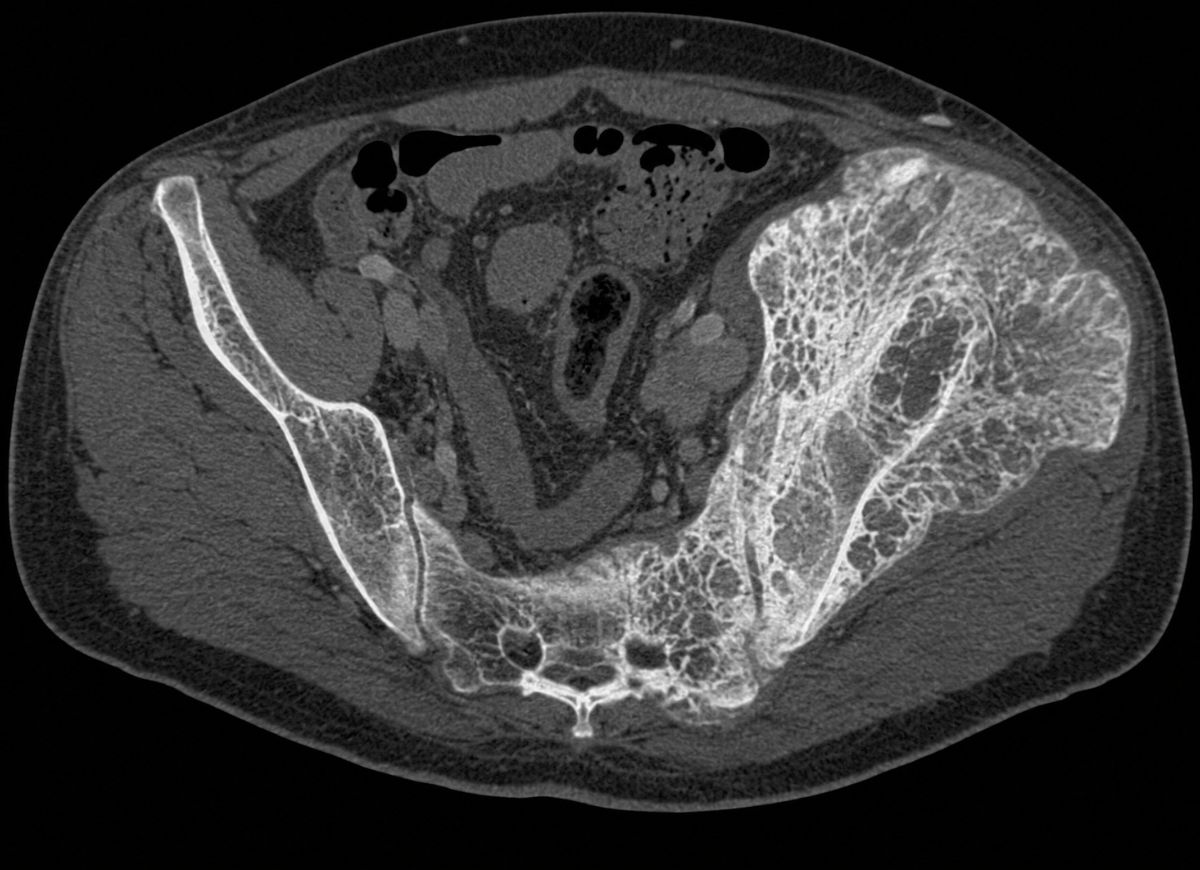
- Pelvis: The most common site, involved in over 70% of cases. It is often symptom-free but can cause hip pain that is mistaken for arthritis. Because it sits next to the hip joint, deformity can change joint mechanics and lead to secondary osteoarthritis.
- Spine: The lower (lumbar) and mid (thoracic) back are common sites. Enlarged vertebrae can cause back pain, stiffness, and kyphosis. More seriously, they can compress nerve roots or the spinal cord, causing sciatica or spinal stenosis.
- Femur: The most commonly affected long bone. Because it carries so much weight, involvement is a particular concern. It can bow, alter the gait, strain the hip and knee, and is a frequent site of chalkstick fractures.
- Skull: Can cause a larger head, frontal bossing, and headaches. The most significant complication is hearing loss from auditory nerve compression or inner ear damage.
- Tibia: Like the femur, it is a weight-bearing bone prone to bowing, which can change alignment and raise the risk of knee and ankle pain and arthritis.
Causes and Risk Factors for Paget’s Disease of the Bone
The exact cause is not fully understood. It appears to be multifactorial: a strong genetic predisposition, certain environmental triggers, and advancing age. Current research points to an interplay between a person’s genes and one or more outside factors that may set the disease off over time. The fact that cases cluster in families and certain regions supports this gene-environment model. Knowing the risk factors helps identify people at higher risk and guides future research on prevention and treatment.
Gene
A family history is one of the biggest risk factors. Up to 40% of people with Paget’s disease have at least one first-degree relative (parent, sibling, or child) with it. In these families the pattern is usually autosomal dominant, so a child of an affected parent has a 50% chance of inheriting the faulty gene.
The SQSTM1 gene makes a protein involved in regulating inflammation and the life cycle of osteoclasts. Mutations in it appear in up to 50% of familial cases and in a smaller share of sporadic (non-familial) cases. The protein is part of the RANK signaling pathway, the master regulator of osteoclast formation and activity. Mutations are thought to make this pathway overly responsive, producing the abnormally large, numerous, overactive osteoclasts typical of the disease.
Other genetic loci have also been identified, so the genetic picture is complex and likely involves several genes interacting with each other and with the environment. Finding them helps researchers understand the disease and develop more targeted treatments.
Age
Risk rises steadily with age. Paget’s disease is extremely rare under 40, begins to climb after that, and is most common over 55. Prevalence in people over 55 may reach 2% to 3% and keeps increasing in older groups. This suggests a long-latency process, in which genetic susceptibility plus a lifetime of environmental exposure is needed to trigger it.
One prominent theory is that a slow-acting virus contracted early in life is the trigger. The leading candidate for many years has been a paramyxovirus, such as measles or canine distemper virus. The idea is that viral particles lie dormant in bone cells for decades and, in genetically susceptible people, later activate abnormal osteoclast behavior as the immune system changes with age. Viral genetic material has been found in Pagetic osteoclasts, but a causal link has not been proven.
Another possibility is that aging itself allows acquired (somatic) mutations to build up in bone precursor cells, eventually disrupting remodeling in localized areas.
Ancestry and geography also play a part. The disease is most common in people of Anglo-Saxon descent, in places like the UK, Australia, New Zealand, and the US, and rare in people from Asia and Africa. This suggests migrating populations carried the predisposing genes with them, and that the disease appears later in life, possibly after exposure to local environmental factors.
Potential Complications of Untreated Paget’s Disease of the Bone
Untreated or severe Paget’s disease can affect more than the skeleton.
- Bowing and fractures: The enlarged but weak bone is prone to bending and breaking, especially in the femur and tibia.
- Joint damage: Distorted bone speeds up cartilage wear and can cause severe osteoarthritis, particularly in the hip and knee, with chronic pain, stiffness, and reduced mobility that may eventually require joint replacement.
- Heart strain: Pagetic bone needs a lot more blood. In extensive disease, the heart may have to work harder, potentially leading to high-output heart failure.
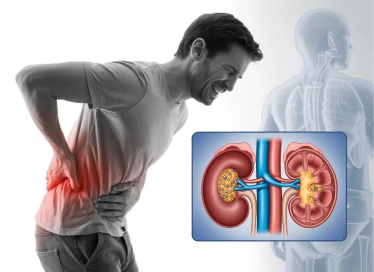
- Metabolic problems: If a person is immobilized, high blood calcium (hypercalcemia) can develop, raising the risk of painful kidney stones.
- Paget’s sarcoma: In fewer than 1% of patients, affected bone can become cancerous. This is extremely rare but the most serious complication, and it is a very aggressive bone cancer.
The Difference Between Paget’s Disease and Osteoporosis
Both raise fracture risk, but they differ in cause, presentation, and scope.
- What happens to the bone: Paget’s disease involves chaotic, accelerated breakdown and rebuilding, producing disorganized, enlarged, weak bone. In osteoporosis, breakdown simply outpaces formation, causing a net loss of bone mass and density.
- Scope: Paget’s disease is localized, affecting specific bones such as the pelvis, skull, spine, and long bones. Osteoporosis is systemic and affects the whole skeleton.
- Appearance: In Paget’s, bones become enlarged and deformed. In osteoporosis, they become thinner and more porous without a major change in shape or size.
- Blood markers: Paget’s disease is linked to notably high serum alkaline phosphatase (ALP), a marker of heavy bone turnover. Markers may be mildly raised in osteoporosis but don’t reach those levels.
Paget’s Disease of the Bone Management
Treatment aims to control the abnormal remodeling, relieve symptoms, stop progression, and lower the risk of complications. It is usually recommended for people with symptoms (pain, nerve compression, deformity) and for people without symptoms whose disease is in high-risk sites like the skull or long bones.
The cornerstone is bisphosphonates, drugs that bind to bone and inhibit osteoclasts. By slowing excess breakdown, they interrupt the chaotic cycle and allow more normal, organized bone to form. Intravenous options like zoledronic acid are often preferred for their potency and lasting effect, and a single infusion can keep the disease in remission for years. Oral options such as alendronate or risedronate also work well.
Supportive care covers symptoms and function:
- Pain relief: Over-the-counter acetaminophen or NSAIDs are commonly used for pain from the disease or secondary osteoarthritis.
- Physical therapy: Therapists can improve muscle strength, joint mobility, and balance to reduce the risk of falls and fractures, and can recommend canes or walkers for safety and independence.
- Surgery: May be needed for fractures (internal fixation), severe joint damage (hip or knee replacement), or deformities that compress nerves.
Three Phases of Paget’s Disease of the Bone
At the cellular level, Paget’s disease moves through three sequential phases, reflecting shifts in activity between osteoclasts and osteoblasts.
1. Lytic phase. Osteoclasts surge in number and activity and break down bone at a rapid rate. This creates wedge-shaped lesions or areas of demineralization on X-rays and leaves weakened bone. This phase often causes the first bone pain, raises the risk of pathological fractures, and releases a lot of calcium into the bloodstream.
2. Mixed phase. Breakdown and formation now happen at the same time. The body sends osteoblasts to repair the damage, but the repair is rushed and disorganized. The matrix is laid down as mosaic-like woven bone instead of strong lamellar bone. Turnover is extremely high, leaving bone that is enlarged, highly vascular, and structurally unsound.
3. Sclerotic phase. Also called the “burned-out” phase. Formation now overtakes breakdown. Osteoclast activity settles down, but overactive osteoblasts keep adding dense, disorganized bone. It is heavily mineralized and looks very dense on X-ray, yet it remains brittle, architecturally weak, and prone to fracture and deformity.
FAQs
1. Is Paget disease painful?
It can be, but not always. Some people have no symptoms and only find out through routine blood tests or imaging. When pain does occur, it is usually a deep, persistent ache in the affected bone that may get worse at night or after physical activity. Complications such as arthritis, nerve compression, or fractures can make discomfort more noticeable. Proper treatment often relieves pain and slows the disease.
2. Who is most at risk for Paget’s disease?
Age is one of the strongest risk factors. It is most often diagnosed after 55, and the risk keeps rising with age. A close relative with the condition also raises the odds, pointing to a genetic role. It is reported more often in people of European ancestry and is slightly more common in men than in women.
3. What can be mistaken for Paget’s disease?
Several conditions cause similar symptoms, so testing is needed to tell them apart. These include osteoarthritis, osteoporosis, bone metastases from cancer, stress fractures, chronic back problems, and other metabolic bone disorders. Providers typically combine medical history, a physical exam, blood tests, X-rays, and sometimes bone scans.
4. How long can you live with Paget disease?
The disease itself usually doesn’t shorten life expectancy. Many people live full, normal lives, especially when it is caught early and managed well. Treatment focuses on reducing symptoms, preventing complications, and preserving mobility, and regular follow-ups let providers track disease activity and catch problems early.
5. What celebrity has Paget’s disease?
I’ve removed the celebrity claim from the original, because I couldn’t verify it. The source called Sir Anthony Hopkins “the late actor” and said he has spoken about Paget’s disease, but as far as I know he is alive and I’m not aware of any such statement. If you want to keep this FAQ, replace it with a verified name or a different question, such as “Can Paget’s disease be prevented?”
6. What kind of doctor treats Paget’s disease?
Care often starts with a primary care physician, who may refer you to a specialist. Depending on the symptoms and bones involved, that could be an endocrinologist, rheumatologist, or orthopedic surgeon. If there is hearing loss, skull involvement, or a neurological complication, an otolaryngologist or neurologist may join the care team.
Conclusion
Paget’s disease of the bone often develops so quietly that it goes unnoticed until complications appear. Knowing the main symptoms (persistent bone pain, enlarged or misshapen bones, joint problems, and nerve-related symptoms) can lead to earlier diagnosis and better treatment.
It can’t be cured, but current therapies can slow abnormal remodeling, ease symptoms, and lower the risk of fractures and other long-term problems. If you have ongoing bone pain or unexplained changes in the shape or function of a bone, don’t ignore them. A timely medical check-up can give you answers and help protect your bone health for years.