Cerebral Amyloid Angiopathy: Causes, Symptoms, and Brain Bleeding Risk
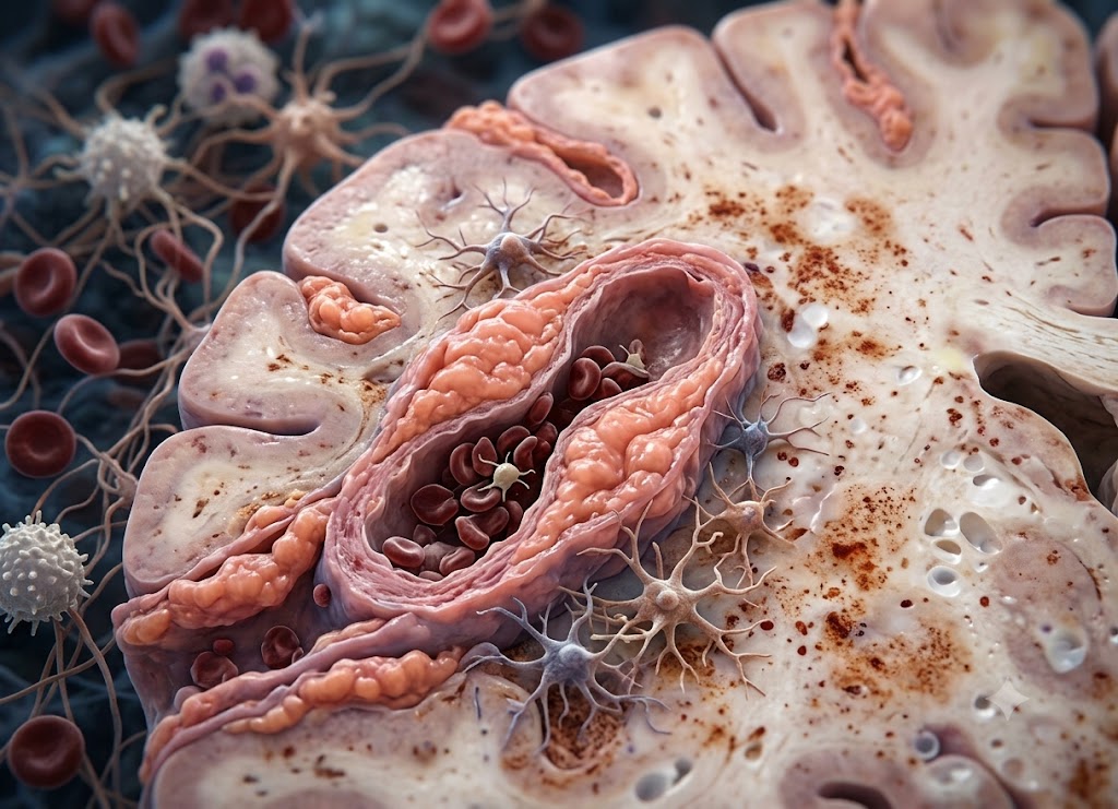
Cerebral amyloid angiopathy (CAA) is a condition in which amyloid protein collects in the walls of small and medium-sized blood vessels in the brain. Over time, these deposits make the vessels fragile and prone to leaking or bursting. It mostly affects older adults and is a leading cause of lobar intracerebral hemorrhage, meaning bleeding in the outer regions of the brain. It can also cause tiny bleeds, changes in thinking, and short stroke-like episodes.
CAA is worth understanding because the first clear sign is sometimes a brain bleed. Symptoms can include sudden weakness, a severe headache, confusion, vision problems, difficulty speaking, seizures, or changes in memory and thinking.
Some people are diagnosed when an MRI shows microbleeds or superficial siderosis. Others are diagnosed after a larger bleed. This article covers the causes, symptoms, and bleeding risk of CAA, and explains why early recognition and careful treatment matter.
Overview of Vascular Protein Accumulation
To understand CAA, it helps to look at how proteins build up in the brain. It is a distinct disorder of the brain’s blood vessels. Amyloid-beta protein gradually collects in the walls of small and medium arteries and capillaries in the cerebral cortex and the brain’s surface membranes (leptomeninges).
How it develops: amyloid-beta is produced → the brain fails to clear it → it deposits in vessel walls → the vessels weaken.
These insoluble deposits change the structure of the vessels, leaving them brittle and likely to rupture. This change is common in the aging brain and is a major cause of cognitive decline and spontaneous bleeding.
CAA also differs from Alzheimer’s disease. In Alzheimer’s, amyloid forms plaques within brain tissue. In CAA, it settles in the vessel walls. Some people have no obvious symptoms, while others develop serious complications, so knowing the warning signs is important for getting timely care.
Pathophysiology and Structural Vessel Damage
The root cause of CAA is a breakdown in the brain’s ability to clear waste. Amyloid-beta peptides accumulate in the middle and outer layers of brain vessels and gradually destroy their structure.
                    [Vascular Pathological Cascade]
                                   │
     ┌─────────────────────────────┴─────────────────────────────┐
     ▼                                                           ▼
[Smooth Muscle Displacement]                        [Fibrinoid Necrosis & Leaks]
 ├── Loss of structural elasticity                   ├── Severe degradation of tissue
 ├── Formation of microaneurysms                     ├── High risk of spontaneous rupture
 └── Impaired pressure autoregulation                └── Blood-brain barrier breakdown
The abnormal protein fibers push aside the healthy smooth muscle cells. The vessels can then no longer widen and narrow safely as blood pressure changes.
As CAA progresses, the damaged wall may be replaced by a weak, uniform material, a process called fibrinoid necrosis. Sometimes the buildup also triggers inflammation, known as CAA-related inflammation, which can cause sudden neurological changes.
Eventually the blood-brain barrier breaks down. Fluid leaks slowly into nearby tissue and causes changes in the deep white matter. Alternatively, a vessel may give way under ordinary pressure and cause a sudden stroke or lobar hemorrhage.
Sporadic vs. Familial Typologies
Doctors divide CAA into two groups by origin: the common sporadic form and the much rarer familial form.
[Sporadic Form]  ──► Linked to advancing age and APOE alleles; non-hereditary patterns
[Familial Form]  ──► Autosomal dominant mutations (APP gene); early onset in 40s or 50s
The sporadic form. This is by far the most common type, and it becomes more likely as people age. It often appears alongside Alzheimer’s changes in the elderly but can occur alone. Age is the main risk factor, and carrying certain APOE variants (ε2 or ε4) raises both the chance and severity of bleeding.
The hereditary form. Familial CAA is very rare and comes from dominant mutations, most often in the amyloid precursor protein (APP) gene. These mutations cause overproduction of amyloid-beta or produce a stickier version that clumps quickly in the vessels. Symptoms therefore start much earlier, often in the 40s or 50s, with a higher risk of repeat bleeds and fast-moving dementia. Well-known examples include the Dutch, Icelandic, and Flemish types.
Modern Diagnostics and Clinical Distinctions
Confirming CAA once required examining brain tissue under a microscope. Today, doctors can diagnose it without surgery using advanced imaging, mainly MRI.
Diagnostic path: specialized MRI (T2*-weighted gradient echo or susceptibility-weighted imaging) → detects microbleeds in the outer brain → supports a diagnosis of probable CAA.
These MRI sequences show tiny dark spots, called microbleeds, along the brain’s outer areas. There is no “cerebral amyloid angioplasty.” Angioplasty treats blockages in large arteries, not fragile small vessels full of protein. Still, identifying CAA on imaging is critical. It lets doctors manage blood pressure carefully and avoid blood thinners that could be dangerous.
Early Symptoms of Cerebral Amyloid Angiopathy
Early CAA symptoms are often mild and vary widely. They usually show up as brief neurological episodes (TFNEs), slowly worsening thinking ability, or new headaches. Catching these signs early is important for prevention, but because they resemble other conditions, diagnosis is often difficult.
Unlike a major stroke, which hits suddenly and hard, CAA tends to creep in. A person may have short-lived tingling or numbness in a limb, or brief weakness that clears within minutes. These “amyloid spells” can repeat in the same way each time. Mental sharpness may also fade slowly, especially planning, organizing, and multitasking, and this is easily blamed on normal aging.
Headaches can also be an early sign, sometimes tied to inflammation around the affected vessels. Because the early signs vary so much, doctors should stay alert when an older person has new, unexplained neurological symptoms.
Early Presentations and “Amyloid Spells”
Early CAA is often subtle, brief, and different from person to person. Unlike a sudden ischemic stroke, its start is usually gradual.
               [TFNEs vs. Standard Ischemic TIAs]
                                       │
     ┌─────────────────────────────────┴─────────────────────────────────┐
     ▼                                                                   ▼
[Amyloid Spells (TFNEs)]                                [Ischemic Mini-Strokes (TIAs)]
 ├── Driven by localized cortical irritation             ├── Driven by temporary arterial blockages
 ├── Features "positive" symptoms (spreading tingling)   ├── Features "negative" symptoms (sudden drop)
 └── High risk of hemorrhage if mistreated               └── Requires immediate antiplatelet therapy
The most typical early warning is the transient focal neurological episode (TFNE), or “amyloid spell.” These are short, repeated episodes of neurological trouble that last a few minutes and then fully resolve.
| Amyloid spells (TFNEs) | Ischemic mini-strokes (TIAs) | |
|---|---|---|
| Cause | Irritation of the brain’s surface | Temporary artery blockage |
| Typical symptoms | “Positive” symptoms, such as spreading tingling | “Negative” symptoms, such as sudden loss of function |
| Treatment concern | Blood thinners may cause bleeding | Usually needs prompt antiplatelet therapy |
Amyloid spells often bring extra sensations rather than lost function. People describe tingling or numbness that slowly spreads across a limb, or flashing or shimmering lights in their vision. These come from irritation of the brain’s surface, which can be caused by tiny bleeds, mini-seizures, or brief spasms in damaged vessels.
Executive Decline and Vascular Headaches
As CAA slowly advances, it brings distinct changes in thinking and physical symptoms that reflect damage to the brain’s tiny vessels.
Gradual decline in executive function. Thinking changes in CAA can look different from typical Alzheimer’s. Memory may be affected, but the bigger problems are with planning, organizing, multitasking, and processing speed. This pattern results from the combined effect of many microbleeds and white matter changes, which disrupt the connections between brain regions.
New headaches and neurological irritation. A new, lasting headache in an older adult can point to vascular changes. In CAA-related inflammation, headaches may become severe and centered in one area. Some people also have unsteady walking, dizziness, or unprovoked seizures. These come from leaked blood products irritating the surface of the brain.
High-Risk Diagnostic Mimics in Older Adults
CAA symptoms are easy to confuse with other conditions. This overlap leads to frequent misdiagnosis, which can lead to risky treatment choices.
                       [The Dangerous Diagnostic Crossroad]
                                          │
    ┌─────────────────────────────────────┼─────────────────────────────────────┐
    ▼                                     ▼                                     ▼
[Transient Ischemic Attacks]     [Late-Onset Migraines]                [Alzheimer's Dementia]
 ├── Mistaken for blood clots     ├── Mistaken for benign aura          ├── Mistaken for pure tau/plaque
 └── Blood thinners prescribed     └── True vascular vulnerability       └── Shared cognitive overlap
          │                                     │                                     │
          └─────────────────────────────────────┼─────────────────────────────────────┘
                                                ▼
                         [High Risk of Major Intracerebral Hemorrhage]
Transient ischemic attacks (TIAs). Amyloid spells cause short-lived symptoms, so they are often mistaken for TIAs. That is dangerous, because TIAs are usually treated with antiplatelet or anticoagulant drugs. In someone with CAA, these medicines can cause fragile vessels to burst and trigger a large bleed.
Migraine with aura. Spreading numbness or flashing lights can look just like a migraine aura. When an older person has these symptoms for the first time, it may be labeled a late-life migraine, particularly if a headache comes with it.
Late-onset epilepsy. Focal seizures can cause brief movement or sensory changes that look the same as TFNEs. Leaking blood and iron deposits on the brain surface irritate the cortex, so CAA itself can cause late-onset epilepsy, which adds to the confusion.
Alzheimer’s disease. It is hard to tell the two apart, partly because they often occur together. CAA tends to affect executive function and processing speed before memory, but a firm diagnosis still needs advanced imaging that shows its typical bleeding markers.
What is the Brain Bleeding Risk with Cerebral Amyloid Angiopathy?
The bleeding risk in CAA is significant. It is a main cause of spontaneous lobar intracerebral hemorrhage (ICH) in older people, and it also produces chronic small bleeds called cerebral microbleeds.
Mechanisms of Vascular Rupture and Lobar Hematomas
CAA is a leading cause of brain bleeds in older adults that are spontaneous, not caused by injury, and not related to high blood pressure.
The risk comes from gradual damage to the vessel walls. As amyloid-beta builds up in the middle layer of cortical and leptomeningeal arteries, it displaces and destroys smooth muscle cells.
Path to rupture: amyloid buildup → smooth muscle loss → fibrinoid necrosis → normal blood pressure pulses → rupture.
Without smooth muscle, arteries lose their stretch and cannot handle blood pressure changes. Fibrinoid necrosis then replaces healthy tissue with brittle material that has almost no strength. Even everyday blood pressure swings can tear these vessels. When one ruptures, blood spills into surrounding brain tissue and forms a mass of blood called a hematoma.
CAA mainly affects small and medium arteries that supply the outer lobes, so these bleeds usually occur near the brain surface. This shallow, lobar location helps neurologists tell CAA apart from the deep bleeds usually caused by long-term high blood pressure.
The Hemorrhagic Spectrum: From Microscopic Leaks to Major Bleeds
CAA damage appears on brain scans as three types of bleeding. Together they help doctors judge how severe the disease is and how likely larger bleeds are in the future.
               [The CAA Hemorrhagic Continuum]
                                 │
     ┌───────────────────────────┼───────────────────────────┐
     ▼                           ▼                           ▼
[Lobar Intracerebral Bleeds]  [Cerebral Microbleeds]     [Cortical Superficial Siderosis]
 ├── Large, acute hematomas   ├── Tiny, chronic dots     ├── Linear iron tracking
 ├── Triggers severe deficits ├── Asymptomatic on check  ├── Traces brain grooves
 └── High recurrence rates    └── Gauges vessel decay    └── Signals extreme bleed risk
Lobar intracerebral hemorrhage (ICH). This is the most serious form. Large bleeds occur in the brain’s lobes (frontal, parietal, temporal, or occipital), usually where grey and white matter meet. They cause sudden, severe problems matching the affected area, and they often happen again.
Cerebral microbleeds (CMBs). These are tiny, long-standing leaks, roughly pinhead-sized (about 2 to 10 mm). They rarely cause symptoms alone, but finding many across the outer lobes on MRI is a classic sign of advanced vessel damage.
Cortical superficial siderosis (cSS). On scans this looks like thin, dark lines of iron tracing the grooves of the brain surface. It marks old, small leaks into the spaces around the brain. Like microbleeds, it is an important clue for diagnosis and points to advanced vessel weakness and a high risk of major bleeding.
Clinical Stratification and Antithrombotic Management
The risk of a major bleed differs from person to person. It rises sharply depending on imaging findings, genetic markers, and medication choices.
Higher-risk combination: existing lobar microbleeds + APOE ε4 allele → severe vessel weakness → high chance of recurrence.
An older person’s risk of a CAA-related stroke or major lobar bleed grows with the number of microbleeds and iron tracks on their MRI. Genes add more information. Carrying the APOE ε2 or ε4 variants is strongly linked with more frequent repeat bleeds.
Antithrombotic drugs, meaning blood thinners and antiplatelets such as aspirin, pose a major challenge. They are widely prescribed to older adults for heart conditions or clot prevention. But in someone with undiagnosed CAA, they can make fragile vessels bleed heavily and turn minor leaks into life-threatening hemorrhages. Spotting these vessel changes early is therefore key to balancing heart protection against brain bleeding risk.
The Diagnostic and Prognostic Considerations for CAA
Diagnosing CAA combines a clinical assessment with advanced imaging. The outlook depends mostly on the risk of repeat bleeding and on how much thinking ability is lost. Knowing how CAA is diagnosed without surgery, how it is managed, and how it differs from related conditions such as Alzheimer’s is essential for good care.
Application of the Modified Boston Criteria
A certain diagnosis once required brain tissue from a biopsy or autopsy to see the protein deposits directly. Because a brain biopsy is risky, doctors now use the modified Boston Criteria for non-invasive diagnosis.
                           [Modified Boston Criteria Protocols]
                                                 │
       ┌─────────────────────────────────────────┼─────────────────────────────────────────┐
       ▼                                         ▼                                         ▼
[Patient Demographic Threshold]        [Advanced MRI Sequences]                  [Key Hemorrhagic Identifiers]
 ├── Age limit strictly > 50            ├── GRE (Gradient-Recalled Echo)         ├── Multiple strictly lobar microbleeds
 └── Spontaneous lobar hemorrhage       └── SWI (Susceptibility-Weighted)        └── Cortical superficial siderosis (cSS)
These guidelines connect the patient’s history with MRI findings. GRE and SWI are especially sensitive to iron and blood products, which makes them ideal for this purpose.
Under the criteria, a person over 50 with a spontaneous lobar hemorrhage, plus multiple strictly outer-lobe microbleeds or cortical superficial siderosis on MRI and no other clear cause, can be diagnosed with “probable CAA.” This lets neurologists spot vessel weakness early and adjust care without surgery.
Clinical Care and Blood Pressure Regulation
There is currently no cure for CAA, and no treatment can remove amyloid once it has settled in the vessel walls. Since the damage can’t be undone, care focuses on reducing the risk of future bleeds and managing complications.
                      [Vascular Management Guidelines]
                                     │
     ┌───────────────────────────────┴───────────────────────────────┐
     ▼                                                               ▼
[Aggressive Autoregulation Support]                  [Antithrombotic Risk Analytics]
 ├── High-precision blood pressure targets            ├── Strict review of anticoagulant use
 ├── Targets strictly below 130/80 mmHg               ├── High risk of transforming minor leaks
 └── Reduces mechanical stress on weak walls          └── Weighs stroke prevention vs. brain bleeds
Strict blood pressure control. This is the most important protective step. High blood pressure adds stress to already brittle vessel walls and raises the chance of rupture. Doctors use blood pressure medicines to reach tight targets, typically below 130/80 mmHg.
Careful review of antithrombotic drugs. Handling blood thinners takes an individual, careful risk assessment. Aspirin or direct oral anticoagulants are often prescribed for conditions like atrial fibrillation or coronary artery disease. In CAA, however, they can turn small, silent leaks into large, life-threatening bleeds. Doctors must weigh the risk of clots against the real danger of a devastating brain bleed.
Managing other symptoms. When leaking blood irritates the brain surface and causes seizures, doctors prescribe anti-seizure medication. After a major bleed, rehabilitation and stroke support help people recover and stay independent.
Structural Comparison: CAA vs. Alzheimer’s Disease
The two conditions are different diagnoses, but they share one process: abnormal buildup of amyloid-beta. The main difference is where in the brain it collects.
[Alzheimer's Disease]  ──► Parenchymal space (extracellular senile plaques) ──► Widespread dementia
[Amyloid Angiopathy]   ──► Cortical/leptomeningeal blood vessel walls       ──► Spontaneous hemorrhages
In CAA, the buildup weakens small and medium arteries until they rupture, causing lobar bleeds, transient spells, and microbleeds.
In Alzheimer’s, amyloid-beta forms extracellular plaques in the brain’s working tissue. Together with tangles of tau protein inside cells, these disrupt communication between neurons and cause inflammation and cell death, leading to progressive memory loss.
Despite the differences, the two diseases overlap a lot. Autopsy studies suggest that up to 90% of people with Alzheimer’s also have some degree of CAA. This can produce a mixed picture with both gradual memory loss and sudden bleeds, which makes diagnosis and long-term care harder.
Apolipoprotein Alleles and Genetic Risk Profiles
Genes strongly influence a person’s baseline risk and the severity of sporadic CAA. The most important is the Apolipoprotein E (APOE) gene, which helps carry cholesterol and clear waste such as amyloid-beta from the brain.
               [APOE Allelic Risk Stratification]
                                  │
     ┌────────────────────────────┴────────────────────────────┐
     ▼                                                         ▼
[The ε4 Allele Variant]                                   [The ε2 Allele Variant]
 ├── Drives high overall amyloid burdens                   ├── Structurally weakens vessel walls
 ├── Increases risk of Alzheimer's tissue plaques          ├── Neutral/protective against Alzheimer's
 └── Accelerates protein build-up in blood vessels         └── Strongly tied to recurrent lobar bleeds
The ε4 allele:
- Leads to a heavy overall amyloid burden.
- Raises the risk of Alzheimer’s plaques in brain tissue.
- Speeds amyloid buildup in blood vessels.
The ε2 allele:
- Weakens vessel walls.
- Is neutral or even protective against Alzheimer’s plaques.
- Is strongly tied to repeated lobar bleeds.
The ε3 allele is considered neutral, while inheriting ε2 or ε4 noticeably changes vascular risk.
The ε4 allele, a known Alzheimer’s risk factor, also raises the risk of vascular amyloid changes by speeding protein buildup in both brain tissue and vessels.
The ε2 allele has a distinct link to bleeding. Although it may protect against Alzheimer’s tissue plaques, it makes vessel walls structurally weak. People with ε2 therefore face a much higher risk of a repeat CAA-related stroke or a large lobar hemorrhage.
Beyond these sporadic risk factors, rare inherited forms are caused by dominant mutations in the amyloid precursor protein (APP) gene. They cause an aggressive, early-onset disease that runs in families and can lead to major brain bleeds in the 40s or 50s.
Conclusion
CAA weakens the brain’s blood vessels and raises the risk of bleeding. It is most common with aging and can be linked to cognitive decline, temporary neurological symptoms, microbleeds, and lobar intracerebral hemorrhage.
Because its symptoms can resemble stroke, dementia, migraine, or seizure disorders, a proper neurological exam and brain imaging are important. Sudden weakness, trouble speaking, severe headache, confusion, vision loss, seizures, or loss of consciousness should be treated as an emergency.
Frequently Asked Questions
1. What is cerebral amyloid angiopathy?
CAA is a condition in which amyloid protein builds up in the walls of the brain’s blood vessels. The deposits make the vessels fragile and more likely to leak or bleed. It mostly affects older adults, though rare inherited forms can appear earlier. It is a recognized cause of brain bleeding and can also contribute to cognitive decline.
2. What causes cerebral amyloid angiopathy?
It is usually caused by an age-related buildup of amyloid-beta in small and medium-sized brain vessels. Why the buildup happens isn’t always clear. Some cases go along with Alzheimer’s disease, while others occur without obvious dementia. Rare hereditary forms may result from specific genetic changes.
3. What symptoms can cerebral amyloid angiopathy cause?
When bleeding occurs, symptoms may include sudden weakness, numbness, confusion, trouble speaking, vision changes, severe headache, seizures, or loss of consciousness. Some people have brief stroke-like episodes that come and go. Others develop gradual memory problems, slower thinking, or attention changes. Sometimes CAA is found on MRI before any clear symptoms appear.
4. Why does cerebral amyloid angiopathy increase brain bleeding risk?
Amyloid deposits weaken the walls of brain vessels. These fragile vessels can leak small amounts of blood or rupture and cause a larger hemorrhage. Bleeding often occurs in the lobar areas of the brain (frontal, parietal, temporal, or occipital). In some patients, blood thinners, uncontrolled blood pressure, and falls add further risk.
5. How is cerebral amyloid angiopathy diagnosed?
Diagnosis usually relies on brain imaging, especially MRI, to look for lobar microbleeds or superficial siderosis. CT scans may be used in emergencies to detect active bleeding. Doctors also review symptoms, age, medical history, medications, and other possible causes of bleeding. In most living patients, diagnosis rests on clinical and imaging criteria rather than a brain biopsy.