Placenta Accreta: Warning Signs, Pregnancy Care, and What to Expect
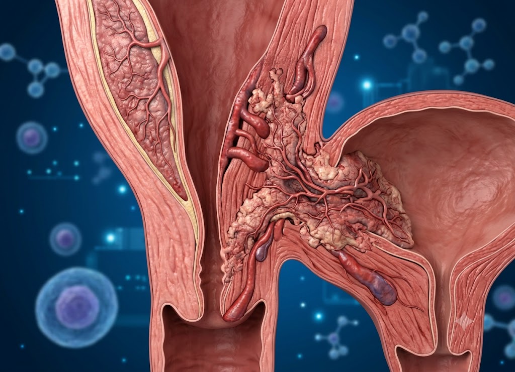
Placenta accreta is a serious pregnancy condition where the placenta attaches too deeply to the uterus, making it difficult to separate after birth and increasing the risk of severe bleeding.
The risk is higher after previous C-sections, uterine surgery, or placenta previa. Because many people have no clear symptoms, prenatal ultrasound and early monitoring are important. Early diagnosis allows a specialist team to plan safer delivery and care.
What is Placenta Accreta and Why Is It a High-Risk Condition?
Placenta Accreta Spectrum (PAS) is a serious pregnancy condition in which the placenta attaches unusually deeply to the uterine wall. One of the greatest concerns is severe postpartum hemorrhage, or heavy bleeding after childbirth.
During a normal delivery, the placenta separates from the uterus after the baby is born. The uterus then contracts, helping close the blood vessels where the placenta was attached. With PAS, the placenta is abnormally attached to or embedded in the uterine tissue, making normal separation difficult. Trying to remove it by force can damage the uterine wall and nearby blood vessels, potentially causing severe bleeding and requiring emergency surgery.
Anatomy of the Spectrum: What Happens to the Placenta?
PAS is often associated with damage or absence of the decidua, the specialized uterine lining that normally provides a protective layer between the placenta and the deeper muscular wall. When this layer is disrupted, often because of previous uterine surgery such as a cesarean section, placental villi can grow abnormally close to or into the myometrium, the muscular layer of the uterus.
A simple way to picture this is to imagine a plant growing in a pot. Normally, its roots stay within the soil and the plant can be removed easily. With PAS, the roots grow through the bottom of the pot and become attached to the surface underneath. Pulling the plant out can damage the structure beneath it.
The term “spectrum” is used because the placenta can invade the uterus to different depths.
1. Placenta Accreta
Placenta accreta is the most common form of PAS. The placental villi attach directly to the surface of the myometrium instead of being separated from it by a normal decidual layer. They do not deeply invade the uterine muscle.
2. Placenta Increta
With placenta increta, the placental tissue grows into the myometrium. The villi extend deeper into the muscular wall of the uterus, making separation after delivery much more difficult.
3. Placenta Percreta
Placenta percreta is the most severe form of PAS. The placental villi grow through the full thickness of the uterine wall and can extend beyond it. In some cases, the placenta may invade nearby organs, particularly the urinary bladder, and less commonly the bowel or rectum.
Normal Attachment vs. Placenta Accreta Spectrum
The difference between normal placental attachment and PAS affects how doctors manage delivery and prepare for possible complications.
| Feature | Normal Placental Attachment | Placenta Accreta Spectrum (PAS) |
|---|---|---|
| Implantation Site | The placenta attaches to the decidua basalis, a specialized layer of the uterine lining. | The placenta attaches directly to or grows into the myometrium, often around a previous uterine scar. |
| Separation Mechanism | The placenta normally separates naturally after the baby is born. | The placenta may remain firmly attached and resist separation. |
| Bleeding Control | Uterine contractions help compress blood vessels and limit bleeding. | Abnormal placental attachment can prevent normal separation and lead to severe bleeding. |
| Standard Delivery Outcome | The placenta is delivered normally with controlled blood loss. | A planned cesarean-hysterectomy may be recommended when PAS is confirmed or strongly suspected. |
The Clinical Takeaway
Placenta Accreta Spectrum has become an important concern in modern maternal care, particularly as cesarean delivery rates have increased. Identifying risk factors early and using appropriate ultrasound monitoring can help doctors recognize PAS and arrange specialized, multidisciplinary care when needed.
Primary Risk Factors for Placenta Accreta
The risk of placenta accreta is strongly associated with previous cesarean deliveries and other forms of uterine scarring. When a placenta implants over a scarred area, the normal protective uterine lining may be disrupted. This can allow the placenta to attach more deeply than it normally would.
The Exponential Multiplier: Previous Cesarean Sections
A history of cesarean delivery is one of the strongest known risk factors for placenta accreta. A C-section creates a scar in the uterus, and the normal tissue in that area may not develop in exactly the same way during a future pregnancy.
The risk can increase as the number of previous cesarean deliveries rises. The combination of previous C-sections and placenta previa is especially important because a low-lying placenta may implant directly over an old scar.
┌─────────────────────────────────────────────────────────────────┐
│ PAS RISK PROFILE: PRIOR C-SECTIONS + PLACENTA PREVIA             │
└─────────────────────────────────────────────────────────────────┘
  1 Prior C-Section:   ▲ 3% Risk of Accreta
  2 Prior C-Sections:  ▲ 11% Risk of Accreta
  3 Prior C-Sections:  ▲ 40% Risk of Accreta
  5+ Prior C-Sections: ▲ >60% Risk of Accreta
When a low-lying placenta grows over a previous scar in the lower part of the uterus, the risk of abnormal invasion can become particularly concerning. In severe cases, placenta percreta can extend through the uterine wall and involve the bladder.
Other High-Risk Uterine Surgeries and Conditions
Cesarean sections are a major risk factor, but other procedures and conditions that affect the uterine lining can also increase the likelihood of placenta accreta.
1. Previous Myomectomy
A myomectomy is a procedure used to remove uterine fibroids. If the fibroid was large or located deep in the uterine wall, surgery may have involved the myometrium. The resulting scar can create an area where normal placental attachment is disrupted.
2. Dilation and Curettage (D&C)
A D&C involves removing tissue from inside the uterus, usually with suction, instruments, or both. It may be performed after a miscarriage, to remove retained pregnancy tissue, or for certain bleeding conditions. Repeated procedures can sometimes cause scarring or damage to the uterine lining.
3. Asherman’s Syndrome (Intrauterine Adhesions)
Asherman’s syndrome involves scar tissue or adhesions forming inside the uterus. It can occur after procedures such as D&C or following uterine infection or surgery. Significant scarring may interfere with normal placental attachment.
4. Advanced Maternal Age and Multiparity
- Age 35 or Older: The likelihood of PAS may increase with maternal age. This can partly reflect the greater chance of previous uterine procedures and other pregnancy-related risk factors.
- High Multiparity (Multiple Pregnancies): Having several previous pregnancies may also be associated with an increased risk of abnormal placental attachment.
Clinical Screening Rule
Because several PAS risk factors can be identified from a person’s medical history, obstetricians review previous uterine surgeries and other pregnancy details early in prenatal care. If a patient has a history of uterine surgery and an ultrasound shows a low-lying placenta, doctors may recommend closer monitoring for Placenta Accreta Spectrum.
Warning Signs of Placenta Accreta Should You Be Aware Of
One of the challenging aspects of placenta accreta is that it may not produce noticeable symptoms during pregnancy. Many cases are suspected because of known risk factors and imaging findings rather than physical symptoms.
Is Third-Trimester Bleeding a Warning Sign?
Painless, bright-red vaginal bleeding during the third trimester can be a warning sign, but it is often associated with placenta previa, which can occur alongside placenta accreta.
- Placenta previa occurs when the placenta lies unusually low and partially or completely covers the cervix. As the lower uterus changes later in pregnancy, bleeding can occur.
- Because placenta previa and placenta accreta can occur together, an episode of bleeding may lead doctors to perform imaging that identifies abnormal placental attachment.
- Crucial Note: Many people with placenta accreta do not experience vaginal bleeding during pregnancy. Not having bleeding does not rule out the condition, particularly when significant risk factors are present.
Technical Markers Caught on Imaging
Because placenta accreta often cannot be identified through symptoms alone, doctors use detailed ultrasound examinations and, when appropriate, MRI to look for specific changes.
1. Placental Lacunae (“Swiss Cheese” Appearance)
On ultrasound, some PAS pregnancies show large, irregular blood-filled spaces within the placenta called lacunae. Multiple prominent lacunae can give the placenta an appearance sometimes described as “Swiss cheese.”
2. Loss of the Retroplacental “Clear Space”
Normally, ultrasound may show a clear or darker area between the placenta and the uterine muscle. With PAS, this space may appear reduced or absent because of abnormal placental attachment.
3. Severe Myometrial Thinning
Deep placental invasion can be associated with significant thinning of the uterine muscle underneath the placenta. Ultrasound and MRI can help doctors evaluate the thickness and appearance of this area.
4. Bladder Wall Irregularity
In severe cases such as placenta percreta, placental tissue can extend beyond the uterus and involve the bladder. Imaging may show abnormal blood vessels or an irregular boundary between the uterus and bladder.
How MRIs Fill the Gaps
Ultrasound is the primary imaging method for evaluating PAS, but MRI can provide additional information in selected cases. It can be particularly helpful when the placenta is located on the back wall of the uterus (posterior placenta) or when ultrasound findings are unclear. MRI can help doctors assess the depth of invasion and possible involvement of nearby organs.
How is High-Risk Pregnancy Care for Placenta Accreta Managed?
Managing Placenta Accreta Spectrum (PAS) requires careful planning because attempting to remove an abnormally attached placenta can cause severe hemorrhage. When PAS is strongly suspected or confirmed, delivery is generally planned at a specialized hospital with a multidisciplinary team.
In many cases, the planned approach involves cesarean delivery followed by hysterectomy, with the placenta left in place rather than forcibly removed.
The Multidisciplinary Care Team
Because PAS can involve major blood loss and complex pelvic surgery, care is often provided at specialized maternal care centers by several medical specialists.
Maternal-Fetal Medicine (MFM) Specialist: The MFM specialist helps monitor the pregnancy, assess the placenta, plan delivery timing, and coordinate care between the different members of the medical team.
Gynecologic Oncologist / Pelvic Surgeon: These surgeons may be involved because of their experience with complex pelvic surgery and difficult anatomy.
Obstetric Anesthesiologist: The anesthesia team prepares for the possibility of major blood loss and helps manage the patient’s circulation, anesthesia, and transfusion needs during surgery.
Urologist: A urologist may be involved when imaging suggests that placenta percreta has reached the bladder or nearby urinary structures.
Interventional Radiologist: In selected cases, an interventional radiologist may assist with procedures intended to control blood flow around the uterus. The use of these techniques depends on the individual case and the hospital’s expertise.
Blood Bank and ICU Teams: The blood bank prepares appropriate blood products when significant bleeding is anticipated. ICU specialists may provide close monitoring after complex surgery when necessary.
The Delivery Protocol
Planning delivery in advance can help reduce unexpected complications. The exact timing and surgical approach are determined by the medical team based on the patient’s condition, imaging findings, bleeding history, and other factors.
1. Late Preterm Timing: Weeks 34+0 to 35+6.
When PAS is confirmed or strongly suspected, delivery may be planned before full term to reduce the possibility of spontaneous labor or emergency bleeding. The exact timing is individualized by the care team.
2. Strategic Uterine Incision: Surgical Entry.
The surgical team plans the uterine incision carefully so it does not cut through the placenta. Depending on the location of the placenta, an incision higher on the uterus may be required.
3. Extracting the Infant: Fetal Delivery.
The baby is delivered through the planned uterine incision and transferred to the newborn care team. If delivery occurs before full term, the baby may require specialized monitoring or NICU care.
4. Hysterectomy with Placenta In Situ: Securing the Organs.
A key part of planned PAS surgery is avoiding forceful removal of the placenta. After the baby is delivered, the placenta is generally left in place while the surgeons proceed with hysterectomy when that is the planned treatment. This approach is intended to reduce the risk of uncontrolled bleeding caused by attempting to separate the placenta.
Operational Contingencies & Radical Nuances
Although cesarean-hysterectomy is a common definitive treatment for PAS, individual cases can require additional surgical planning and specialized techniques.
1. Advanced Interventional Mapping
In particularly complex cases, surgical teams may use additional planning tools and blood-conservation techniques. Cell-saver technology, for example, can collect and process blood lost during surgery for possible reinfusion when appropriate.
2. Conservative (Uterine-Sparing) Management
In carefully selected cases, doctors may discuss conservative or uterus-preserving approaches, particularly when future fertility is an important consideration. One approach may involve delivering the baby, leaving the placenta in place, and allowing it to gradually resolve under close medical monitoring.
⚠️ Critical Risk Warning
Uterine-sparing treatment is not appropriate for everyone and carries significant risks. Possible complications include delayed severe bleeding, infection, sepsis, and the eventual need for emergency hysterectomy. Any such approach requires careful selection, specialist supervision, and extensive discussion of the risks and benefits.
Diagnostic and Post-Delivery Aspects of Placenta Accreta
Successfully managing Placenta Accreta Spectrum (PAS) involves both accurate diagnosis before delivery and careful support after diagnosis and treatment. Doctors need to determine how deeply the placenta may have invaded the uterus, distinguish PAS from other placental conditions, and prepare the patient and family for the possible delivery plan.
Technical Diagnostics: Mapping the Invasion Before Birth
Doctors use imaging and medical history to evaluate the placenta and look for signs of abnormal invasion. This information helps the surgical team prepare for delivery and potential complications.
1. Frontline Ultrasound & Color Doppler
High-resolution transabdominal and transvaginal ultrasound is the main imaging method used to assess possible PAS. Doctors may look for several features, including:
- Loss of the Retroplacental “Clear Zone”: A missing or irregular area between the placenta and uterine muscle.
- Placental Lacunae: Irregular vascular spaces within the placenta that may be associated with PAS.
- Color Doppler Flow: Abnormal or increased blood flow patterns around the placenta and uterus can provide additional clues about placental invasion.
2. The Role of Pelvic MRI
MRI can provide additional information when ultrasound findings are uncertain or when the placenta is located in a difficult-to-visualize position, such as the posterior uterus. It can help assess the uterine wall and determine whether the placenta may have extended beyond the uterus or toward nearby organs.
The Three Tiers of the Spectrum
The type of PAS depends on how deeply the placenta has attached to or invaded the uterine wall.
- Placenta Accreta (75% of cases): The placental villi attach directly to the uterine muscle without deeply invading it.
- Placenta Increta (15% of cases): The placental villi extend into the muscular layer of the uterus.
- Placenta Percreta (5–10% of cases): The placenta grows through the uterine wall and may involve nearby organs, particularly the bladder or bowel.
Placenta Accreta vs. Placenta Previa
These conditions are sometimes confused because both involve the placenta and can increase the risk of bleeding, but they describe different problems.
- Placenta Previa is a problem of POSITION. The placenta implants unusually low in the uterus and may partially or completely cover the cervix. Painless, bright-red bleeding during the third trimester is a common warning sign.
- Placenta Accreta is a problem of DEPTH. The placenta attaches too deeply to the uterine wall. It may not cause bleeding during pregnancy, but it can lead to severe bleeding when the placenta cannot separate normally after delivery.
The “Perfect Storm”
Placenta previa and placenta accreta can occur together. When a low-lying placenta implants over a previous C-section scar, the combination can significantly increase concern for abnormal placental invasion. This is why patients with these risk factors often receive specialized ultrasound monitoring.
Navigating the Psychological Impact
A PAS diagnosis can be emotionally difficult for expectant parents. The diagnosis may involve discussions about preterm delivery, major surgery, possible NICU care, blood transfusions, and hysterectomy. These issues can create significant anxiety and emotional stress.
Building a strong support system can be an important part of managing the experience.
- Perinatal Mental Health Counseling: A therapist or psychologist experienced in pregnancy complications or medical trauma can help patients cope with anxiety, uncertainty, and changes to their birth plans.
- Patient Advocacy Foundations: Organizations such as the National Accreta Foundation and the Hope for Accreta Foundation provide educational resources and support for families affected by PAS.
- Peer-to-Peer Support Networks: Support groups can connect expectant parents with people who have experienced PAS. Speaking with others who understand the condition can provide reassurance and emotional support.
Conclusion
Placenta accreta is a serious pregnancy condition that requires careful monitoring and specialized medical care. It may not cause obvious symptoms during pregnancy, so recognizing risk factors and using appropriate prenatal imaging are important for early detection.
When PAS is suspected or confirmed, doctors can develop a detailed delivery plan designed to reduce the risk of severe bleeding and protect both the mother and baby. Care may involve maternal-fetal medicine specialists, surgeons, anesthesiologists, blood bank staff, neonatal specialists, and other healthcare professionals.
Early identification and coordinated care can help the medical team prepare for potential complications and provide safer, more organized care throughout pregnancy and delivery.
Frequently Asked Questions
1. What is placenta accreta?
Placenta accreta is a condition in which the placenta attaches too deeply to the uterine wall and does not separate normally after childbirth. In more severe cases, the placenta can grow through the uterine wall and involve nearby organs. This increases the risk of heavy bleeding during delivery.
2. What are the warning signs of placenta accreta?
Many people with placenta accreta have no noticeable symptoms during pregnancy. Painless vaginal bleeding can occur, particularly when placenta previa is also present. Risk factors and ultrasound findings are often more important for detecting PAS than symptoms alone.
3. What causes placenta accreta?
The exact cause is not fully understood. However, previous cesarean sections, uterine surgery, D&C procedures, placenta previa, and other conditions that cause uterine scarring can increase the risk. Some people develop placenta accreta without an obvious risk factor.
4. How is placenta accreta diagnosed during pregnancy?
Ultrasound is the main method used to evaluate suspected placenta accreta. MRI may be used in selected cases to provide additional information about placental location and possible invasion. Doctors also consider the patient’s medical and surgical history.
5. What happens during pregnancy care for placenta accreta?
Care usually includes closer monitoring, additional imaging, and delivery planning with a team of specialists. Delivery is often planned at a hospital equipped to manage major bleeding, complex surgery, anesthesia needs, and newborn care. The exact plan depends on the severity of PAS and the patient’s individual circumstances.
6. Can placenta accreta be treated?
Treatment focuses on preventing severe bleeding and safely managing delivery. In many cases, a planned cesarean delivery followed by hysterectomy is recommended, with the placenta left in place rather than forcibly removed. Selected patients may be considered for uterus-preserving approaches, but these carry significant risks and require specialist care.