10 Things to Know About Polymyositis and Muscle Inflammation
Polymyositis is a rare inflammatory muscle disease that causes gradual, progressive muscle weakness. It most often affects muscles close to the center of the body, including the shoulders, upper arms, hips, thighs, and neck. Many people first notice problems when climbing stairs, getting up from a chair, lifting objects, reaching overhead, or keeping their head upright for extended periods.
Because the weakness usually develops slowly, early symptoms can sometimes be mistaken for normal aging, physical overuse, tiredness, or a simple muscle strain.
Understanding polymyositis and muscle inflammation can make it easier to recognize when ongoing weakness needs medical attention. The condition is generally considered part of a group of inflammatory myopathies involving an abnormal immune response against muscle tissue. In some cases, it can also affect swallowing, breathing, joints, and everyday mobility.
This article covers 10 important things to know about polymyositis, including its symptoms, causes, diagnosis, treatment, physical therapy, and long-term management.
What Is Polymyositis?
Polymyositis is a rare inflammatory muscle disease that causes persistent weakness in muscles involved in movement. It commonly affects muscles near the center of the body, including the shoulders, upper arms, hips, thighs, and neck.
Because these muscles are essential for many everyday activities, people with polymyositis may have difficulty climbing stairs, standing up from a chair, lifting objects, reaching overhead, or holding their head upright for long periods.
The condition occurs when the immune system becomes involved in attacking or inflaming healthy muscle tissue. Muscle weakness often develops gradually over several weeks or months, which can make the condition difficult to recognize at first. Some people may also experience muscle tenderness, joint discomfort, swallowing difficulties, or breathing problems when other muscles become involved.
Polymyositis requires medical evaluation because untreated inflammation can lead to worsening weakness. Doctors may use blood tests, muscle enzyme testing, MRI scans, electromyography, or a muscle biopsy when evaluating the condition. Treatment commonly involves medicines that control inflammation, along with physical therapy to help maintain strength, movement, and independence.
Symptoms of Polymyositis: What Are the Primary Signs of Muscle Weakness?
The main signs of polymyositis are symmetrical and progressive muscle weakness, especially in muscles located close to the trunk. Profound fatigue and difficulty with everyday activities may occur alongside the weakness.
Unlike an acute muscle injury, polymyositis usually develops gradually over weeks or months. This slow progression can make the early symptoms easy to overlook or confuse with ordinary fatigue or physical strain.
The severity can vary from person to person, but a consistent pattern of weakness in the proximal muscles on both sides of the body is an important clinical clue.
Understanding the specific pattern of weakness and the other symptoms associated with polymyositis can help people recognize when medical evaluation may be needed.
Clinical Overview: What is Polymyositis?
Polymyositis is a rare, chronic inflammatory muscle condition associated with an abnormal autoimmune response involving the body’s skeletal muscles. In simple terms, the immune system becomes involved in inflammation that can damage muscle tissue and gradually reduce muscle strength.
The typical pattern includes two important features:
Proximal Muscle Distribution
- Muscles closest to the torso are primarily affected.
- Hip and pelvic muscles may make standing difficult.
- Shoulder muscles may make lifting or reaching harder.
Symmetrical Presentation
- Weakness generally affects both sides of the body.
- The pattern is often similar on the right and left sides.
- This distribution can provide an important clue during clinical evaluation.
Recognizing the early symptoms of polymyositis is important because the condition can resemble other neuromuscular disorders. Identifying the characteristic pattern of weakness can help guide further testing and support an accurate polymyositis diagnosis.
Proximal and Symmetrical Muscle Involvement
One of the defining features of polymyositis is weakness in proximal muscles, which are located closest to the center of the body. The weakness commonly affects both sides in a similar way.
- Hip and Thigh Muscles: Weakness around the hips and thighs can make lower-body movements increasingly difficult. Getting up from a low chair or from the floor may require using the arms for support. Climbing stairs, getting into a vehicle, or standing from a low toilet may also become tiring.
- Shoulder and Upper Arm Muscles: Weakness in the shoulder area can make lifting and reaching difficult. A person may struggle to lift heavier household items, reach high shelves, or raise their arms while washing or brushing their hair.
- Neck Muscles: Weakness in the neck muscles may make it difficult to keep the head upright. This can contribute to neck discomfort and make activities such as driving, reading, or working at a computer more difficult.
The Nature of Progressive Weakness
Muscle weakness associated with polymyositis generally develops slowly over weeks or months. The gradual progression is an important part of the overall clinical pattern.
- Initial Stages: Early symptoms may be subtle. Someone might notice unusual tiredness after light activity or find that climbing stairs takes more effort than usual. They may also start using their arms to push themselves up from a sofa.
- Intermediate Stages: As the condition progresses, weakness can become more noticeable. Using handrails, taking breaks while carrying groceries, or avoiding deep chairs because they are harder to get out of may become common.
- Advanced Stages: Without appropriate treatment, ongoing inflammation can lead to significant loss of strength. Some people may eventually have difficulty turning over in bed, raising their arms above their shoulders, or walking without assistance.
Systemic and Extra-Muscular Signs
Although muscle weakness is the main concern, polymyositis symptoms can sometimes involve other parts of the body.
- Debilitating Fatigue: Severe fatigue may occur alongside muscle weakness. It can be related to ongoing inflammation as well as the additional effort needed to move weakened muscles.
- Difficulty Swallowing (Dysphagia): When muscles involved in swallowing are affected, a person may cough or choke while eating or feel as though food is getting stuck. Significant swallowing problems can increase the risk of poor nutrition or aspiration pneumonia.
- Joint Pain and Stiffness (Arthralgia): Some people experience aching, stiffness, or mild swelling in areas such as the wrists, hands, knees, and ankles.
- Shortness of Breath (Dyspnea): Breathing difficulties may occur when respiratory muscles are affected. Polymyositis can also be associated with interstitial lung disease (ILD), which involves inflammation and scarring of lung tissue.
Diagnostic Impact: During a clinical polymyositis diagnosis, doctors may consider these symptoms along with blood tests, including creatine kinase levels, electromyography (EMG), imaging, and muscle biopsy findings.
Causes of Polymyositis: What Triggers This Autoimmune Muscle Disease?
The exact cause of polymyositis is not fully understood. The condition is associated with an autoimmune response in which the immune system mistakenly targets muscle tissue, resulting in inflammation and damage.
Researchers believe that a combination of genetic susceptibility and environmental influences may contribute to this abnormal immune response. However, the specific event that triggers the condition is not always known.
The autoimmune nature of polymyositis helps explain how the disease develops and why treatment often focuses on controlling inflammation and reducing abnormal immune activity.
The Cellular Mechanisms of Immune System Aggression
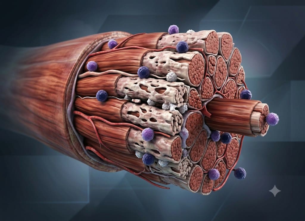
In a healthy immune system, cytotoxic T-cells help identify and destroy infected or abnormal cells. In polymyositis, these immune cells can become incorrectly activated and attack healthy muscle tissue.
Loss of Self-Tolerance: For reasons that remain unclear, the immune system may lose its ability to properly distinguish healthy muscle tissue from harmful targets. An external factor may alter how muscle proteins are recognized, contributing to an abnormal immune response.
Endomysial Inflammation: Once activated, CD8+ cytotoxic T-lymphocytes can enter muscle tissue and surround or invade individual muscle fibers. This type of inflammation within the muscle tissue is an important finding considered during a polymyositis diagnosis and can help distinguish it from other inflammatory muscle diseases.
Cellular Destruction and Fibrosis: Immune cells can release substances that damage muscle fibers. As muscle tissue is repeatedly injured, normal repair may become impaired. Over time, muscle fibers can shrink and some damaged tissue may be replaced by connective tissue, contributing to persistent weakness.
Risk Factors and Demographic Profiles
Polymyositis is considered a rare condition and can develop at different ages. Understanding the populations in which it occurs more often can provide clues about the disease.
Age and Gender Distribution: Polymyositis most commonly develops in adults, with many cases occurring between the ages of 30 and 60. Women are affected more often than men.
Genetic Predisposition: Polymyositis does not generally follow a simple parent-to-child inheritance pattern. However, certain Human Leukocyte Antigen (HLA) genes have been associated with an increased susceptibility to inflammatory muscle diseases. These genes play an important role in helping the immune system distinguish the body’s own tissues from foreign substances.
Environmental Triggers and Overlap Syndromes
Current understanding suggests that environmental influences may interact with genetic susceptibility to contribute to polymyositis.
| Risk Category | Key Biological Associations | Clinical Impact |
|---|---|---|
| Autoimmune Overlap | Lupus, Scleroderma, Sjögren’s, Rheumatoid Arthritis | May be associated with overlap syndromes involving more than one autoimmune condition. |
| Environmental Triggers | Coxsackie B virus, Influenza, Retroviruses, toxic exposures | May contribute to abnormal immune activation in susceptible individuals. |
| Malignant Paraneoplastic | Lung, bladder, breast, or ovarian cancers | In some cases, an immune response associated with a tumor may also affect healthy muscle tissue. |
Cancer Screening Importance: Because inflammatory muscle diseases can sometimes be associated with underlying cancer, adults diagnosed with polymyositis may undergo appropriate, age-related cancer screening based on their individual risk factors and medical history.
Diagnosis of Polymyositis: How Do Doctors Confirm the Condition?
Doctors diagnose polymyositis through a combination of medical history, physical examination, blood tests, electrical studies of the muscles and nerves, imaging, and sometimes a muscle biopsy.
There is no single test that confirms every case. Instead, doctors consider findings from multiple sources while also looking for other conditions that could explain progressive muscle weakness.
This approach is important because neurological disorders, medication-related muscle problems, genetic muscle diseases, and other inflammatory conditions can produce similar symptoms.
The Comprehensive Multi-Faceted Evaluation Process
A polymyositis diagnosis cannot always be established with one test. Because several other disorders can cause similar muscle weakness, doctors may use multiple diagnostic tools.
These may include:
- Blood Tests: Help identify elevated muscle enzymes and certain antibodies associated with inflammatory muscle disease.
- Electromyography: Evaluates the electrical activity of muscles and can help determine whether weakness originates from muscle tissue.
- Muscle MRI: Can identify areas of active inflammation and help doctors select an appropriate muscle for further evaluation.
- Muscle Biopsy: Provides a tissue sample that can be examined under a microscope for patterns of inflammation and muscle damage.
By combining laboratory results, electrical testing, imaging, and tissue findings, specialists can develop a clearer picture of the condition. This process also helps rule out other disorders before long-term immunosuppressive treatment is started.
Advanced Laboratory Panels, Electrical Tests, and Muscle Imaging
To investigate whether progressive weakness may be caused by an inflammatory muscle disorder, doctors can use several important polymyositis diagnosis tools.
Comprehensive Blood Profiles: When muscle fibers are damaged, substances inside the cells can enter the bloodstream. Creatine kinase (CK) is an important muscle enzyme that may become significantly elevated. Doctors may also test for Myositis-Specific Antibodies (MSAs). Other tests, such as an Antinuclear Antibody (ANA) test, may provide additional information about autoimmune activity.
Electromyography (EMG): An EMG records the electrical activity of muscles during rest and contraction. In inflammatory muscle disease, the test can show patterns consistent with a myopathy. This can help doctors distinguish muscle-related weakness from weakness caused primarily by nerve problems.
Targeted Magnetic Resonance Imaging (MRI): MRI scans can show areas of active inflammation in large muscle groups such as the thighs or pelvis. They may also show muscle wasting or fatty replacement in more chronic disease. MRI can help identify an area that may be appropriate for biopsy.
The Biopsy: The Uncontested Gold Standard for Certainty
Blood tests, imaging, and electrical studies can provide valuable evidence, but a muscle biopsy may be used to examine muscle tissue directly when confirmation is needed.
Proving Endomysial Inflammation: Under a microscope, doctors can examine the pattern of immune-cell activity within the muscle. Findings may include CD8+ cytotoxic T-lymphocytes surrounding and invading muscle fibers, a pattern associated with endomysial inflammation.
Differentiating Myositis Mimics: A biopsy can help distinguish polymyositis from other conditions that may require different treatments. Dermatomyositis, for example, has a different distribution of inflammation. Inclusion Body Myositis (IBM) can show distinctive findings such as rimmed vacuoles.
Excluding Non-Inflammatory Disorders: Muscle biopsy can also help doctors investigate genetic muscular dystrophies and other non-inflammatory causes of weakness. These conditions may produce muscle weakness and elevated CK levels but have different tissue findings.
The role of physical therapy in managing Polymyositis
Physical therapy plays an important role in managing Polymyositis. It can help maintain muscle strength, improve physical function, protect joint movement, and support quality of life.
Medication is used to control the underlying inflammation, while physical therapy focuses on the weakness and loss of conditioning that can result from the disease. The program should be carefully adjusted according to disease activity, muscle strength, and the individual’s overall condition.
The goals of physical therapy can change as the condition improves.
During Active Inflammation: When inflammation is high, therapy may focus on gentle or passive range-of-motion exercises. These movements can help reduce stiffness and prevent contractures caused by inactivity. Intense strengthening exercises may be avoided during periods of active inflammation. Therapists can also teach energy-conservation strategies and ways to balance rest with light activity.
As Inflammation Subsides: Once treatment begins controlling inflammation and muscle enzyme levels improve, a more active rehabilitation program may be introduced. This can include gradual strengthening exercises, isometric movements, gentle resistance training, and functional exercises such as standing from a chair or practicing stairs. Progress should be gradual to avoid excessive fatigue or strain.
Long-Term Maintenance and Function: As strength improves, therapy may include aerobic activities such as walking or stationary cycling to improve endurance. Balance, coordination, and gait training can also support safer mobility. Occupational therapy may help people find easier ways to perform daily tasks and determine whether assistive devices would be useful. For swallowing difficulties, speech-language professionals may provide swallowing exercises and dietary guidance.
Broader Implications and Distinctions of Polymyositis?
The broader implications of Polymyositis include its relationship with other inflammatory muscle diseases, possible complications involving organs such as the lungs and heart, and the importance of long-term rehabilitation and lifestyle support.
Understanding these differences and potential complications can help patients and healthcare professionals create a more complete approach to managing the condition.
Comparing Polymyositis and Dermatomyositis
Both polymyositis and dermatomyositis are inflammatory muscle diseases that can cause progressive weakness. However, they have important differences in their physical findings and patterns of immune involvement.
Dermatological Manifestations: One of the main distinguishing features of dermatomyositis is its characteristic skin involvement. This can include a heliotrope rash, which causes purplish discoloration around the eyelids, and Gottron’s papules, which are raised reddish areas over the knuckles, elbows, or knees. Other patterns may include a “shawl sign” across the shoulders or a “V-sign” on the upper chest.
In contrast, polymyositis does not typically have these characteristic skin findings. Its primary effects involve skeletal muscle tissue.
Muscle Involvement: Both conditions can cause symmetrical weakness in proximal muscles, including the hips, thighs, shoulders, and neck. As a result, people with either condition may have difficulty standing from a chair, climbing stairs, or lifting their arms.
Distinct Biopsy Signatures: A polymyositis diagnosis may rely heavily on muscle biopsy findings when the clinical picture is unclear. Polymyositis is associated with an endomysial inflammatory pattern involving cytotoxic T-cells. Dermatomyositis typically has a different pattern, with inflammation concentrated around blood vessels.
Systemic Comorbidities and Organ Involvement
Polymyositis can have effects beyond skeletal muscles. The immune process involved in inflammatory muscle disease may also be associated with complications affecting other organs.
Interstitial Lung Disease (ILD): ILD is an important potential complication of inflammatory muscle disease. It can cause inflammation and scarring within lung tissue, leading to symptoms such as a persistent dry cough and shortness of breath. Doctors may use pulmonary function tests and chest imaging to monitor lung health when appropriate.
Malignancy Risk: Inflammatory myopathies have been associated with an increased risk of certain cancers. Depending on age, symptoms, risk factors, and the specific inflammatory muscle disease involved, doctors may recommend appropriate cancer screening.
Cardiovascular and Esophageal Strain: In some cases, inflammatory muscle disease can involve the heart muscle and contribute to heart-related complications. Weakness affecting the throat and upper esophagus can also cause dysphagia, increasing the risk of aspiration and nutritional problems.
Long-Term Prognosis and Rehabilitation
The long-term outlook for someone with polymyositis can vary depending on factors such as age, the severity of muscle involvement, associated complications, and how well the condition responds to treatment.
Although there is no definitive cure, medical treatment and rehabilitation can help many people control inflammation and improve or maintain function.
Long-term management may include:
- Medical Treatment: Corticosteroids and other immune-suppressing medicines may be used to control inflammation.
- Physical Therapy: A structured rehabilitation program can help rebuild strength, improve endurance, and maintain mobility.
- Comorbidity Surveillance: Regular monitoring may be needed for lung, heart, swallowing, and other health concerns.
First-line treatment may include corticosteroids such as prednisone to reduce inflammation and control active disease. Doctors may later add steroid-sparing medicines, such as methotrexate or azathioprine, when appropriate.
A personalized physical and occupational therapy program is also an important part of long-term care. Although rest can be necessary during an active flare, prolonged inactivity can contribute to muscle loss. Gradual, supervised exercise can help restore strength, maintain joint movement, and improve endurance.
Evidence-Based Lifestyle Support Strategies
Lifestyle changes can provide supportive benefits alongside medical treatment. They may help with nutrition, fatigue management, physical activity, and overall daily function.
Implementing an Anti-Inflammatory Diet
A balanced diet can support overall health while a person receives treatment for polymyositis. Meals can emphasize nutrient-rich whole foods while limiting highly processed foods and excess added sugars.
| Recommended Anti-Inflammatory Foods | Foods to Avoid/Limit |
|---|---|
| Omega-3 Fatty Acids: Salmon, mackerel, walnuts, chia seeds | Refined Sugars: Soda, pastries, processed sweets |
| Antioxidant-Rich Produce: Berries, leafy greens, tomatoes | Simple Carbohydrates: White bread, processed snacks |
| Healthy Fats: Extra virgin olive oil, avocados | Inflammatory Fats: Trans fats and excessive red meat |
Nutritional Supplementation: Long-term corticosteroid treatment can affect bone health. Doctors may recommend nutrients such as Calcium and Vitamin D when appropriate to help support bone density.
Energy Conservation and Pacing
Severe fatigue can be an important part of living with polymyositis. Pacing daily activities can help prevent a cycle in which doing too much on a good day results in significant exhaustion afterward.
Try breaking larger household tasks into smaller steps. Sitting while cooking or grooming can help conserve energy, and assistive tools may reduce the physical effort required for certain activities.
Stress Reduction and Rest
Long-term physical and emotional stress can make it harder to manage chronic illness and fatigue. Healthy stress-management habits, including mindfulness, deep breathing, relaxation techniques, or meditation, may support overall well-being.
Adequate rest is also important, particularly when fatigue is significant. The goal is to find a sustainable balance between rest and appropriate activity.
Conclusion
Polymyositis is more than temporary muscle soreness because it involves ongoing inflammation that can cause progressive weakness in important skeletal muscles. The condition commonly affects both sides of the body and can gradually make everyday movements more difficult.
Diagnosis may involve blood tests, muscle enzyme testing, imaging, electromyography, and, in some cases, a muscle biopsy. Treatment generally focuses on controlling inflammation while supporting muscle function and mobility.
With appropriate medical care, physical therapy, lifestyle support, and regular monitoring, many people can manage polymyositis and work to reduce the risk of long-term muscle weakness and complications.
Frequently Asked Questions
1. What is polymyositis?
Polymyositis is an inflammatory muscle disease that causes weakness in the skeletal muscles used for movement. It commonly affects muscles close to the center of the body, including the shoulders, hips, thighs, upper arms, and neck.
The weakness often develops gradually over weeks or months. The condition is associated with an abnormal immune response that causes inflammation in muscle tissue.
2. What are the early symptoms of polymyositis?
Early symptoms often include gradual muscle weakness, fatigue, and difficulty with everyday movements.
A person may have trouble climbing stairs, getting up from a chair, lifting objects, or reaching overhead. Muscle pain or tenderness may occur, but weakness is usually the main concern. The weakness commonly affects both sides of the body rather than a single isolated muscle.
3. How is polymyositis diagnosed?
Doctors diagnose polymyositis by combining information from symptoms, a physical examination, blood tests, imaging, muscle and nerve studies, and sometimes a muscle biopsy.
Blood tests may show elevated muscle enzymes such as creatine kinase. MRI and electromyography can provide additional information about muscle inflammation and function. A muscle biopsy may be used to examine tissue directly and help rule out other muscle disorders.
4. Can polymyositis affect breathing or swallowing?
Yes. Polymyositis can sometimes affect muscles involved in swallowing or breathing.
Swallowing problems may cause coughing or choking during meals or make food feel as though it is stuck. Breathing difficulties may occur when respiratory muscles or the lungs are affected. These symptoms should be discussed with a healthcare professional because they can become serious.
5. How is polymyositis treated?
Treatment generally focuses on reducing inflammation and supporting muscle strength and function.
Corticosteroids are often used to control inflammation, while other immune-suppressing medicines may be added when needed. Physical therapy can help maintain mobility, rebuild strength, and reduce disability.
Treatment should be individualized and monitored by appropriate healthcare professionals, which may include specialists such as a rheumatologist or neurologist.