Graft vs. Host Disease: Causes, Symptoms, and Treatment Explained
Graft vs. host disease (GVHD) is an unusual, sometimes serious complication that can follow certain transplants, particularly stem cell or bone marrow transplants. Transplantation can be a life-saving treatment for many conditions, but occasionally the donor’s immune cells mistake the recipient’s own body for a foreign threat and begin attacking healthy tissue.
This happens because the transplanted donor cells carry immune system components, especially T cells, whose job is to identify and fight off threats. In GVHD, these cells wrongly target the recipient’s organs, causing inflammation and damage. The condition ranges from mild to severe and can involve different parts of the body.
GVHD generally falls into two categories: acute GVHD, which tends to show up within the first few months after transplant, and chronic GVHD, which can appear later and linger for a longer stretch of time. How severe the symptoms are, and what form they take, depends on things like the type of transplant, how closely donor and recipient are matched, and the person’s overall health.
Common signs include skin changes like rash or itching, digestive issues such as diarrhea or abdominal discomfort, and liver problems that can affect the whole body. Because symptoms vary so much from person to person, spotting early warning signs and getting timely care matters a great deal.
Doctors have several tools for reducing risk and treating GVHD, including medications that dial down the immune response. Treatment is tailored to how severe the symptoms are and which organs are involved, and advances in transplant medicine have improved both prevention and outcomes over time.
This article covers the causes of GVHD, its common symptoms, risk factors, how it’s diagnosed, treatment options, and what patients should know about managing this transplant-related complication.
What Is Graft vs. Host Disease and Why Does It Occur?
Graft vs. Host Disease is a systemic immune disorder that happens when transplanted donor immune cells (the graft) mistake the recipient’s body (the host) for foreign tissue and launch an inflammatory attack against it. It’s essentially the reverse of transplant rejection, where the host’s immune system is the one attacking the transplant instead.
To understand why this happens, it helps to look at how allogeneic transplantation works. The goal of this type of transplant is to replace a patient’s faulty or cancerous blood-forming system with a healthy one from a donor. But that process doesn’t just transfer stem cells; it also brings along mature, functional immune cells, particularly T-lymphocytes, from the donor.
These donor T-cells are built to identify and eliminate foreign threats. Once they enter the recipient’s body, they start surveying their new surroundings, and that’s where the trouble can begin.
How Does a Donated Graft Recognize the Recipient’s Body as Foreign?
A donated graft recognizes the recipient’s body as foreign mainly through differences in cell-surface proteins called Human Leukocyte Antigens (HLA), which function like molecular ID cards for the immune system. These proteins sit on nearly all cells in the body and let the immune system tell self from non-self.
When donor T-cells encounter the recipient’s cells, they check these HLA markers. If the HLA proteins on the recipient’s cells don’t match the patterns the donor T-cells learned to recognize as “self” back in the donor’s own body, they read the recipient’s tissue as a threat and kick off a powerful immune response.
The recognition process is actually more sophisticated than that. T-cells don’t just look at the HLA molecule itself; they recognize a complex formed by the HLA molecule presenting a small fragment of protein, a peptide, from inside the cell. In GVHD, donor T-cells are scrutinizing the recipient’s HLA molecules as they present the recipient’s own peptides.
Even with a full match on the major HLA genes, subtle differences can still exist in what are called minor histocompatibility antigens, different versions of normal body proteins that can look foreign to donor T-cells even when presented by an identical HLA molecule. This is why GVHD can still occur in transplants between HLA-identical siblings.
Once recognition happens, donor T-cells activate. They multiply rapidly and release a flood of inflammatory signaling molecules called cytokines. This “cytokine storm” drives a widespread attack, pulling in other immune cells and directly damaging the host’s tissue, producing the clinical symptoms of GVHD.
Types of Transplants Where GVHD Typically Happens
GVHD typically shows up as a major complication of allogeneic hematopoietic stem cell transplantation, which includes bone marrow, peripheral blood stem cell, and umbilical cord blood transplants. This is the classic setting for GVHD, since the entire purpose of the transplant is to introduce a new, functional immune system into the recipient.
The transplanted material is rich in immune cells, especially T-lymphocytes, the main drivers of the GVHD reaction. Meanwhile, the recipient has usually gone through intensive chemotherapy or radiation beforehand to wipe out their own bone marrow and immune system, leaving them unable to reject the donor cells. That combination creates the perfect conditions for the new donor immune system to turn against a defenseless host.
While HSCT is the most common setting, GVHD can occasionally occur elsewhere. Solid organ transplantation is one, though it’s much rarer. For GVHD to develop after a solid organ transplant, two things have to line up: the transplanted organ has to contain a significant number of viable donor immune cells, and the recipient has to be immunosuppressed enough that they can’t reject those cells. This shows up most often with organs rich in lymphoid tissue, like the liver, small bowel, and lung. Liver transplants, for instance, carry a large population of donor lymphocytes along with them, and if the recipient’s immune system is compromised, those cells can take hold and attack.
Another rare setting is transfusion of non-irradiated blood products into an immunocompromised patient. The lymphocytes in that transfused blood can attack the recipient, a condition called transfusion-associated GVHD, extremely rare but often fatal.
Symptoms and Signs of Graft vs. Host Disease
The main symptoms of GVHD most commonly show up in the skin, liver, and gastrointestinal tract, and are categorized as either acute or chronic based on timing and clinical presentation. Symptoms can range from mild and manageable to severe and life-threatening, depending on the disease’s grade and which organs are involved.
Recognizing these signs matters for prompt diagnosis, since early intervention can significantly reduce the long-term impact of the disease. The clinical picture varies not just by organ, but also by whether the process is acute (rapid, direct inflammatory damage) or chronic (slower, progressive inflammation and scarring more similar to autoimmune disease).
Common Signs Grouped by Affected Organ
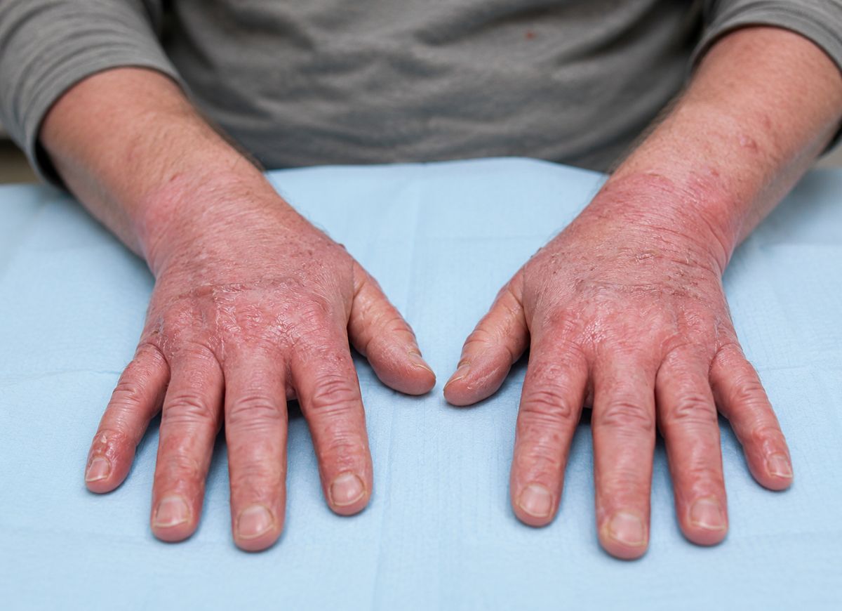
Skin is the most frequently affected organ. The first sign is usually a rash combining flat, discolored spots with small raised bumps, typically starting on the palms, soles, and head and neck before spreading to the trunk and limbs. It can be itchy and painful. In more severe cases, the rash can progress to widespread redness across the entire skin surface, or develop blisters that rupture and cause skin sloughing resembling a severe burn. Severity is graded by the percentage of body surface area involved.
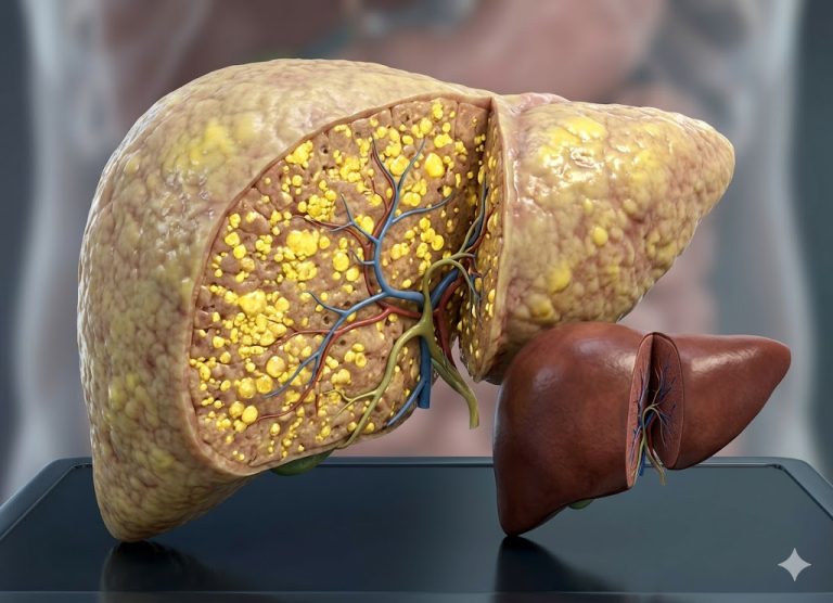
Liver GVHD shows up as a rise in liver function tests, particularly elevated bilirubin. Clinically, this appears as jaundice, a yellowing of the skin and the whites of the eyes. Patients may also feel pain in the upper right abdomen from an enlarged liver. The underlying damage comes from donor lymphocytes attacking the small bile ducts, a condition called cholestasis that impairs bile flow.
GI tract GVHD can affect both upper and lower portions. Upper GI involvement brings persistent nausea, vomiting, loss of appetite, and a feeling of fullness. Lower GI involvement causes profuse, watery diarrhea that can turn bloody in severe cases, often along with significant cramping and pain. The fluid loss from diarrhea can lead to severe dehydration, electrolyte imbalances, and malnutrition, making it one of the more dangerous aspects of GVHD.
Acute GVHD vs. Chronic GVHD
Acute GVHD is marked by rapid-onset, aggressive inflammation that typically shows up within 100 days of transplant. Chronic GVHD usually develops later and presents with a broader, more complex set of symptoms resembling autoimmune and fibrotic disease. The distinction matters a great deal, since diagnosis and management differ significantly between the two.
Acute GVHD centers on the classic triad of skin, liver, and GI tract, with symptoms directly caused by T-cell-mediated tissue destruction. The skin rash is inflammatory and can progress quickly. Liver involvement leads to cholestatic jaundice. GI symptoms show up as high-volume, watery diarrhea from damage to the intestinal lining. Onset is typically abrupt, and severity is graded across these three organs. The underlying mechanism is a direct immune assault.
Chronic GVHD can either evolve from a resolved case of acute GVHD or appear without any prior acute symptoms at all. Its onset tends to be more gradual and shows up later, often more than 100 days post-transplant. It can affect virtually any organ system, and its symptoms are more varied, often mimicking autoimmune conditions. Skin changes can resemble scleroderma (tightening and thickening) or lichen planus (purplish, itchy flat bumps), along with pigmentation changes and hair loss.
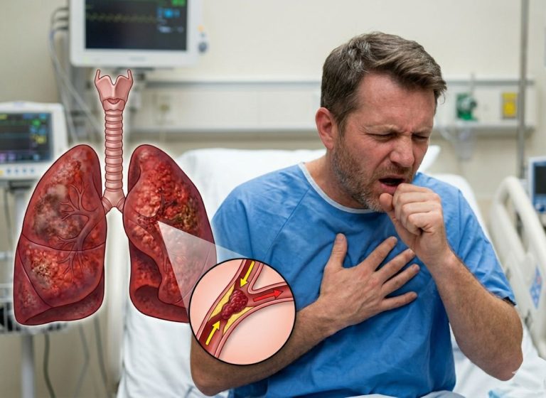
Sicca syndrome, severe dryness of the mouth and eyes, is common, along with painful mouth ulcers and inflammation. Bronchiolitis obliterans syndrome can cause progressive scarring and narrowing of the small airways, leading to shortness of breath and a persistent cough. Patients may also develop joint stiffness and contractures from inflammation and fibrosis of connective tissue. The liver, GI tract, and genitals can be affected too, but here the pathology often involves scarring alongside inflammation, making chronic GVHD a more complex, long-term challenge to manage.
Causes and Risk Factors for Graft vs. Host Disease
The core cause of GVHD is the immunological mismatch between donor and recipient. Key risk factors that amplify this risk include the degree of HLA mismatch, older age in either the donor or recipient, and the source of the transplanted stem cells. At its heart, GVHD is a predictable consequence of introducing an outside immune system into a body that doesn’t recognize it as its own.
While the central trigger stays constant, immune recognition of foreignness, how likely GVHD is to occur, and how severe it becomes, depends on a specific set of clinical and biological factors that can often be assessed before the transplant even happens.
The Primary Trigger
The primary trigger for GVHD is the recognition of mismatched HLA and minor histocompatibility antigens on the recipient’s cells by the donor’s T-lymphocytes. This sets off a complex, destructive cascade of immune activation. HLA proteins act as gatekeepers of self-recognition, and any perceived mismatch signals an alarm to the patrolling T-cells of the newly engrafted immune system.
The development of GVHD is often described in three phases:
1. Initial Tissue Damage: This starts even before donor cells are infused. The pre-transplant conditioning regimen, high-dose chemotherapy and/or radiation, damages host tissue, particularly in the gut, liver, and skin. This releases inflammatory cytokines like TNF-α and IL-1, creating a cytokine storm that primes the recipient’s body for a heightened immune response and makes tissues more visible and vulnerable to incoming donor T-cells.
2. Donor T-Cell Activation: Once the donor stem cell graft is infused, mature donor T-cells encounter the host’s antigen-presenting cells, which display the host’s HLA and minor histocompatibility antigens. The donor T-cells read these as foreign, triggering activation, rapid multiplication, and differentiation into effector cells, all amplified by the inflammatory environment already set up in phase one.
3. Effector Phase and Tissue Destruction: In the final phase, activated donor T-cells, along with other recruited immune cells like natural killer cells and macrophages, migrate to target organs. They launch a direct attack on host cells and release more inflammatory cytokines, perpetuating a cycle of inflammation and damage. This cellular assault produces the clinical symptoms of GVHD seen in the skin, liver, and GI tract.
Key Risk Factors
Risk factors for GVHD generally fall into categories related to the donor-recipient match, patient and donor characteristics, and the specifics of the transplant itself.
Histocompatibility mismatch is the single most important risk factor. Risk correlates directly with the number of mismatched HLA loci between donor and recipient. A transplant from an HLA-identical sibling carries the lowest risk; a matched unrelated donor carries higher risk; and a mismatched unrelated donor or half-matched family member carries the highest risk. Even with a perfect HLA match, differences in other cell surface proteins can still trigger GVHD.
Older recipients and older donors are independently linked to higher risk of both acute and chronic GVHD, possibly due to age-related immune changes and reduced capacity for tissue repair.
A female donor who has previously been pregnant, donating to a male recipient, raises risk as well. During pregnancy, a mother can become sensitized to male-specific minor antigens encoded on the Y chromosome, and her immune cells may later recognize and attack the male recipient’s cells.
The type of graft matters too. Peripheral blood stem cells contain far more T-cells than bone marrow, so using them is linked to faster engraftment but a higher chance of chronic GVHD. Umbilical cord blood contains naive, immature T-cells and carries the lowest risk of severe GVHD, though engraftment is slower.
More intensive conditioning regimens cause greater initial tissue damage, leading to a stronger release of inflammatory cytokines and higher risk of acute GVHD.
Types of GVHD Treatment and Management
Treatment for both acute and chronic GVHD centers on suppressing the overactive donor immune system, with systemic corticosteroids as the universal first-line therapy, followed by a range of second-line immunosuppressive agents for cases that don’t respond. The goal is to stop the immune attack on host tissue while preserving the beneficial graft-versus-leukemia effect, where donor cells help eliminate any remaining cancer.
Management is tailored to how severe the disease is and whether it’s acute or chronic. Prevention is also a cornerstone of post-transplant care, with most patients receiving immunosuppressive drugs starting before the transplant to reduce initial risk. Once GVHD develops, though, a more aggressive approach is needed to bring the immune response under control and prevent irreversible organ damage.
Main Treatment Strategies
Treatment generally follows a stepwise ladder: first-line, second-line, and targeted therapies, escalating in intensity based on how the patient responds and how severe the disease is.
First-line therapy relies on high-dose systemic corticosteroids, such as methylprednisolone or prednisone, the undisputed standard of care for newly diagnosed acute and chronic GVHD. These drugs broadly inhibit T-cell activation and cytokine production, calming the immune storm. For localized GVHD, like a mild skin rash, topical steroids may be enough; for disease affecting internal organs, higher intravenous or oral doses are needed. About half of patients respond well to this initial treatment.
Second-line therapies come into play for steroid-refractory GVHD, cases that don’t improve or that worsen despite adequate steroid treatment, a situation associated with a worse prognosis. A variety of second-line agents provide additional immunosuppression in these cases.
Targeted and novel therapies have expanded treatment options in recent years. A JAK inhibitor that blocks key inflammatory signaling pathways is now an FDA-approved standard second-line treatment for steroid-refractory acute GVHD. Another approach involves drawing the patient’s blood, separating out white blood cells, treating them with a photosensitizing agent and UV light, then returning them to the body, a process thought to induce immune tolerance and particularly effective for chronic GVHD affecting the skin and other organs. Other targeted agents, including ibrutinib for chronic GVHD and abatacept, are also changing the treatment landscape.
Is Graft vs. Host Disease Always Treatable?
Mostly, yes, but with real caveats. Most cases of GVHD are treatable and can be managed effectively, but severe or steroid-refractory forms can be extremely difficult to control and may lead to irreversible organ damage, long-term disability, or death. Treatment success depends heavily on how severe the initial presentation is, which organs are involved, and how the patient responds to first-line therapy.
Most patients with mild to moderate acute GVHD respond well to standard corticosteroid treatment, and with appropriate management, symptoms can be controlled and the immune reaction suppressed. The prophylaxis regimens given to nearly all allogeneic transplant recipients have also significantly reduced the incidence of severe GVHD overall. For chronic GVHD, while it may need long-term therapy, many patients reach a stable condition and maintain good quality of life with treatments like low-dose steroids or the photopheresis procedure described above.
That said, treatment isn’t always successful. The biggest challenges come with severe acute GVHD and steroid-refractory disease, both of which are life-threatening. When high-dose steroids fail to control the disease, prognosis worsens dramatically. Later lines of therapy have limited success rates and carry their own risks, most notably a much higher susceptibility to opportunistic infections from prolonged immunosuppression, itself a leading cause of death in this population.
Chronic GVHD can also be relentless, leading to progressive scarring and permanent organ damage, such as lung failure from bronchiolitis obliterans, that may not reverse even if the underlying immune activity is brought under control. GVHD is treatable, but it remains a serious and sometimes fatal complication of transplantation.
Graft vs. Host Disease Diagnosis
Diagnosing GVHD relies on a combination of clinical observation, lab tests, and, most definitively, examination of affected tissue under a microscope. Physicians first look for characteristic signs, which often show up in the skin (rashes, redness, blistering), GI tract (nausea, vomiting, diarrhea, cramping), and liver (jaundice, elevated liver enzymes).
The timing of these symptoms after transplant helps distinguish acute from chronic forms. These clinical signs are strong indicators, but they can also mimic other post-transplant complications like infections or drug toxicities, so further investigation is needed for a conclusive diagnosis.
Blood tests help support the clinical picture, particularly liver function tests measuring bilirubin and alkaline phosphatase; a significant rise can suggest liver involvement. But the gold standard for confirming GVHD is a biopsy of affected tissue, most commonly from the skin or gut, examined under a microscope by a pathologist.
The pathologist looks for evidence of donor T-lymphocytes that have infiltrated the host’s tissue, along with characteristic damage from this immune attack, such as programmed cell death in the skin or intestinal lining. The biopsy also helps rule out other possible causes of the symptoms, like viral infections or medication reactions, ensuring the right treatment gets used.
The Difference Between GVHD and Transplant Rejection
The fundamental difference between GVHD and transplant rejection comes down to the direction of the immune attack, who’s attacking whom.
In GVHD, immune cells from the donated tissue recognize the recipient’s body as foreign and attack it. In transplant rejection, it’s the opposite: the recipient’s own immune system identifies the new organ as foreign and tries to destroy it. This distinction shapes everything from which type of transplant is involved to the clinical presentation and treatment approach.
GVHD is almost exclusively a complication of allogeneic hematopoietic stem cell transplantation, where a whole new immune system from a donor gets introduced, and the mature T-cells in that new graft are the aggressors. Solid organ transplant rejection, like a kidney, liver, or heart transplant, is the classic opposite scenario, where the host’s immune system attacks the donated organ.
How to Prevent Graft vs. Host Disease Before a Transplant
Significant steps are taken to prevent, or at least reduce the severity of, GVHD both before and immediately after a transplant. This preventative strategy, known as prophylaxis, is a standard, critical part of the allogeneic stem cell transplant process.
The goal is to suppress the donor T-cells responsible for attacking the recipient’s tissue, without completely wiping out their beneficial graft-versus-leukemia effect, which helps eliminate remaining cancer cells.
One of the most important preventative steps is careful donor selection through HLA matching. The closer the HLA match between donor and recipient, the lower the odds that donor T-cells will perceive the recipient’s body as foreign, which reduces the risk of severe GVHD. Siblings have the best chance of being a perfect match, though unrelated donors from registries can also be closely matched.
Alongside HLA matching, immunosuppressive drugs form another cornerstone of prevention. These are typically started before the transplant and continued for several months afterward.
Long-term Outlook for Patients Who Develop GVHD
The long-term outlook for GVHD patients varies widely, depending on the type (acute vs. chronic), severity, organs involved, and how the patient responds to treatment.
Mild cases can often be managed effectively with minimal long-term impact, while severe or treatment-resistant GVHD can lead to significant illness, reduced quality of life, and higher mortality risk. Prognosis is formally assessed using grading systems that quantify how severely different organs are involved.
Mild acute GVHD limited to the skin, and responsive to initial steroid treatment, generally carries a good prognosis. Severe acute GVHD involving multiple organs, particularly the gut or liver, is linked to a much poorer outcome and higher risk of non-relapse mortality.
Chronic GVHD, which can develop months or even years after transplant, brings its own set of challenges. It can look a lot like autoimmune disease, causing skin thickening, dry eyes and mouth, joint stiffness, and lung damage. Managing it often requires long-term immunosuppression, which itself carries risks like increased susceptibility to infections and potential organ toxicity. Quality of life can be significantly affected, often requiring ongoing care from specialists across dermatology, gastroenterology, pulmonology, and physical therapy.
FAQs
1. What is the survival rate for GVHD?
Survival rates vary widely depending on whether the disease is acute or chronic, how severe the symptoms are, which organs are affected, how quickly treatment starts, and the person’s overall health. Mild cases often respond well to treatment, while severe GVHD involving multiple organs can be harder to manage. Advances in transplant care, prevention, and treatment have improved outcomes for many patients.
2. Does GVHD mean the transplant failed?
No. GVHD happens because donor immune cells recognize the recipient’s tissue as different and mount a response, it doesn’t mean the transplant itself failed. In some cases, donor immune cell activity can even help fight remaining cancer cells, a benefit known as the graft-versus-tumor effect. Uncontrolled GVHD, though, can cause complications that need treatment.
3. Which organ has the highest chance of GVHD?
The skin, gastrointestinal tract, and liver are most commonly affected. Skin symptoms may include rash, redness, or itching; digestive symptoms can include diarrhea, abdominal pain, or nausea; and liver involvement can affect liver function. The severity and combination of affected organs varies from person to person.
4. Can GVHD happen years after transplant?
Yes. Chronic GVHD can show up months or even years after a transplant. Acute GVHD usually develops earlier, but chronic GVHD may appear much later and cause long-term immune-related issues, which is why ongoing follow-up care matters.
5. Is GVHD terminal?
Not always. Many people improve with treatment, especially when the condition is caught and managed early. Severe GVHD, though, can become life-threatening, particularly if it causes serious organ damage or increases infection risk due to weakened immunity.
6. How do you get GVHD?
GVHD occurs after a transplant, usually bone marrow or stem cell, when donor immune cells attack the recipient’s tissue. The donor cells may see the recipient’s cells as foreign due to genetic differences between the two. Risk depends on factors like donor matching, transplant type, and immune system interactions.
Conclusion
Graft vs. host disease is a complex immune complication that can follow certain transplants, especially bone marrow or stem cell transplants. It happens when donor immune cells mistakenly attack the recipient’s healthy tissue, leading to inflammation and potential organ damage.
GVHD can be serious, but it doesn’t automatically mean a transplant has failed or that recovery is impossible. Many patients respond to treatments that help control the immune reaction and ease symptoms. Recognizing warning signs early, staying on top of regular monitoring, and keeping close communication with a healthcare team are all key parts of managing GVHD.
Understanding the causes, symptoms, risks, and treatment options for GVHD can help patients and families feel more prepared after transplantation. As transplant medicine continues to advance, prevention and treatment approaches keep improving outcomes and supporting a better quality of life.