The Dangerous Link Between COVID-19 and Pulmonary Embolism You Shouldn’t Ignore
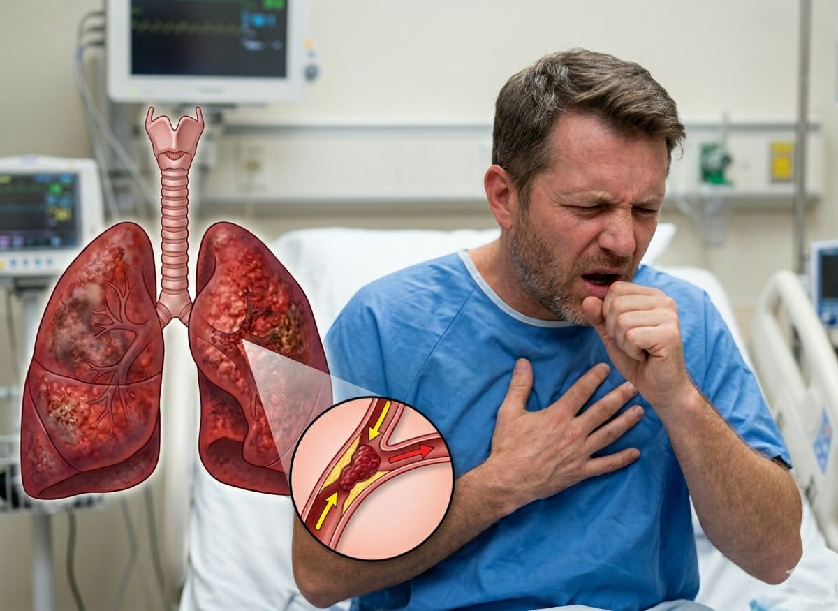
At the height of the pandemic, most people worried about COVID-19 mainly because of what it did to the lungs. What caught many doctors off guard was something happening deeper in the body, inside the bloodstream. COVID-19 isn’t just a respiratory illness. It can trigger abnormal blood clotting, raising the risk of life-threatening conditions like pulmonary embolism.
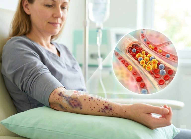
Research has shown that as many as 20 to 30% of hospitalized COVID-19 patients developed blood clots, even while on preventive treatment. In severe cases, these clots travel to the lungs, blocking major arteries and cutting off oxygen supply, a condition known as pulmonary embolism. It can escalate quickly and turn fatal without much warning. What’s even more concerning is that these clots can form during the infection itself, or weeks after recovery, right when many people assume the danger has passed.
The connection between COVID-19 and clotting comes down to how the virus affects the body. It can damage blood vessels, trigger inflammation, and disrupt normal clotting processes, creating exactly the conditions clots need to form quietly. For some, the first warning sign is sudden shortness of breath or chest pain. For others, there’s no warning at all until things become critical. That’s why understanding this link matters so much. COVID-19 may come and go, but its effects can stick around in ways many people don’t expect. In the sections ahead, you’ll learn about the connection between COVID-19 and pulmonary embolism, the warning signs worth watching for, and steps that can help protect your health.
The Link Between COVID-19 and Pulmonary Embolism
The main mechanism connecting COVID-19 to a higher risk of pulmonary blood clots involves a complex combination of direct viral damage to blood vessel cells, an intense and dysregulated inflammatory response (often called a cytokine storm), and the resulting activation of the body’s clotting system. This multi-pronged attack on the vascular system throws off the natural balance between clotting and bleeding, tipping things heavily toward clot formation, a condition known as COVID-19-associated coagulopathy (CAC). SARS-CoV-2 doesn’t just damage the lungs, it mounts a body-wide assault on the circulatory system, making dangerous clots a defining feature of severe disease. This process is often explained through Virchow’s triad, a century-old concept identifying three broad contributors to clot formation: injury to blood vessel walls, abnormal blood flow (stasis), and an increased tendency to clot. COVID-19 significantly affects all three.
What is a Pulmonary Embolism and Its Danger
A pulmonary embolism (PE) is a life-threatening condition that occurs when a blood clot, usually formed in a deep leg vein (deep vein thrombosis, or DVT), breaks loose, travels through the bloodstream, and lodges in one of the pulmonary arteries in the lungs. This blockage cuts off blood flow to part of the lung tissue, setting off a chain of dangerous physiological effects.
The main danger of a PE lies in the strain it places on the heart and the sharp drop it causes in the body’s oxygen levels. The consequences can be severe and even fatal without immediate treatment.
Restricted Blood Flow and Lung Tissue Damage: When an artery is blocked, the lung tissue it feeds can’t get oxygen-rich blood and may begin to die, a condition called pulmonary infarction. This doesn’t just impair the lung’s ability to exchange gases, it can also cause inflammation and fluid buildup.
Decreased Blood Oxygen Levels (Hypoxemia): With blood flow to parts of the lungs blocked, the body’s ability to oxygenate blood drops sharply. This leads to low oxygen throughout the body, which can damage vital organs like the brain and heart, and cause symptoms such as severe shortness of breath, confusion, and cyanosis (a bluish tint to the skin).
Increased Strain on the Heart: The heart’s right ventricle is responsible for pumping blood into the pulmonary arteries. When those arteries are blocked by a clot, pressure inside them rises sharply (pulmonary hypertension), forcing the right ventricle to work much harder to push blood past the obstruction. This intense strain can weaken, enlarge, and eventually cause the right ventricle to fail, a condition called acute right heart failure, one of the leading causes of death from PE. In severe cases, this can progress to obstructive shock and cardiac arrest.
Ways SARS-CoV-2 Virus Promotes Blood Clot Formation
SARS-CoV-2 promotes blood clot formation through three connected pathways: it directly attacks and damages the cells lining blood vessels, it triggers an overactive immune response that causes body-wide inflammation, and it directly activates platelets and other parts of the clotting system. Together, these create a uniquely clot-prone environment throughout the body.
1. Direct Damage to the Endothelium
SARS-CoV-2 enters human cells by binding to the ACE2 receptor, which is abundant on endothelial cells, the thin layer of cells lining every blood vessel. When the virus infects these cells, it causes direct injury and cell death. A healthy endothelium is smooth and releases substances that prevent clotting. A damaged one, though, becomes a strong trigger for coagulation, it exposes underlying tissue factor and collagen, which immediately kicks off the clotting cascade, and it loses its ability to regulate local blood flow and prevent platelets from clumping together. This widespread endothelial dysfunction, sometimes called endotheliitis, is a core feature of severe COVID-19.
2. Overactive Immune Response (Cytokine Storm)
In severe cases, COVID-19 triggers a hyperinflammatory state known as a cytokine storm, where the immune system floods the body with inflammatory signaling molecules (cytokines like IL-6 and TNF-alpha). This systemic inflammation further damages the endothelium, making blood vessels leaky and more prone to clotting. These cytokines also push the liver to produce more clot-promoting factors, like fibrinogen, and activate immune cells such as neutrophils, which can release neutrophil extracellular traps (NETs), web-like structures of DNA and protein that trap pathogens but also provide a scaffold clots can form around.
3. Activation of Platelets and Clotting Factors
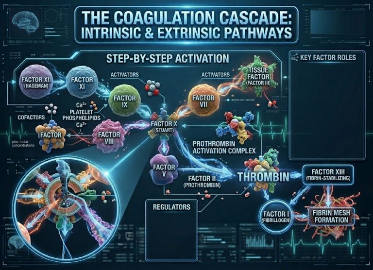
Both the direct viral infection and the inflammatory cytokines can activate platelets, the small cell fragments responsible for forming initial plugs at injury sites. In COVID-19, platelets become hyperactive, stickier and more prone to clumping, forming microthrombi (tiny clots) in small vessels, particularly in the lungs and other organs. On top of that, the inflammatory state activates the whole coagulation cascade, driving excess production of thrombin, the key enzyme that converts fibrinogen into fibrin, the protein mesh that forms the structural backbone of a stable clot.
The Risk of Blood Clots Higher with COVID-19 Compared to Other Viral Infections
Yes, the risk of both venous and arterial blood clots is significantly and consistently higher with COVID-19 than with other severe viral respiratory infections, influenza included. Any severe illness requiring hospitalization and causing immobility can raise clotting risk, but the scale and mechanisms behind clot formation in COVID-19 appear unique and far more pronounced. This has held up consistently across numerous large studies since the pandemic began.
Higher Incidence Rates: Studies published in journals like The Lancet and the BMJ have compared large groups of hospitalized COVID-19 and influenza patients. These analyses consistently show that venous thromboembolism (VTE), which includes both DVT and PE, occurs several times more often in COVID-19 patients. Among critically ill ICU patients, thrombotic complication rates have been reported as high as 30 to 40% in some COVID-19 groups, substantially higher than rates seen in ICU patients with influenza or other forms of acute respiratory distress syndrome (ARDS).
Different Clotting Profiles: The nature of the clotting in COVID-19 also looks distinct. Beyond typical VTE, COVID-19 is linked to a high rate of immunothrombosis, where inflammation and clotting become pathologically intertwined. This leads to microthrombi forming in the small blood vessels of the lungs and other organs, something far less commonly seen with influenza. COVID-19 has also been linked to a higher risk of arterial clots, leading to strokes and heart attacks, at rates above what’s typically seen with influenza.
Unique Pathophysiological Drivers: The main reason for this difference comes down to the specific way SARS-CoV-2 behaves. Its ability to directly infect and cause widespread endothelial damage via the ACE2 receptor is a key distinguishing factor. Influenza can cause systemic inflammation too, but it doesn’t typically trigger the same degree of severe, widespread endotheliitis. The combination of direct endothelial injury, an intense cytokine storm, and platelet hyperactivation in COVID-19 creates a uniquely strong clot-promoting state beyond what’s typically seen with other viral infections.
Symptoms of Pulmonary Blood Clots Following a COVID-19 Infection
Common Warning Signs of a Pulmonary Embolism
The most common warning signs of a pulmonary embolism involve a sudden, dramatic shift in respiratory and cardiovascular status, abrupt shortness of breath, severe chest pain, and a fast heart rate among them. These classic symptoms should never be brushed off, especially in someone recovering from or currently dealing with COVID-19.
Sudden Shortness of Breath (Dyspnea): This is the most common symptom. Unlike the gradual breathlessness that can come with a lung infection, dyspnea from a PE usually appears suddenly and without an obvious trigger. It can happen at rest or with very little activity and often feels disproportionate to what’s actually being exerted. The person may feel like they can’t get enough air no matter how deeply they breathe.
Sharp Chest Pain (Pleuritic Pain): This is another hallmark sign. The pain is often described as sharp, stabbing, or knife-like, and it’s usually located on one side of the chest. A key detail: the pain gets noticeably worse with a deep breath, coughing, sneezing, or even moving the upper body. This happens because of inflammation in the pleura, the lining of the lungs, which becomes irritated by the nearby clot and lack of blood flow.
Rapid or Irregular Heartbeat (Tachycardia/Palpitations): The heart tries to compensate for reduced oxygen and the blocked pulmonary arteries by beating faster. Someone might feel their heart racing, pounding, or fluttering. A resting heart rate consistently over 100 beats per minute is a significant warning sign.
Coughing Up Blood (Hemoptysis): Less common than the other symptoms, but coughing up blood or blood-streaked mucus is a very serious indicator of a PE. It suggests the lack of blood flow has already caused damage, or infarction, to lung tissue.
Dizziness, Lightheadedness, or Fainting (Syncope): These can occur if the clot is large enough to significantly block blood flow, causing a drop in blood pressure and reduced oxygen to the brain. Fainting alongside other PE symptoms points to a massive, life-threatening embolism.
Blood Clot Symptoms vs. COVID-19 Recovery Symptoms
Symptoms of a pulmonary blood clot can easily be mistaken for ordinary COVID-19 recovery symptoms, creating real potential for delayed diagnosis and treatment. This overlap is one of the biggest challenges in post-infection care. Many people recovering from COVID-19, particularly moderate to severe cases, experience lingering issues that mirror PE warning signs, making it hard to tell the difference between normal recovery and a medical emergency without a proper clinical evaluation.
1. Overlapping Symptoms
The most prominent overlapping symptoms are shortness of breath and fatigue. Long COVID often involves persistent breathlessness, especially during activity, as the lungs continue healing. Profound fatigue and general malaise are also common to both conditions. A lingering cough can show up during COVID recovery too. If someone’s already experiencing these symptoms, they might write off new or worsening signs of a PE as just part of the ongoing recovery process.
2. Key Differentiating Factors
Despite the overlap, there are important differences worth watching for. The most important one is how the symptoms start and feel.
Sudden Onset: While post-COVID breathlessness tends to be chronic or worsen gradually with activity, shortness of breath from a PE is usually very sudden and can happen even at rest. Someone might go from feeling relatively fine to acutely breathless within minutes.
Pleuritic Chest Pain: The sharp, stabbing pain that worsens with a deep breath is highly characteristic of a PE and isn’t typical of COVID-19 recovery. COVID-19 can cause chest pain too, but it’s usually described as a dull ache or tightness, whereas PE pain is distinctly sharp and tied to breathing.
Unilateral Leg Swelling: Swelling, pain, tenderness, and redness in one leg is a classic sign of DVT, the precursor to most pulmonary embolisms. This symptom has nothing to do with COVID-19 recovery and is a major red flag that other respiratory symptoms might actually be a PE.
Risk Factors for Developing Pulmonary Blood Clots from COVID-19
The main risk factors for developing pulmonary blood clots from COVID-19 include how severe the infection is, hospitalization (ICU admission in particular), older age, a personal or family history of blood clots, and existing medical conditions.
Generally speaking, the more intense the body’s inflammatory response to the virus, the higher the risk of clotting complications. Patients who need hospitalization face substantially higher risk than those with mild, outpatient cases, not just because their illness is more severe, but also because of prolonged immobility, which is its own powerful, independent risk factor for clots. Other significant contributors include obesity, active cancer, recent surgery or trauma, and underlying cardiovascular conditions like hypertension and heart failure.
Groups At Risk for COVID-19-associated Blood Clots
The people considered most at risk for COVID-19-associated blood clots are those with severe or critical disease requiring hospitalization, especially ICU admission, older adults, and people with certain pre-existing health conditions. This risk profile comes from the combination of intense inflammation, immobility, and underlying vascular vulnerability.
Patients with Severe COVID-19: This is the single biggest risk factor. People who develop pneumonia, ARDS, or need supplemental oxygen or mechanical ventilation experience the most extreme levels of systemic inflammation and endothelial damage. Inflammatory markers in these patients, like C-reactive protein (CRP) and D-dimer, are often highly elevated, directly tracking with a clot-prone state. Prolonged immobility from being bedridden in a hospital or ICU setting adds to this risk further by letting blood pool in the deep veins of the legs.
Older Adults: Advanced age (typically over 60 or 65) is its own independent risk factor for VTE. Older individuals often have less resilient vascular systems, higher baseline inflammation, and more coexisting health conditions. Their response to COVID-19 tends to be more severe, and they’re more likely to experience prolonged immobility during illness, all of which compound to significantly raise clotting risk.
Individuals with Pre-existing Conditions: Several underlying health issues dramatically raise the likelihood of developing a clot during a COVID-19 infection, including:
- Obesity: Fat tissue is a known source of chronic, low-grade inflammation and clot-promoting factors.
- Cardiovascular Diseases: Conditions like hypertension, coronary artery disease, and heart failure mean the vascular system is already compromised and more vulnerable to the damage caused by the virus and inflammation.
- Cancer: Active cancer is a well-known clot-promoting state on its own, and combined with a severe infection like COVID-19, the risk climbs even higher.
- Prior History of Blood Clots: Anyone with a personal or family history of DVT or PE carries an inherently higher risk of a new clot, especially when faced with a major trigger like severe COVID-19.
- Diabetes: This condition is linked to endothelial dysfunction and a pro-inflammatory state, adding to overall risk.
Long COVID to Long-term Risk of Blood Clots
Growing evidence strongly suggests that Long COVID, also called Post-Acute Sequelae of COVID-19 (PASC), can raise the long-term risk of blood clots for months, and potentially over a year, after the initial infection resolves. This risk isn’t confined to the acute phase of illness. The lingering biological disruptions from the virus can keep the body in a clot-prone, hypercoagulable state long after someone tests negative.
The mechanisms behind this extended risk are still being actively researched, but several factors tied to Long COVID appear to play a role.
Persistent Inflammation: One core feature of Long COVID is ongoing, low-grade systemic inflammation. The immune system doesn’t fully return to baseline, and inflammatory markers can stay elevated for months. This chronic inflammation keeps irritating the endothelial lining of blood vessels, preventing full healing and maintaining a state of endothelial dysfunction that promotes clotting.
Endothelial Dysfunction: The widespread endothelial damage that occurs during acute infection may not fully resolve in people with Long COVID. These blood vessel linings can stay dysfunctional, unable to properly regulate blood flow or prevent inappropriate clotting. This ongoing vascular issue is a key driver of long-term clotting risk.
Autoimmunity and Persistent Viral Reservoirs: Some theories suggest Long COVID may involve an autoimmune component, where the body produces antibodies that mistakenly attack its own cells, including endothelial cells. Others propose that fragments of the virus or its genetic material may linger in certain tissues, acting as an ongoing trigger for inflammation. Either scenario could contribute to a sustained clot-promoting environment.
Clinical Evidence: Large population studies back up this long-term risk. A notable study published in the BMJ tracked millions of people and found the risk of DVT stayed significantly elevated for up to three months post-infection, while PE risk remained elevated for up to six months. Another study in Circulation showed an increased risk of cardiovascular events, including blood clots, up to a year after acute infection, even among people who’d had mild cases. This points to a profound, long-lasting impact on the vascular system.
Pulmonary Blood Clots Diagnosis In COVID-19 Patients
Diagnosing a PE in a COVID-19 patient involves a mix of clinical assessment, blood tests, and advanced imaging, since symptoms like shortness of breath and chest pain can overlap with the viral pneumonia itself. The first step is usually a blood test measuring D-dimer levels, a protein fragment produced when a blood clot dissolves in the body. Elevated D-dimer isn’t specific to PE (inflammation from COVID-19 can raise it too), but a normal level can help rule out a clot with a fairly high degree of confidence.
If D-dimer is elevated and clinical suspicion is high, the gold standard for diagnosis is a CT pulmonary angiography (CTPA). This specialized CT scan uses an injected contrast dye to let radiologists visualize the pulmonary arteries and identify any blockages or clots with real precision. If CTPA isn’t feasible, a ventilation/perfusion (V/Q) scan may be used instead. This structured diagnostic pathway is essential for accurately identifying and treating these life-threatening clots.
Initial Screening: The D-dimer test is a sensitive but non-specific screening tool. In COVID-19, where inflammation is widespread, its main value often lies in ruling things out, a normal result makes a significant clot less likely, letting clinicians explore other causes for symptoms without jumping straight to more invasive imaging.
Confirmatory Imaging: CTPA is the definitive diagnostic test. It provides a detailed, three-dimensional map of the lung’s blood vessels, clearly showing the location, size, and extent of any clots, which is critical for determining severity and guiding treatment.
Supportive Assessments: Alongside these primary tests, doctors may use an ECG to check for signs of heart strain caused by the PE, along with an echocardiogram to assess right ventricle function, since it can become enlarged and strained by a large clot.
Consequences of Surviving a COVID-19-related Pulmonary Embolism
Recovery often extends well beyond the initial hospital stay, with many people facing long-term health consequences that affect their quality of life. One of the most common is post-PE syndrome, marked by persistent symptoms like shortness of breath (especially with exertion), chronic chest discomfort, and reduced capacity for physical activity. These symptoms can linger for months or even years, making it hard to return to previous levels of function.
In a more severe, rarer subset of cases, the clots don’t fully dissolve. Instead, they organize into fibrous, scar-like tissue within the pulmonary arteries, leading to a serious condition called Chronic Thromboembolic Pulmonary Hypertension (CTEPH). This scarring blocks blood flow, causing dangerously high blood pressure in the lungs and straining the right side of the heart, which can eventually lead to heart failure if left untreated. Many survivors also need long-term management to prevent recurrence. These long-term effects highlight why follow-up care and monitoring matter so much after a PE diagnosis.
Ongoing Anticoagulation: To prevent new clots, most survivors are prescribed anticoagulant therapy (blood thinners) for at least three to six months, sometimes indefinitely. This requires regular monitoring and lifestyle adjustments to manage the added bleeding risk.
Functional and Psychological Impact: Beyond physical symptoms, post-PE syndrome can take a real psychological toll, contributing to anxiety, depression, and fear of recurrence. Full recovery often involves cardiac rehabilitation programs designed to slowly and safely rebuild exercise tolerance and confidence.
Management of CTEPH: For patients who develop CTEPH, treatment is complex, potentially involving specialized medications to lower pulmonary artery pressure, and in some cases, a major surgical procedure called pulmonary thromboendarterectomy to remove the chronic, organized clots from the arteries.
Blood Clot Caused by COVID-19 vs. One Caused by Prolonged Inactivity
The fundamental difference between a clot caused by COVID-19 and one caused by prolonged inactivity comes down to the underlying mechanism: a flight-related clot is primarily a mechanical issue involving blood flow, while a COVID-19 clot stems from a systemic, inflammatory disease process. A clot formed during a long flight is a classic example of venous stasis, where blood in the deep leg veins pools and becomes stagnant from lack of movement. This sluggish flow lets clotting factors accumulate, leading to DVT.
If part of this clot breaks off, it can travel to the lungs and cause a PE. The main driver here is mechanical, localized to the veins. COVID-19, by contrast, causes severe systemic inflammation and an increased tendency to clot throughout the body. SARS-CoV-2 can directly infect and damage the endothelium (the inner lining of blood vessels), triggering an aggressive inflammatory response and activating the clotting cascade throughout the entire circulatory system, not just in the legs.
This widespread, inflammation-driven clotting is far more aggressive and can form clots directly within the small vessels of the lungs (immunothrombosis) in addition to causing traditional DVTs. This difference in underlying mechanism explains why COVID-19-associated clots tend to be more severe and widespread.
Primary Cause: A flight-related clot comes from stasis (impaired blood flow due to immobility). A COVID-19-related clot comes from a combination of endothelial injury (viral damage to vessel walls), hypercoagulability (an overactive clotting system), and inflammation (the cytokine storm).
Location of Formation: Both can lead to a PE, but flight-related clots almost always start as a DVT in the legs. COVID-19 can cause DVTs too, but it’s also linked to clotting in unusual locations, including the arteries, brain, and the tiny blood vessels within the lungs themselves.
Systemic vs. Localized: Immobility creates a localized problem in the deep veins. COVID-19 creates a systemic problem where the entire circulatory system is primed for clotting due to viral attack and an overblown immune response, making the risk much higher and harder to predict.
Does COVID-19 Vaccination Reduce The Risk of Developing These Blood Clots?
Yes, COVID-19 vaccination significantly reduces the risk of developing blood clots from a SARS-CoV-2 infection by preventing severe disease, the primary driver behind these clotting events. The severe inflammatory response, endothelial damage, and hypercoagulable state that lead to pulmonary emboli are hallmarks of moderate to severe COVID-19 illness, often requiring hospitalization.
Vaccination works by training the immune system to recognize and neutralize the virus quickly upon exposure. This prevents the virus from replicating out of control and triggering the widespread, dysregulated immune reaction (the cytokine storm) responsible for systemic inflammation and blood vessel damage. By keeping infection severity down, often to a mild, cold-like illness or even no symptoms at all, vaccination effectively removes the conditions needed for dangerous clots to form.
The risk of developing a PE or other major blood clot is substantially higher in unvaccinated individuals who contract COVID-19 compared to vaccinated individuals. The protective benefit of vaccination far outweighs any associated risks, which puts the danger of infection-related clots into clear perspective.
Mechanism of Protection: Vaccination reduces the risk of severe COVID-19, hospitalization, and ICU admission. Since severe disease is the strongest predictor of developing COVID-19-associated coagulopathy, preventing severe illness is the most effective way to prevent related blood clots.
Comparative Risk: Studies have consistently shown that DVT and PE occur far more often following a SARS-CoV-2 infection than in the weeks following vaccination. For example, the risk of cerebral venous sinus thrombosis (a rare brain clot) is estimated to be 8 to 10 times higher after infection than after an mRNA vaccine.
Clarifying Vaccine-Associated Risks: While extremely rare cases of clotting disorders (like VITT with certain viral vector vaccines) were identified early on, these events are exceedingly uncommon. The danger posed by the virus itself, given its tendency to cause widespread, life-threatening clots, remains a far greater and more common threat to health overall.
FAQs
1. What are the pulmonary complications of COVID-19? COVID-19 can affect the lungs in several ways, ranging from mild irritation to serious, long-term damage. Common complications include pneumonia, where the air sacs fill with fluid, and ARDS, a severe condition that makes breathing extremely difficult. Blood clot formation is another major concern, which can lead to pulmonary embolism. Some people also experience long-term inflammation and scarring (fibrosis), which can reduce lung function even after recovery. These complications vary depending on how severe the infection was and the person’s overall health.
2. How long after COVID are you susceptible to blood clots? Blood clot risk can stay elevated for several weeks to months after a COVID-19 infection. Research suggests the highest risk falls within the first 2 to 6 weeks, though in some cases it may persist longer, especially for people who had severe illness or were hospitalized. This lingering risk is exactly why symptoms like sudden shortness of breath or chest pain shouldn’t be ignored, even after recovery.
3. Does COVID permanently damage the lungs? Not always, but in some cases, COVID-19 can cause long-term or permanent lung damage. Severe infections may lead to scarring of lung tissue (fibrosis), which can affect breathing capacity. Many people recover fully, though, especially with mild to moderate illness. The outcome often depends on how severe the infection was and how quickly treatment started.
4. How long does it take for lungs to heal from COVID? Recovery time varies quite a bit. For mild cases, lung function often returns to normal within a few weeks. For more severe infections, healing can take several months, with symptoms like shortness of breath or fatigue sometimes lingering. Rehabilitation, breathing exercises, and proper medical follow-up can all support recovery and improve lung function over time.
Conclusion
COVID-19 changed how people think about respiratory illness, but its impact reaches well beyond the lungs. The connection between COVID-19 and pulmonary embolism reveals a hidden layer of risk, one that can develop quietly, even after the initial infection has passed. Understanding this link matters. Blood clots don’t always show up immediately, and symptoms can appear when you least expect them. That’s why ongoing awareness is so important, especially during recovery. Paying attention to your body, recognizing warning signs, and seeking timely care can genuinely make a difference.
The reassuring part is that knowledge puts you in a better position. Understanding how COVID-19 affects both the lungs and the bloodstream means you’re better equipped to protect your health and respond quickly if something feels off.