8+ Key Symptoms of Sarcoidosis Disease Explained Clearly
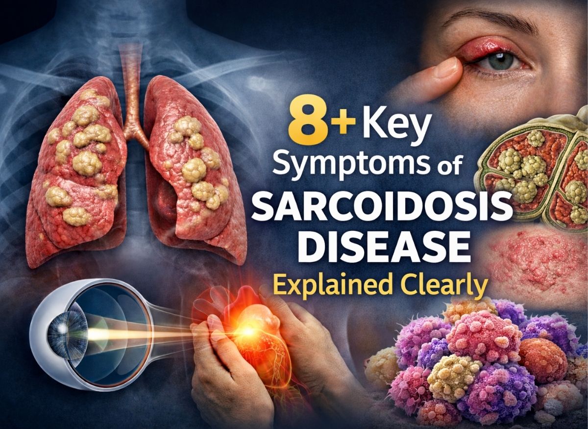
Sarcoidosis often develops quietly, yet its symptoms can affect multiple organs and gradually disrupt everyday life. Many people search for clear answers because the early signs are easy to overlook or confuse with common conditions. Understanding the key symptoms is essential for recognizing when something’s not right and seeking timely evaluation. The condition causes inflammation leading to tiny clusters of cells called granulomas, most often in the lungs, skin, eyes, and lymph nodes.
Below, we break down the key symptoms of sarcoidosis so you can connect what you’re experiencing with what may be happening inside the body, an important step toward proper diagnosis, monitoring, and long-term management.
What Is Sarcoidosis?
Sarcoidosis is a complex, multi-system inflammatory condition of unknown origin. It’s clinically defined by noncaseating granulomas, abnormal clusters of inflammatory cells, forming in one or more organs. This represents an exaggerated, dysregulated immune response that causes persistent inflammation and can significantly alter an organ’s structure and function.
While it can affect nearly any part of the body, there’s a strong tendency toward involvement in the lungs and associated lymph nodes, seen in over 90% of patients. The outlook is highly variable: for many people it’s a temporary, acute condition that resolves on its own, while for others it becomes chronic and debilitating. A major challenge in medicine is predicting which patients will experience a severe, progressive form requiring long-term intervention to prevent irreversible organ damage.
How Granulomas Form and Cause Harm
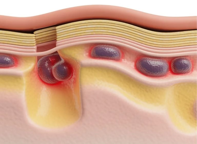
The disease progresses through granuloma formation, compact, organized clusters of immune cells that interfere with normal organ function. This begins when the immune system perceives a foreign trigger and launches a defense. T-lymphocytes activate and recruit macrophages to the site, which clump together and transform into specialized cells, forming the core of a granuloma.
The physical presence of these clusters causes several effects:
- Respiratory disruption. In pulmonary sarcoidosis, granulomas can stiffen lung tissue, making breathing harder, reducing oxygen exchange efficiency, and potentially leading to pulmonary fibrosis.
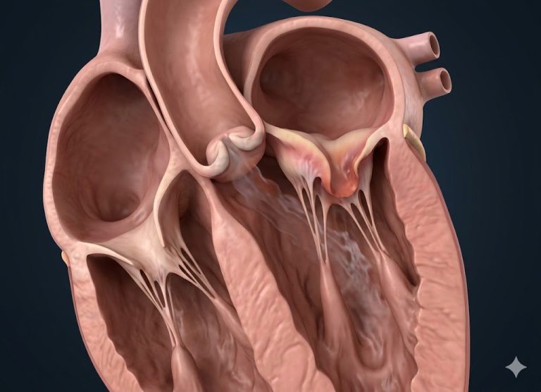
- Cardiac and liver interference. When granulomas infiltrate the heart muscle (cardiac sarcoidosis), they disrupt electrical conduction and can cause arrhythmias. Similarly, liver involvement (hepatic sarcoidosis) can disrupt normal liver function.
- Systemic inflammation and flare-ups. Ongoing immune activity releases inflammatory chemicals into the bloodstream, causing systemic symptoms like profound fatigue and fever. A “sarcoid flare” signals a period of heightened immune activity and potential new organ involvement.
Who’s Most at Risk
Sarcoidosis most often affects young adults between 20 and 40. In the U.S., it’s about three to four times more common in African Americans than Caucasian Americans, and African Americans often experience a more severe, chronic form with a higher likelihood of multi-organ involvement.
- Race and ethnicity: Beyond the higher incidence in African Americans, people of Scandinavian, German, and Irish descent also show increased prevalence, suggesting ties to ancestral lineage.
- Age and sex: Peak onset is between 20 and 40, with a secondary peak often seen in women over 50; women are slightly more likely to develop the condition than men.
- Genetics: Having a first-degree relative with sarcoidosis significantly raises risk, pointing to a hereditary component. Researchers have identified specific genes in the human leukocyte antigen (HLA) system that regulate immune sensitivity.
For some, the disease appears as Lofgren syndrome, a specific, often acute form; for others, it shows up as skin involvement (subcutaneous sarcoidosis) or a distinctive nose and facial rash known as lupus pernio.
The Key Symptoms
Symptoms vary widely depending on which organ systems are affected, and the condition often mimics other illnesses, which can complicate diagnosis. Some people experience a sudden onset, as with Lofgren syndrome (fever, swollen lymph nodes, joint pain, and a specific skin rash); others face a slow, gradual progression over years. Many people remain entirely symptom-free, with the disease discovered incidentally on a chest X-ray.
1. Persistent cough and shortness of breath. The respiratory system is the most frequently affected area, with some degree of lung involvement in over 90% of patients. Symptoms range from mild irritation to serious complications:
- Shortness of breath is the most common respiratory sign, as inflammation impairs oxygen transfer from the lungs into the bloodstream
- A dry, chronic cough, distinct from an infection-related cough, stems from irritation of the bronchial tubes
- Chest pain, usually from swollen lymph nodes near the lungs, which over time can contribute to irreversible scarring (fibrosis)
2. Skin rashes and sores. These affect roughly 25% of patients and serve as important visual clues. They occur when inflammatory granulomas form within the skin’s deeper layers:
- Erythema nodosum: tender, red bumps typically on the shins, which, paired with swollen chest lymph nodes, indicates Lofgren syndrome, a generally favorable acute form
- Lupus pernio: a chronic, disfiguring condition with purplish plaques on the nose, cheeks, and ears, often signaling a more severe disease course
- Other skin changes: rash-like eruptions, nodules under the skin, or swelling and discoloration in old tattoos and scars
3. Extreme fatigue, fever, and flare-ups. These constitutional symptoms result from a widespread inflammatory battle driven by an overactive immune system:
- Profound fatigue, an overwhelming exhaustion, similar to a severe flu but lasting months, not relieved by rest
- Fever and night sweats, a classic sign of flare activity, particularly early on
- Unexplained weight loss, from a chronic inflammatory state that can cause the body to break down tissue for energy
When these coincide with organ-specific symptoms, like heart palpitations or liver discomfort, it confirms the systemic nature of the disease, and monitoring for flares is essential for adjusting treatment.
4. Eye problems. Ocular involvement occurs in up to 50% of patients and, for many, is the very first sign of a systemic issue. Because it can lead to glaucoma, cataracts, and permanent vision loss, comprehensive eye exams are important:
- Uveitis (inflammation of the eye’s middle layer): anterior uveitis causes redness and light sensitivity, while posterior uveitis can cause visual floaters
- Conjunctival granulomas: small yellowish bumps on the white of the eye, often symptom-free but useful for diagnosis through biopsy
- Dry eye syndrome: inflammation of the tear glands reduces tear production, causing a gritty or itchy feeling
- Optic nerve involvement: rare but serious, potentially causing sudden vision loss requiring immediate attention
5. Joint pain and stiffness. This affects 10% to 35% of patients and often accompanies a flare:
- Acute sarcoid arthritis, often part of Lofgren syndrome, involves sudden, symmetric swelling and redness in multiple joints, particularly the ankles
- Chronic sarcoid arthritis, a rarer form in long-standing disease, causes persistent swelling and potential joint damage, sometimes resembling rheumatoid arthritis
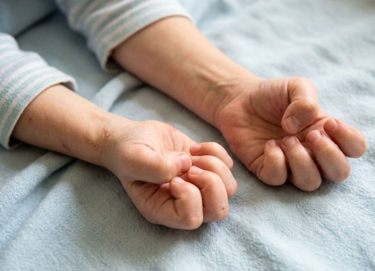
- Dactylitis and bone cysts: in chronic cases, granulomas can affect the small bones of the hands and feet, causing swollen “sausage digits,” visible on X-ray as “punched-out” cystic lesions
6. Cardiac involvement. This is one of the most life-threatening forms. It’s clinically diagnosed in about 5-10% of patients, though autopsy studies suggest granulomas may be present in the heart in up to 25% of those with sarcoidosis:
- Arrhythmias and heart block, from damaged electrical pathways, causing a slow heart rate, dizziness, or fainting, and carrying a risk of sudden cardiac death if unmanaged
- Heart failure, if widespread scarring weakens the heart’s pumping ability
- Chest pain, less common, but sometimes mimicking angina from inflammation of the heart muscle or small vessels
7. Swollen lymph nodes. This is an extremely common, classic sign:
- Bilateral hilar lymphadenopathy, symmetric swelling of the nodes where the bronchi enter the lungs, is the most frequent finding, present in the vast majority of patients, and highly suggestive of sarcoidosis in a young adult
- Superficial lymphadenopathy: painless, firm lumps in the neck, armpits, or groin, often biopsied to confirm diagnosis
- Mediastinal lymphadenopathy: enlargement of other central chest nodes, helping distinguish sarcoidosis from similar lung conditions
8. Neurological symptoms (neurosarcoidosis). Occurring in only 5-10% of cases, but potentially severe when it does:
- Cranial neuropathy, most commonly facial nerve palsy (resembling Bell’s palsy), causing sudden weakness on one side of the face
- Meningitis, inflammation of the membranes around the brain, causing chronic headaches, neck stiffness, and confusion
- Brain and spinal cord lesions, granulomas forming masses in the central nervous system that can cause seizures, cognitive changes, or limb weakness, significantly affecting overall outlook
What Causes Sarcoidosis
The exact cause is still under intense research, but the leading theory is that it results from an abnormal immune response to an unknown environmental trigger in people who are genetically susceptible. It isn’t caused by one isolated factor, but by a complex interplay between genetics and exposure to specific external substances.
When a genetically vulnerable person encounters a specific trigger, a bacterium, virus, or inorganic chemical particle, their immune system launches an exaggerated, prolonged inflammatory response instead of clearing the substance and returning to normal. This persistent response leads to granuloma formation. This model explains why sarcoidosis isn’t contagious yet often clusters within families and certain ethnic groups.
Is It an Autoimmune Condition?
Not in the classic sense. It’s considered an immune-mediated inflammatory disease with autoimmune features, rather than a true autoimmune disease like lupus, where the immune system attacks healthy self-cells directly.
In sarcoidosis, evidence suggests the immune system is mounting an overly aggressive response against a persistent, poorly cleared external substance rather than attacking the body’s own tissue. Still, it shares features with autoimmune disorders: a dysregulated immune system, chronic inflammation, and a strong genetic link to HLA genes. People with sarcoidosis also have a higher rate of coexisting autoimmune conditions like Sjögren’s syndrome or celiac disease, suggesting a shared predisposition to general immune dysregulation.
Potential Environmental Triggers
Suspected triggers fall into a few categories, supported by case clusters linked to specific occupations or major exposure events:
- Infectious agents: DNA and proteins from certain bacteria, particularly Mycobacteria (related to tuberculosis) and Propionibacterium acnes, have been found in granuloma samples
- Inorganic and environmental particles: occupational exposure to certain dusts and chemicals raises risk, including beryllium (which causes a mimicking condition called berylliosis), aluminum, silica, and agricultural dusts containing insecticides
- Notable exposure events: a well-documented cluster occurred among first responders exposed to dust and debris from the World Trade Center collapse on September 11, 2001
Regardless of the trigger, the result is often a flare requiring careful symptom monitoring and long-term management.
Sarcoidosis vs. Tuberculosis
Both conditions cause granulomas, but they’re fundamentally different. Sarcoidosis is an immune-mediated inflammatory response, while tuberculosis (TB) is a strictly infectious disease caused by Mycobacterium tuberculosis.
Because TB is contagious and spreads through airborne droplets, it requires immediate isolation and public health reporting, while sarcoidosis is not transmissible. Even though the two can share overlapping symptoms like a dry cough and fever, they’re pathologically worlds apart.
The most critical difference is microscopic: sarcoidosis produces non-caseating granulomas, solid, organized cell clusters without a “cheese-like” dead center, while TB typically produces caseating granulomas with significant tissue death at the core, which is why biopsy is often necessary for an accurate diagnosis. Standard TB tests (PPD skin test or IGRA blood tests) consistently come back negative in sarcoidosis.
Treatment approaches are opposite. Sarcoidosis, especially during a flare, is usually treated with corticosteroids or other immunosuppressants to calm the overactive immune response. But giving these same medications to someone with tuberculosis could be fatal, letting the bacterial infection spread unchecked. So while sarcoidosis treatment focuses on immune modulation, TB recovery depends entirely on a rigorous, multi-month course of specialized antibiotics to eradicate the bacteria.
How Sarcoidosis Is Diagnosed
Diagnosis is primarily a process of exclusion, ruling out other potential causes or infections before confirming sarcoidosis. It combines clinical evaluation, imaging, and lab analysis.
Because over 90% of patients have respiratory involvement, a chest X-ray is almost always the first step, potentially revealing bilateral hilar lymphadenopathy or specific lung infiltrates. A high-resolution CT (HRCT) scan provides a clearer picture of granulomas and potential scarring. Blood tests may show elevated angiotensin-converting enzyme (ACE), though this isn’t a standalone diagnostic test, and other labs check calcium levels and liver function to assess liver involvement.
The gold standard for confirmation is a tissue biopsy, typically taken from the lungs (via bronchoscopy), skin lesions, or accessible lymph nodes. A pathologist examines the sample to:
- Identify granulomas: the definitive finding is the presence of non-caseating granulomas
- Exclude other conditions: ruling out mimics like tuberculosis or fungal diseases
- Support comprehensive assessment: combining imaging, labs, and biopsy to confirm diagnosis and begin targeted treatment
The Siltzbach Staging System
“Staging” in sarcoidosis specifically refers to the radiographic appearance of the lungs, and it doesn’t always correlate with symptom severity or heart involvement. A patient at an early stage may still have a heavy systemic symptom burden.
- Stage 0: Normal chest X-ray, found in 5-10% of patients whose disease affects other organs while sparing the lungs
- Stage I: Bilateral hilar lymphadenopathy only, with the highest rate of spontaneous remission
- Stage II: Both swollen lymph nodes and lung tissue abnormalities
- Stage III: Lymph node enlargement has resolved, but lung infiltrates remain
- Stage IV: Permanent lung scarring (pulmonary fibrosis), representing advanced, irreversible damage and a more chronic disease course
Long-Term Outlook
The outlook is highly variable. For roughly two-thirds of patients, sarcoidosis is a temporary health challenge that resolves spontaneously within two to five years, especially common with Lofgren syndrome.
For about 30% of people, though, it becomes chronic and can lead to permanent organ damage:
- Pulmonary and cardiac risks: about 5-15% of patients develop pulmonary fibrosis, and monitoring for cardiac involvement is essential since it can be life-threatening
- Ongoing monitoring: because of flare risk, regular check-ups, breathing tests, and imaging are needed even during quiet periods
- Chronic disease management: for progressive cases, long-term corticosteroids or immunosuppressants are often necessary to manage flares and maintain quality of life
While a minority face severe complications like neurosarcoidosis or serious cardiac involvement, most people with sarcoidosis lead full lives by staying attentive to their symptoms and following a professional care plan.
Frequently Asked Questions
What is sarcoidosis and how does it affect the body?
A multi-system inflammatory condition where small clusters of immune cells (granulomas) form in different organs, most commonly the lungs, lymph nodes, skin, and eyes, though they can appear almost anywhere. Over time, this inflammation can interfere with normal organ function, especially if it becomes chronic.
What are the most common symptoms?
A persistent dry cough, shortness of breath, fatigue, and skin rashes are most common, along with fever, night sweats, or swollen lymph nodes for many people. Because it can affect multiple organs, symptoms vary widely, and it’s sometimes discovered incidentally during routine imaging.
Is it serious or life-threatening?
It can range from mild to severe depending on which organs are involved. Many people have a temporary form that resolves on its own within a few years, but in some cases it becomes chronic and can lead to complications like lung scarring, heart rhythm problems, or vision loss. Early recognition plays an important role in preventing long-term damage.
What causes it?
The exact cause remains unknown, but it’s believed to result from an abnormal immune response, possibly triggered by environmental factors like bacteria, viruses, or chemical exposure in people with a genetic predisposition. Instead of resolving the trigger, the immune system keeps reacting, leading to chronic inflammation and granuloma formation.
How is it diagnosed?
Usually through a combination of imaging, blood work, and biopsy. A chest X-ray or CT scan often shows enlarged lymph nodes or lung involvement, blood tests may indicate inflammation (though not definitively), and a biopsy confirming non-caseating granulomas is the most reliable way to establish the diagnosis.
Can it go away on its own?
Yes, many cases resolve without long-term treatment, especially in the early stages, with a significant number of patients experiencing spontaneous remission within a few years. Others develop a chronic form requiring ongoing monitoring and management, and because the course is unpredictable, regular follow-up is essential.
The Bottom Line
Recognizing the symptoms of sarcoidosis early can make a meaningful difference in how the condition is managed over time. Some symptoms may seem mild or unrelated at first, but they often reflect underlying inflammation worth paying attention to. Watching for patterns like persistent cough, unexplained fatigue, skin changes, or vision issues can help guide timely medical conversations and prevent complications. Because sarcoidosis varies so much from person to person, staying informed, tracking symptoms, and seeking professional evaluation when needed are key steps toward maintaining overall health, and with the right understanding and support, many people manage sarcoidosis effectively and continue their daily routines with confidence.