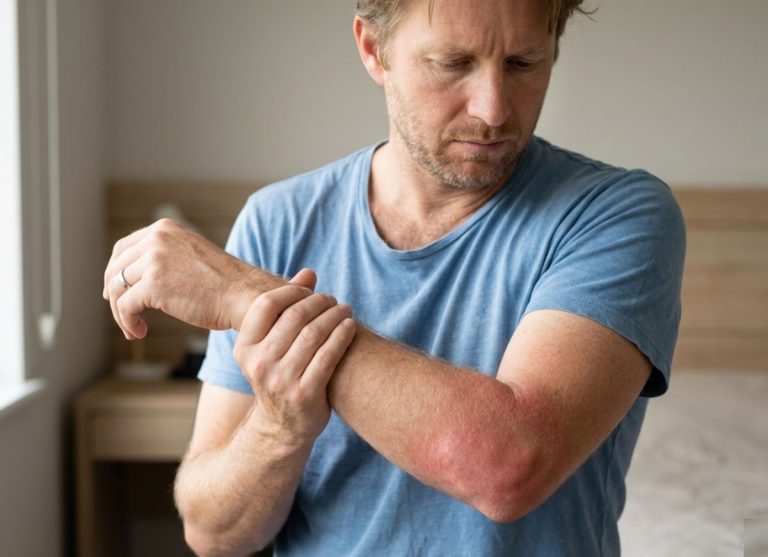
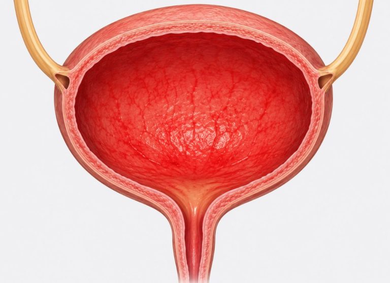
10 Causes of Erythema Nodosum and What They May Reveal
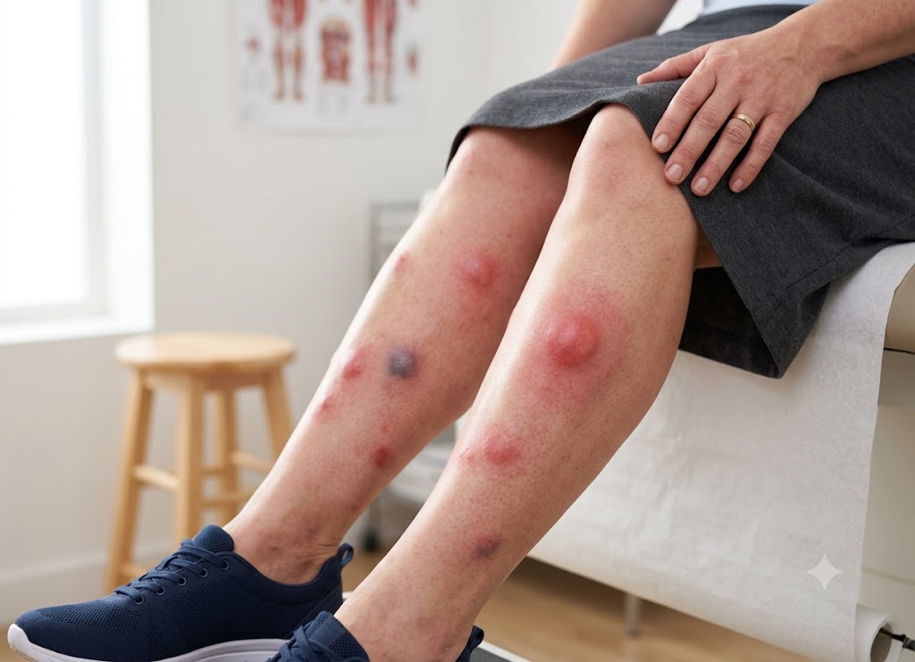
Erythema nodosum is a skin problem that produces sore, red or purplish lumps, usually on the shins. As the lumps fade they can look like bruises. They come from inflammation in the layer of fat under the skin. The condition matters because it is often the body’s reaction to something else, such as an infection, a medicine, an inflammatory disease, or pregnancy.
Knowing the possible causes explains why treating only the skin is sometimes not enough. The lumps can follow strep throat, tuberculosis, inflammatory bowel disease, sarcoidosis, certain drugs, or other immune triggers. Sometimes no cause turns up, but doctors may still look for hidden conditions based on your symptoms and history. This article covers 10 causes and what each one can tell you about your health.
What is Erythema Nodosum and What Are Its Key Symptoms?
Erythema nodosum is a type of panniculitis, meaning inflammation of the fat beneath the skin. It shows up as tender red nodules, most often on the shins. It is a delayed-type hypersensitivity reaction, so it acts as a skin signal of some other process, whether an infection, a systemic disease, or a drug, rather than a disease in its own right. Recognising how it looks and feels is the first step toward finding the cause.
1. Defining the Inflammatory Skin Pathology
Erythema nodosum is a reactive skin disorder marked by the sudden appearance of very tender red nodules, typically on the shins. It is the most common form of panniculitis, a group of conditions that inflame the fat layer under the skin.
Ordinary rashes sit in the top layers of skin (the epidermis or dermis). This condition starts deeper, in the fibrous walls (septa) that divide the fat lobules. Because of that depth, the lesions feel like firm lumps under the skin instead of surface changes.
It is an immune-driven reaction. The immune system targets something specific, such as a bacterial component, a drug molecule, or a self-antigen in an autoimmune disease, and sets off local inflammation. The small blood vessels in the fatty septa become inflamed, which produces the painful red nodules.
During an exam, doctors often compare the lesions with reference photos of erythema nodosum to check their shape and their symmetrical spread.
                 [Deep Tissue Hypersensitivity Profile]
                                    │
       ┌────────────────────────────┴────────────────────────────┐
       ▼                                                         ▼
[Epidermal / Dermal Rashes]                             [Erythema Nodosum]
- Superficial surface scaling                           - Deep septal panniculitis
- Flat or raised plaques                                - Located in subcutaneous fat
- Confined to top skin layers                           - Firm, deep, nodular architecture
2. Investigating the Underlying Causes
Erythema nodosum is rarely a stand-alone disease. It works more like a warning light pointing to something else. Finding the cause is essential to identify the illness driving the immune response.
                      [Primary ETIOLOGICAL TRIGGERS]
                                    │
       ┌────────────────────────────┼────────────────────────────┐
       ▼                            ▼                            ▼
[Infectious Agents]        [Systemic Illnesses]        [Pharmacological Inputs]
- Streptococcal pharyngitis - Sarcoidosis (Löfgren's)   - Oral contraceptive pills
- Tuberculosis exposure     - Inflammatory Bowel (IBD)  - Sulfonamide antibiotics
- Fungal/Deep lung spores  - Behçet's disease profile  - Bromides & Iodides
Infectious triggers.
In children and young adults, the most common cause is a recent bacterial infection. Strep throat is a leading trigger, and the nodules usually appear two to three weeks after the throat infection clears. Other infectious causes include tuberculosis, Yersinia gut infection, and deep fungal infections such as coccidioidomycosis and histoplasmosis.
Systemic and autoimmune conditions.
The nodules can also point to a hidden inflammatory or autoimmune disease:
- Sarcoidosis: There is a strong link between the two. When nodules appear with fever, joint pain, and swollen lymph nodes in the chest (hilar adenopathy), the pattern is called Löfgren’s syndrome.
- Inflammatory bowel disease (IBD): The nodules can be an outside sign of Crohn’s disease or ulcerative colitis. Skin flares often follow the level of inflammation in the gut.
Medication and hormonal triggers.
Some prescription drugs can set off this reaction. The most frequent are oral contraceptive pills, sulfonamide antibiotics, and bromides. Hormonal shifts, such as those in pregnancy, can also trigger an episode by changing how the immune system reacts. If extensive testing finds no cause, the condition is called idiopathic.
3. Cutaneous and Systemic Symptoms
Symptoms fall into two groups: changes in the skin and effects on the whole body. Whole-body symptoms often begin one to three weeks before the nodules appear and serve as an early warning.
Skin symptoms and how the lesions progress.
The hallmark is the sudden appearance of very tender nodules. They start as bright red, warm, firm, painful lumps about 1 to 5 centimetres across. They are most common on the front of the lower legs and often appear on both legs at once.
Over three to six weeks the colour changes the way a healing bruise does, going from bright red to deep purple, then yellowish-brown, before disappearing. A key feature is that the nodules heal completely, without opening, ulcerating, or leaving scars. Doctors compare real cases with reference photos to tell these non-scarring lumps from more destructive types of panniculitis.
Whole-body symptoms.
Since the underlying driver is systemic inflammation, the skin changes come with wider symptoms:
- Fever and malaise: A low-grade fever, ongoing tiredness, and a general unwell feeling are common, and can resemble the start of a bad viral illness.
- Joint pain and swelling (arthralgia): More than half of patients have joint pain, most often in the ankles, then the knees and wrists. It ranges from mild aching to visible swelling, redness, and heat (arthritis). Sometimes it lasts for months after the skin has cleared.
- Other signs: Depending on the cause, people may also have a lasting cough (often tied to sarcoidosis or lung infections), severe headaches, or stomach cramps and long-term diarrhoea (linked to IBD).
10 Common Triggers for Erythema Nodosum and the Diseases They Signal
The 10 common triggers are infections (Streptococcus, tuberculosis, yersiniosis, coccidioidomycosis), systemic inflammatory diseases (sarcoidosis, IBD, Behçet’s disease), medications, pregnancy, and idiopathic cases with no known cause. Each one sets off a hypersensitivity reaction that inflames the fat under the skin and points to a different underlying condition. Pinning down the specific cause matters most, because treatment and outlook depend on the underlying disease.
1. Pathogenic Triggers: Bacterial and Fungal Sources
Infection is a frequent trigger. When the body meets certain bacteria or fungi, the immune system can mount a delayed hypersensitivity reaction that causes deep, painful inflammation in the fat layer. Identifying the cause is what guides proper care.
                [Infectious Hypersensitivity Inflow]
                                    │
       ┌────────────────────────────┴────────────────────────────┐
       ▼                                                         ▼
[Bacterial Antigens]                                    [Fungal Antigens]
- Group A Streptococcus (Pharyngitis)                   - Coccidioides immitis (Spore Inhalation)
- Mycobacterium tuberculosis (Primary Tuberculid)        - Triggers localized "Desert Rheumatism"
- Yersinia enterocolitica (Undercooked Pork/Water)       - Indicates robust, protective immunity
1. Streptococcal Infections
Reactions after strep are the most common identifiable cause, especially in children and young adults. Nodules usually show up two to three weeks after Group A strep throat.
As the immune system fights the bacteria, it forms immune complexes that settle in the small blood vessels of the fat. Doctors confirm this trigger with a rapid strep test, a throat culture, or a blood test for raised Antistreptolysin O (ASO) titres.
2. Tuberculosis (TB)
Sudden tender nodules can be an outside sign of a first-time infection with Mycobacterium tuberculosis. Here the skin lesions are a reactive change called a tuberculid, showing that the body is mounting a strong cell-mediated defence against the bacteria.
Where TB is common, the nodules call for prompt tests, including a chest X-ray and either an Interferon-Gamma Release Assay (IGRA) or a PPD skin test.
3. Yersiniosis
This trigger comes from a gut infection with Yersinia enterocolitica, usually caught from contaminated water or undercooked pork. It causes gastroenteritis with fever, severe stomach pain, and diarrhoea.
The red nodules usually appear one to two weeks after the digestive symptoms begin. The link is most often diagnosed in European countries, using stool cultures or antibody tests.
4. Coccidioidomycosis (Valley Fever)
This fungal infection comes from breathing in Coccidioides spores, found in the dry soil of the southwestern United States and parts of Central and South America. It usually begins in the lungs as a mild, flu-like illness.
When nodules appear together with fever and severe joint pain, the pattern is known as “desert rheumatism.” The nodules are actually a good sign, because they show the immune system is strong enough to contain the fungus and stop it spreading.
2. Systemic Inflammatory and Autoimmune Indicators
When infection is not the cause, an overactive internal immune response is often responsible. The tender nodules can be an early clue to a systemic autoimmune or autoinflammatory disease.
5. Sarcoidosis (Löfgren’s Syndrome)
Sarcoidosis is a whole-body disease in which small clusters of inflammatory cells, called non-caseating granulomas, form in organs, most often the lungs and lymph nodes. Erythema nodosum is one of its most common outward signs.
When the nodules occur together with high fever, severe ankle pain, and swollen chest lymph nodes visible on X-ray, the combination is called Löfgren’s syndrome. This acute form usually has a good outlook and often clears by itself without long-term immune-suppressing treatment.
6. Inflammatory Bowel Disease (IBD)
These painful lumps are the most common skin sign of IBD, affecting up to 15% of people with Crohn’s disease and 10% of those with ulcerative colitis.
The skin lesions track the activity of the bowel disease. A sudden flare of nodules often means the gut is flaring too. So when nodules appear with stomach cramps, ongoing diarrhoea, or rectal bleeding, a quick check for IBD is needed.
Flow: active Crohn’s/UC flare → gut wall inflammation → widespread vessel hypersensitivity → skin nodules.
7. Behçet’s Disease
Behçet’s is a rare, long-term inflammatory disorder in which blood vessels throughout the body become inflamed (vasculitis). Its classic signs are repeated painful mouth sores, genital ulcers, and severe eye inflammation (uveitis).
The nodules in Behçet’s can look just like ordinary ones on the surface. Under a microscope, though, they often show real damage to the blood vessel walls. Spotting them alongside recurring mouth or genital ulcers is an important step in diagnosis.
3. Pharmacological, Physiological, and Idiopathic Triggers
Often there is no infection or disease behind the condition. It may instead follow a medicine, a hormonal change, or nothing identifiable at all.
8. Medications
Many prescription drugs can cause this reaction, usually within one to three weeks of starting the medicine:
- Oral contraceptive pills: Estrogen-containing pills are among the most common drug triggers. The synthetic hormones are thought to change how the immune system reacts, and the nodules usually clear once the pill is stopped.
- Sulfonamides: This antibiotic class is well known for causing drug-related skin reactions, including this one.
- Other agents: Some cough syrups, imaging contrast dyes, and specialised cancer drugs such as BRAF inhibitors have also been linked.
9. Pregnancy
The condition can develop in pregnancy, most often in the second trimester. The likely trigger is the sharp rise in natural hormones, especially estrogen, which shifts the body’s immune baseline.
The nodules can be very painful and upsetting for the mother, but they do not harm the baby. They usually clear on their own within a few weeks after delivery.
10. Idiopathic Manifestations
Even after thorough testing, 30% to 60% of cases are idiopathic, meaning no infection, disease, or drug can be found.
This is a diagnosis of exclusion, made only after a doctor has ruled out everything else. It can be frustrating not to have an answer, but the outlook is excellent. The condition clears by itself and is managed with rest, leg elevation, and nonsteroidal anti-inflammatory drugs (NSAIDs) for pain and swelling.
The Process for Diagnosing and Managing Erythema Nodosum
Care has two parts. First, the doctor confirms the condition through an exam and selected tests. Then the focus shifts to finding and treating the trigger while easing the painful nodules. The doctor must also rule out look-alike skin conditions. The long-term outlook is generally good, though the nodules can return if the root cause is not dealt with.
1. The Clinical Diagnostic Framework
Diagnosis takes two steps. The doctor first recognises the lesions by sight and touch, then orders targeted tests to find the underlying trigger. The exam focuses on the condition’s typical location: painful, firm, red nodules on the front of both shins, appearing symmetrically.
By pressing on the nodules, the doctor confirms that the inflammation lies deep in the fat and not on the skin’s surface. The doctor also takes a detailed history, asking about recent respiratory infections, new medicines, or unexpected digestive changes that could point to an internal cause.
A standard set of tests then helps confirm the diagnosis and find the trigger. A complete blood count checks for active infection, while Erythrocyte Sedimentation Rate (ESR) and C-Reactive Protein (CRP) measure overall inflammation.
An Antistreptolysin O (ASO) titre checks for a recent strep infection that may have gone unnoticed. If the patient reports a recent sore throat, a throat culture looks for Group A beta-hemolytic streptococci. A chest X-ray is also a core part of the workup, to look for enlarged lymph nodes on both sides of the chest (bilateral hilar lymphadenopathy), which suggests sarcoidosis, or lung infiltrates that suggest active TB.
If the picture is unusual, a deep skin biopsy reaching into the fat is done. Under the microscope, the tissue typically shows septal panniculitis without vasculitis. This means immune cells are gathered only in the connective-tissue walls between fat lobules, and the nearby blood vessels are not destroyed.
               [Diagnostic Diagnostic Investigative Flow]
                                      │
     ┌────────────────────────┬───────┴────────┬────────────────────────┐
     ▼                        ▼                ▼                        ▼
[Serological Assays]   [Microbiology]   [Chest Radiography]      [Deep Tissue Biopsy]
- CBC Evaluation       - Throat Swab    - Rules out Sarcoidosis  - Confirms Septal
- Elevated ESR & CRP   - Tracks Strep     (Hilar Adenopathy)       Panniculitis
- High ASO Titers        Pharyngitis    - Screens for Active TB  - Rules out Vasculitis
2. Differential Diagnostics: Distinguishing Look-Alike Lesions
Many inflammatory conditions cause painful red swelling on the lower legs, so doctors must separate this one from its look-alikes before treating. Cellulitis is a deep bacterial infection of the dermis and the tissue beneath it, usually caused by Streptococcus or Staphylococcus.
It can look similar at first glance, but cellulitis almost always affects one leg only, spreads quickly, and has poorly defined edges. Instead of firm, deep nodules, it produces a flat, spreading area of warm, swollen skin. It is often accompanied by high fever, chills, and a visible break in the skin where bacteria got in.
The difference from other fat inflammations, such as erythema induratum of Bazin, is subtler but just as important. Erythema induratum centres on the fat lobules themselves and involves direct inflammation of blood vessels. It has a strong historical link to tuberculosis. Erythema nodosum, by contrast, is a septal panniculitis that appears symmetrically on the front of the shins and heals without ulcers or scars. Erythema induratum produces long-lasting purplish nodules, typically on the back of the calves.
Those lobular lesions often break down and ulcerate, leaving deep permanent scars. Rarer forms, such as alpha-1 antitrypsin deficiency panniculitis, also cause tender nodules, but they ulcerate heavily and leak an oily discharge. A deep skin biopsy is needed to tell them apart by their tissue features.
3. Comprehensive Treatment and Management Strategies
The main goal is to find and resolve the cause. If a medicine is responsible, it should be stopped right away. If a bacterial infection is found, targeted antibiotics are given. While the cause is being handled, other measures reduce local pain and inflammation.
Non-drug measures come first. Patients are advised to rest in bed and raise their legs above heart level, which lowers pressure in the lower legs, reduces swelling, and eases tissue tension. Once the painful phase settles, graduated compression stockings can support the veins and help control longer-term swelling.
           [Symptomatic Management Protocol]
                                    │
       ┌────────────────────────────┼────────────────────────────┐
       ▼                            ▼                            ▼
[Mechanical Rest]          [First-Line Medications]    [Advanced Interventions]
- Strict bed rest          - High-dose NSAIDs          - Potassium Iodide drops
- Elevate legs above heart   (Ibuprofen / Naproxen)    - Systemic Corticosteroids
- Reduces edema/pressure   - Inhibits prostaglandins     (Only after ruling out TB)
Drug treatment relies mainly on NSAIDs such as ibuprofen, naproxen, or indomethacin as the first choice. They block prostaglandin production and quickly ease joint pain and nodule tenderness. For persistent or recurring cases, a short course of oral potassium iodide solution can work well, because it helps stabilise cell membranes and limits the release of inflammatory oxygen radicals from white blood cells.
In severe, non-infectious cases that do not respond to first-line treatment, a short course of oral prednisone may be used to bring inflammation down fast. Before starting steroids, however, doctors must be sure there is no underlying infection such as TB or a deep fungal infection, because dampening the immune system could let these spread dangerously. To monitor progress, doctors often compare against baseline photos to track how the nodules shrink and fade.
4. Prognosis and Recurrence Pathways
The long-term outlook is generally excellent. The condition is self-limiting, so the painful nodules usually clear by themselves within three to six weeks, even without intensive treatment. As they heal, they change colour like a fading bruise, from bright red to deep purple and finally to a faint yellowish-brown.
Reassuringly, the nodules heal without breaking open or leaving permanent scars. Some people notice temporary darkening of the skin (post-inflammatory hyperpigmentation), but this fades over the following months.
Flow: persistent or repeated trigger → immune system reactivated → nodules return on the shins.
Ultimately, though, the long-term picture depends on the cause. If a chronic disease such as Crohn’s or sarcoidosis is behind it, new crops of nodules often appear alongside flare-ups of that illness. Likewise, if a medicine such as an oral contraceptive or a sulfonamide antibiotic caused the reaction, taking that drug again will often bring it back. Idiopathic cases rarely recur, but avoiding known triggers remains the best way to prevent future episodes.
Conclusion
Erythema nodosum is often more than a simple rash, because it can reflect the body’s response to an infection, inflammation, a medicine, or another medical condition. Common causes include strep infections, tuberculosis, sarcoidosis, inflammatory bowel disease, pregnancy, and certain drugs. In many cases the lumps improve once the underlying cause is treated or controlled. If painful nodules appear on your legs, especially with fever, joint pain, cough, digestive symptoms, or a recent change in medication, see a doctor to find out what is behind them.
Frequently Asked Questions
1. What is erythema nodosum?
Erythema nodosum is an inflammatory skin condition affecting the fat layer beneath the skin. It typically causes tender, red, swollen bumps on the shins, though other areas can be involved. The bumps may turn purple or bruise-like as they heal. It is usually a reaction to another trigger, not an infection of the skin itself.
2. What causes erythema nodosum?
Causes include infections, medications, pregnancy, autoimmune conditions, inflammatory bowel disease, and other inflammatory disorders. Strep throat is one of the most common infectious triggers. Sarcoidosis, tuberculosis, Crohn’s disease, and ulcerative colitis may also be linked. Sometimes no specific cause is found even after evaluation.
3. Can erythema nodosum reveal an infection?
Yes, it can sometimes point to an infection that needs attention. Strep is a well-known cause, especially after a sore throat. Tuberculosis, certain fungal infections, and other bacterial infections may also be considered, depending on where you live, your symptoms, and your risk factors. A doctor may order tests if you have fever, cough, sore throat, or other warning signs.
4. Is erythema nodosum linked to inflammatory bowel disease?
Yes, it can be linked to inflammatory bowel disease, including Crohn’s disease and ulcerative colitis. It may appear during a digestive flare or alongside stomach pain, diarrhoea, weight loss, or blood in the stool. The skin bumps often improve when the bowel inflammation is treated. This link is one reason persistent or unexplained erythema nodosum should be medically reviewed.
5. How is erythema nodosum treated?
Treatment depends on the cause and how severe the symptoms are. Mild cases may improve with rest, leg elevation, cold compresses, and pain relief recommended by a healthcare provider. If an infection, drug reaction, or inflammatory disease is responsible, treating that trigger is important. Do not stop prescribed medicines without talking to a doctor first.