5 Warning Signs of Esophageal Varices You Shouldn’t Ignore
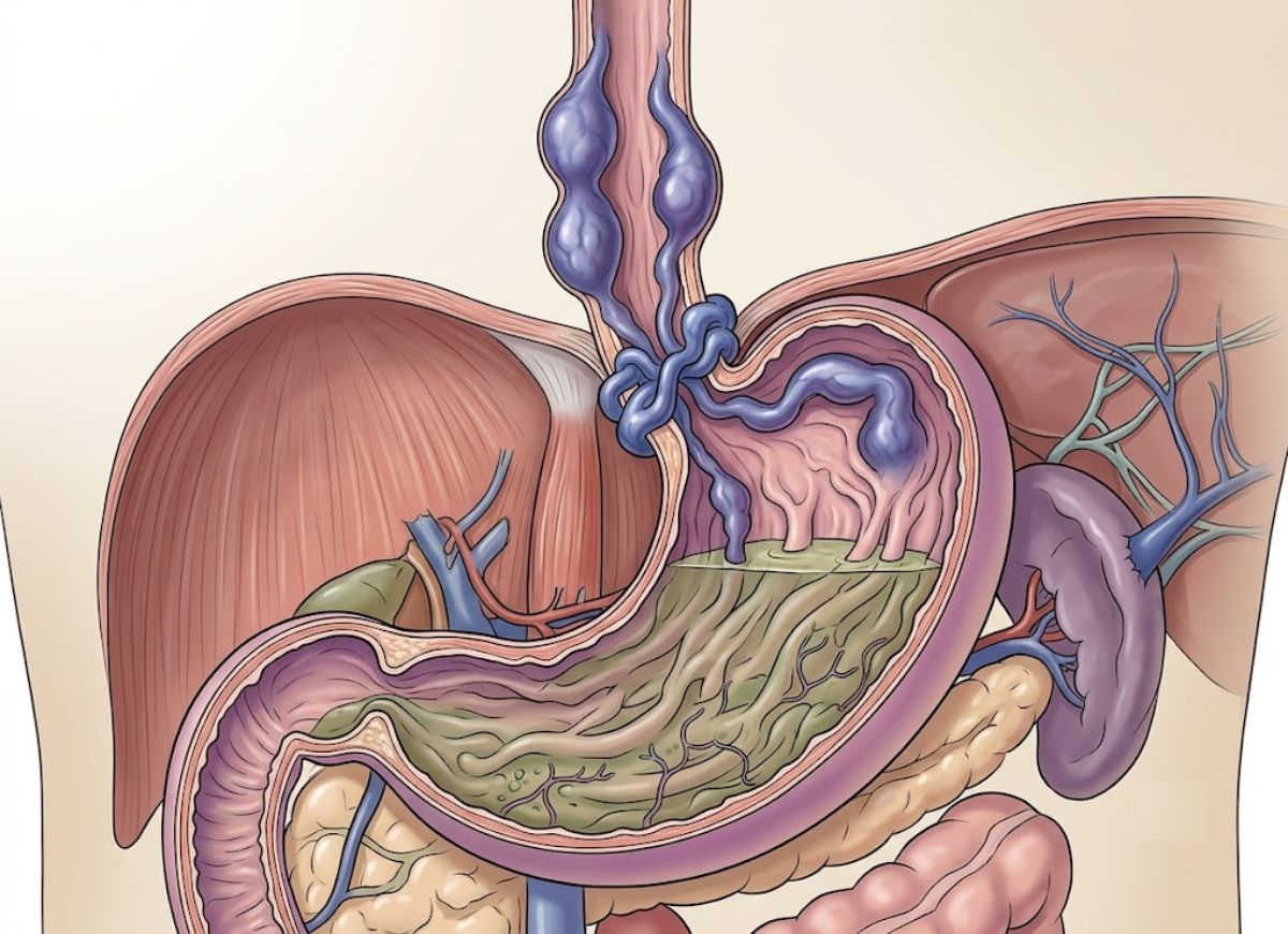
The digestive system depends on a network of blood vessels working correctly, and when pressure builds up inside certain veins, serious problems can follow. Esophageal varices are veins in the esophagus that swell up when blood flow through the liver gets blocked or restricted. They often stay quiet in the early stages, but if they tear open, they can cause dangerous internal bleeding.
These varices are usually linked to portal hypertension a rise in pressure inside the portal vein system, which is the network of veins carrying blood into the liver. Liver disease, especially cirrhosis (long-term scarring of the liver), is the leading cause. Once blood can’t move through the liver normally, it gets pushed into smaller veins nearby, including the ones lining the esophagus. Over months or years, those veins stretch, thin out, and become prone to rupture.
Most people don’t realize they have esophageal varices until something goes wrong, which is exactly why it helps to know the warning signs. How noticeable the symptoms are depends on how large the varices have grown and whether bleeding has already started. Some people notice small changes; others develop symptoms that call for emergency care right away.
Research shows that esophageal varices are common in people with cirrhosis roughly 30–40% of those with early-stage (compensated) cirrhosis have them, and that number climbs to as high as 85% in people with advanced liver disease. Because of this strong connection, doctors keep a close eye on anyone with chronic liver disease, watching for changes that raise the odds of a bleed.
This guide walks through five warning signs of esophageal varices, explains why they form in the first place, and covers when it’s time to get medical help.
What Are Esophageal Varices?
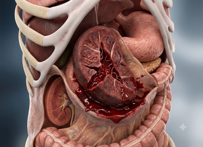
Esophageal varices are veins in the lower esophagus that have become abnormally swollen and stretched because of high pressure in the portal venous system. Think of them as the esophagus’s version of varicose veins.
They show up as a complication of advanced liver disease cirrhosis being the most frequent cause. When blood can no longer pass easily through a scarred, damaged liver, the body reroutes it through smaller veins that offer less resistance. The esophagus happens to sit along one of these detour routes.
The problem is that these rerouted veins were never built to carry high-pressure blood. Their walls are thin, which makes them fragile and prone to bursting and when that happens, it can trigger life-threatening bleeding. Their presence is essentially a red flag pointing to serious liver damage and portal hypertension underneath.
How Does Liver Disease Lead to Esophageal Varices?
Chronic liver disease cirrhosis in particular causes heavy scarring inside the liver that blocks normal blood flow. This blockage raises pressure in the portal vein system, forcing blood to squeeze through smaller alternate veins, including those in the lower esophagus. Under that pressure, those veins swell and become varices.
Here’s the chain of events in more detail. It typically starts with ongoing liver injury from heavy alcohol use, chronic hepatitis B or C infection, or fatty liver disease. Over time, healthy liver tissue gets replaced by scar tissue, and that scarring is what defines cirrhosis. This scarred tissue behaves like a dam sitting inside the liver, slowing the flow of blood trying to pass through.
As blood backs up against that “dam,” pressure builds much the way water pressure rises behind a dam wall. This is portal hypertension. To relieve the pressure and keep blood moving, the body opens up new pathways that bypass the liver altogether.
Blood gets diverted into smaller veins around the esophagus, stomach, and rectum vessels that were never designed to handle this kind of volume or pressure. The esophageal veins, sitting close to the surface with thin walls, are especially vulnerable. They swell, twist, and stretch under the extra blood flow, turning into esophageal varices.
Are Esophageal Varices Always Dangerous?
Yes esophageal varices are always treated as dangerous, because their biggest threat is the possibility of sudden, severe, life-threatening bleeding. Even small varices that aren’t causing symptoms signal serious liver disease and unstable portal hypertension underneath. The real danger isn’t the varices sitting quietly it’s the risk that they could rupture without warning.
Their walls are thin and already under constant strain from high blood pressure passing through them. A rupture can be triggered by something as ordinary as a coughing fit, straining, or acid reflux wearing down the vessel wall. Once a varix tears, the bleeding that follows can escalate fast.
Because this bleeding comes from a high-pressure vein system, blood loss can happen quickly and in large amounts, pushing the body toward hypovolemic shock a state where the heart can’t pump enough blood to keep organs supplied. The death rate from a first variceal bleed sits somewhere between 15% and 25%, and without treatment, the chances of it happening again are high.
That’s why even silent, symptom-free varices need active medical management regular endoscopy checkups and treatments to lower portal pressure before a first bleed ever occurs.
5 Common Symptoms of Esophageal Varices
Vomiting Significant Amounts of Blood (Hematemesis)
Vomiting a large amount of blood is one of the most alarming signs that a varix has ruptured. It usually shows up as bright red blood, which means the bleeding is fresh, fast, and coming directly from a high-pressure vessel in the upper digestive tract.
When a swollen esophageal vein tears, blood rushes rapidly into the esophagus and stomach. This irritates the stomach lining and triggers vomiting. Bright red blood specifically tells doctors the bleed is happening fast, with no time for stomach acid to change its color.
The amount can range from streaks in the vomit to large clots or pools of liquid blood this isn’t a subtle sign. Blood loss can add up quickly, pushing the body toward shock in a short window of time.
If bleeding is slower, blood may sit in the stomach longer before being vomited. During that time, stomach acid breaks down the blood’s hemoglobin, turning it dark brown or black often described as looking like coffee grounds.
Coffee-ground vomit still points to a serious bleed, but bright red blood usually means the bleeding is more acute and faster-moving more typical of a ruptured varix. Either way, vomiting blood is always a medical emergency requiring immediate hospital care.
Black, Tarry, or Bloody Stools (Melena)
Black, sticky, tar-like stools known medically as melena are a classic sign of bleeding somewhere in the upper digestive tract, including from ruptured varices. This happens when blood from the bleed travels through the stomach, small intestine, and colon.
Along the way, digestive acids, enzymes, and gut bacteria break down the blood’s hemoglobin and turn it into a black pigment called hematin. That’s what gives the stool its dark, sticky texture and strong, unpleasant odor.
Melena typically requires a fairly significant blood loss around 50 to 100 milliliters, or roughly 3-4 tablespoons and takes several hours to appear as the blood moves through the digestive system. So melena often signals that bleeding has been going on for a while, even before more dramatic symptoms like vomiting blood show up. It’s an important clue that helps doctors pinpoint an upper GI bleed.
It’s worth distinguishing melena from hematochezia, which is fresh, bright red blood passed from the rectum usually a sign of bleeding lower in the digestive tract, like the colon.
That said, in cases of extremely heavy, fast upper GI bleeding, blood can move through the intestines too quickly to be broken down fully, resulting in red or maroon stools instead of black ones. Whatever the color, any blood in the stool needs urgent medical evaluation.
Lightheadedness, Dizziness, or Fainting (Syncope)
Feeling dizzy, lightheaded, or actually passing out are serious warning signs tied to major blood loss and the brain not getting enough blood flow.
When a varix bursts, the sudden loss of blood volume causes blood pressure to drop sharply a condition called hypotension. The brain is extremely sensitive to drops in blood pressure and oxygen.
As blood pressure falls, less oxygen-rich blood reaches the brain, a state called cerebral hypoperfusion. This is what causes the dizzy, weak, lightheaded feeling especially noticeable when standing up too quickly (known as orthostatic hypotension).
As bleeding continues and blood pressure keeps dropping, the body’s backup systems start to fail. The heart speeds up (tachycardia) to try to compensate, but if too much blood has been lost, that’s not enough to keep up. Once blood flow to the brain drops below a critical point, brain function is briefly disrupted, leading to fainting. This is actually the body’s protective response lying flat helps restore blood flow to the brain by removing gravity’s effect.
When these symptoms appear alongside vomiting blood or black stools in someone with known liver disease, it’s a clear sign of major bleeding and possible shock a serious medical emergency.
Jaundice (Yellowing of Skin and Eyes)
Jaundice a yellow tint to the skin and the whites of the eyes signals the underlying liver damage behind esophageal varices, rather than being a direct symptom of the bleeding itself. When it shows up alongside signs of GI bleeding, it’s a strong clue to doctors that the bleeding is connected to chronic liver disease.
Jaundice happens when bilirubin, a yellow pigment created when red blood cells break down, builds up in the bloodstream. A healthy liver normally processes bilirubin and clears it out through bile.
In advanced liver disease like cirrhosis, damaged liver cells can’t process bilirubin properly anymore. It accumulates in the blood, and its yellow color becomes visible in the skin and eyes. So when someone shows signs of GI bleeding vomiting blood, black stools along with jaundice, doctors immediately suspect bleeding esophageal varices.
Jaundice essentially acts as an outward sign of the liver failure and portal hypertension driving the whole condition. It shows the liver is struggling to do its job, which makes managing the bleeding more complicated and usually points to a worse overall outlook.
Rapid Heart Rate and Low Blood Pressure (Shock)
A fast heartbeat paired with low blood pressure are hallmark signs of hypovolemic shock a life-threatening state caused by heavy, rapid blood loss from a ruptured varix. Shock means the body’s organs and tissues aren’t getting enough oxygen and nutrients because there simply isn’t enough blood circulating.
When bleeding starts, the body kicks off compensatory responses to protect vital organs like the brain and heart. One of the earliest is a faster heartbeat, trying to circulate whatever blood remains more efficiently. That’s why a rapid pulse is often an early clue of serious bleeding.
But as blood loss continues, these compensations start to fail. Blood volume drops too low to maintain pressure in the arteries, and blood pressure begins to fall. A fast, weak pulse combined with dropping blood pressure is the textbook picture of shock setting in.
Other signs can include cool, clammy skin (as blood gets diverted away from the extremities), fast shallow breathing, confusion, and reduced urination.
This is a top-priority medical emergency. Without immediate IV fluids, blood transfusions, and control of the bleeding source, the lack of oxygen can quickly lead to organ damage, organ failure, and death. This progression from bleeding to shock is exactly what makes ruptured esophageal varices so dangerous.
When to Seek Medical Help?
Bleeding Esophageal Varices
Bleeding esophageal varices are always a life-threatening emergency with a high risk of death if not treated immediately. The danger comes from how quickly and heavily blood loss can happen. Varices are already swollen, thin-walled veins sitting under high pressure from portal hypertension.
Once one ruptures, the bleeding isn’t a slow trickle it’s often a heavy gush into the esophagus and stomach. A person can lose a large share of their blood volume within minutes to hours, which quickly leads to hypovolemic shock the heart no longer has enough blood to maintain pressure and deliver oxygen to organs.
Without fast intervention, this can progress to organ failure and death. The death rate for a single episode of acute variceal bleeding can reach 20% or higher, even with modern treatment available. In the emergency room, the priorities are restoring blood volume with IV fluids and transfusions, protecting the airway from blood, and stopping the bleeding usually with emergency endoscopy.
The underlying liver disease makes things harder too, since it interferes with blood clotting and raises the risk of infection and other complications. Every minute matters delaying care sharply increases the risk of a fatal outcome.
Type of Doctor Treating Esophageal Varices
Esophageal varices are primarily managed by gastroenterologists and hepatologists, usually working as a team. Each brings a different piece of expertise from handling an active bleed to managing the liver disease behind it long-term.
A gastroenterologist specializes in the digestive system esophagus, stomach, and liver and performs the key diagnostic and treatment procedures. During active bleeding, they’ll perform an emergency upper endoscopy to locate the varices and stop the bleeding, most often using variceal band ligation, where small elastic bands are placed around the bleeding vein to cut off its blood supply.
A hepatologist focuses specifically on liver, gallbladder, and bile duct disease, and takes the lead on long-term management. They treat the underlying liver disease and portal hypertension, prescribe medications like beta-blockers to lower portal pressure and reduce the risk of a first or repeat bleed, manage other cirrhosis complications, and evaluate patients for liver transplant when needed the only real cure for the underlying disease.
In complicated cases, an interventional radiologist may step in to perform a TIPS procedure (transjugular intrahepatic portosystemic shunt), which creates a new pathway to relieve portal pressure.
Esophageal Varices Diagnosis
The gold-standard way to diagnose esophageal varices is an upper endoscopy, also called an EGD (esophagogastroduodenoscopy). It gives doctors a direct look inside the upper digestive tract. During the procedure, a gastroenterologist passes a thin, flexible tube with a light and camera through the mouth and down into the esophagus, stomach, and the start of the small intestine.
This lets the doctor closely examine the lining for problems. They’re specifically looking for swollen, twisted, bluish veins in the lower esophagus the telltale sign of varices.
The procedure not only confirms varices are present but also allows doctors to grade their size (small, medium, or large) and spot warning markers like red streaks or spots on the vein surface that suggest a higher bleeding risk. Patients are usually sedated and asked to fast beforehand so the stomach is empty for a clear view.
During the exam, doctors classify the varices by bleeding risk. Small, straight varices are considered lower risk, while large, twisted ones taking up a lot of space in the esophagus are high risk and may need immediate preventive treatment.
People diagnosed with cirrhosis are generally advised to get screening endoscopies regularly typically every one to three years depending on how advanced their liver disease is and whether varices were already found.
While EGD remains the standard, imaging tests like CT or MRI scans can sometimes pick up indirect signs of portal hypertension, such as an enlarged spleen or widened portal vein. A newer, non-invasive option capsule endoscopy, where a patient swallows a tiny camera pill can also spot varices, though it doesn’t allow doctors to treat anything during the exam.
Main Risk Factors for Developing Bleeding Varices
Whether a varix is likely to bleed depends on a mix of factors: its physical size and appearance, how severe the underlying liver disease is, and direct pressure readings within the portal system.
Not every varix carries the same risk, so doctors weigh several things together. Size is the single biggest predictor larger varices have thinner, more stretched walls that can’t withstand high blood pressure as well, making them far more likely to rupture than smaller ones.
Specific warning marks on the vein surface, called stigmata, also matter. These include red streaks (called “red wale marks”) and cherry-red spots both signs that the vessel wall has become especially thin and fragile in that spot.
Finally, how advanced the underlying liver disease is often measured using the Child-Pugh or MELD score is a strong predictor too. A higher score means more advanced liver failure, which usually means higher portal pressure and a greater overall risk of bleeding.
Esophageal Varices vs. Peptic Ulcers
Esophageal varices and peptic ulcers are two very different conditions, even though both can cause serious upper GI bleeding. The key difference is what causes them. Varices develop because of portal hypertension, almost always tied to advanced liver disease like cirrhosis.
High pressure in the portal vein forces blood to find new routes back to the heart, and that’s what creates these swollen, fragile veins in the esophagus. Peptic ulcers, on the other hand, are open sores in the lining of the digestive tract. They’re usually caused by H. pylori bacterial infection or long-term use of NSAIDs like ibuprofen or aspirin.
These factors break down the protective lining of the stomach or duodenum, letting stomach acid damage the tissue underneath. This difference in cause also explains their location varices form in the esophagus, while ulcers typically show up in the stomach or duodenum. Treatment differs completely too.
Varices are treated by lowering portal pressure with beta-blockers or procedures like TIPS while ulcers are treated by clearing the H. pylori infection with antibiotics and reducing stomach acid with proton pump inhibitors.
How to Prevent Esophageal Varices
Bleeding from esophageal varices can often be prevented by lowering the dangerously high pressure in the portal vein system an approach known as prophylaxis.
The first line of defense is usually medication non-selective beta-blockers like propranolol or nadolol. These drugs reduce cardiac output and narrow the blood vessels feeding into the portal system, together lowering portal pressure.
By easing that pressure, the strain on the varices’ thin walls drops too, significantly cutting the risk of rupture. Doctors typically aim to bring the hepatic venous pressure gradient (HVPG) below 12 mmHg, or reduce it by at least 20% from baseline. If beta-blockers aren’t tolerated or don’t work well enough alone, an endoscopic procedure is usually the next step.
Endoscopic variceal ligation (banding) is a very effective option, where a doctor uses an endoscope to place small elastic bands at the base of each varix. This cuts off blood flow, causing the vein to shrink and eventually fall off removing the immediate bleeding risk from that vessel.
Alongside these treatments, addressing the root cause matters just as much. Managing the condition driving cirrhosis and portal hypertension antiviral treatment for hepatitis B or C, complete abstinence from alcohol, or treating autoimmune or metabolic liver disease can slow or even reverse liver damage, naturally easing portal pressure.
For anyone who’s already had a variceal bleed, prevention becomes even more critical, since a repeat bleed carries a high death rate. This usually means a combination of beta-blockers and ongoing endoscopic band ligation sessions until all varices are gone.
Doctors often recommend a low-sodium diet to help manage ascites (fluid buildup linked to portal hypertension), along with avoiding heavy lifting or straining during bowel movements, which can sharply raise abdominal pressure and increase rupture risk.
FAQs
1. Do esophageal varices go away?
Not usually on their own they’re caused by changes in blood flow, often from portal hypertension. Treatment can lower the bleeding risk and help manage the condition, but it typically doesn’t make them disappear entirely.
2. Can you feel esophageal varices in your throat?
Most people can’t feel them. They sit inside the esophagus and usually don’t cause pain or a noticeable lump. Many people only find out they have them through testing or once bleeding symptoms appear.
3. How long is life expectancy with esophageal varices?
It largely depends on the severity of the underlying liver disease. Many people manage the condition for years with proper monitoring and treatment, while others face higher risk if the varices bleed or the liver disease is advanced. Regular care improves outcomes.
4. What are stage 1 esophageal varices?
This usually refers to small varices with a lower immediate bleeding risk compared to larger ones. They’re typically monitored through follow-up visits, especially in people with cirrhosis, to track any changes in size or risk.
5. Can you fly if you have esophageal varices?
Many people with stable varices can fly, but it depends on the severity of their condition. Anyone with recent bleeding, unstable liver disease, or major complications should talk to their doctor before booking travel.
6. What to avoid if you have esophageal varices?
Generally, avoid heavy alcohol use, certain medications (like blood thinners, unless prescribed), and habits that could worsen liver damage or raise bleeding risk. Always follow your doctor’s specific guidance.
7. What is the biggest complication of esophageal varices?
Bleeding is by far the most serious risk. A rupture can cause vomiting blood, black stools, dizziness, weakness, or signs of shock all of which need urgent medical attention since bleeding can start suddenly.
Conclusion
Esophageal varices are swollen veins in the esophagus that usually develop from rising pressure in the portal vein system, most often tied to liver disease. They may stay silent for a long time, but a rupture can turn dangerous fast.
Knowing the warning signs, understanding what raises the risk, and staying on top of recommended medical care can go a long way toward preventing serious complications. Regular monitoring is especially important for anyone with liver disease or other causes of portal hypertension.
Esophageal varices are a serious condition, but proper evaluation and treatment can keep the risk of bleeding under control. If symptoms like vomiting blood, black stools, or sudden weakness show up, getting medical help right away is essential.