NUT Carcinoma: Causes, Symptoms, Diagnosis, and Treatment Options
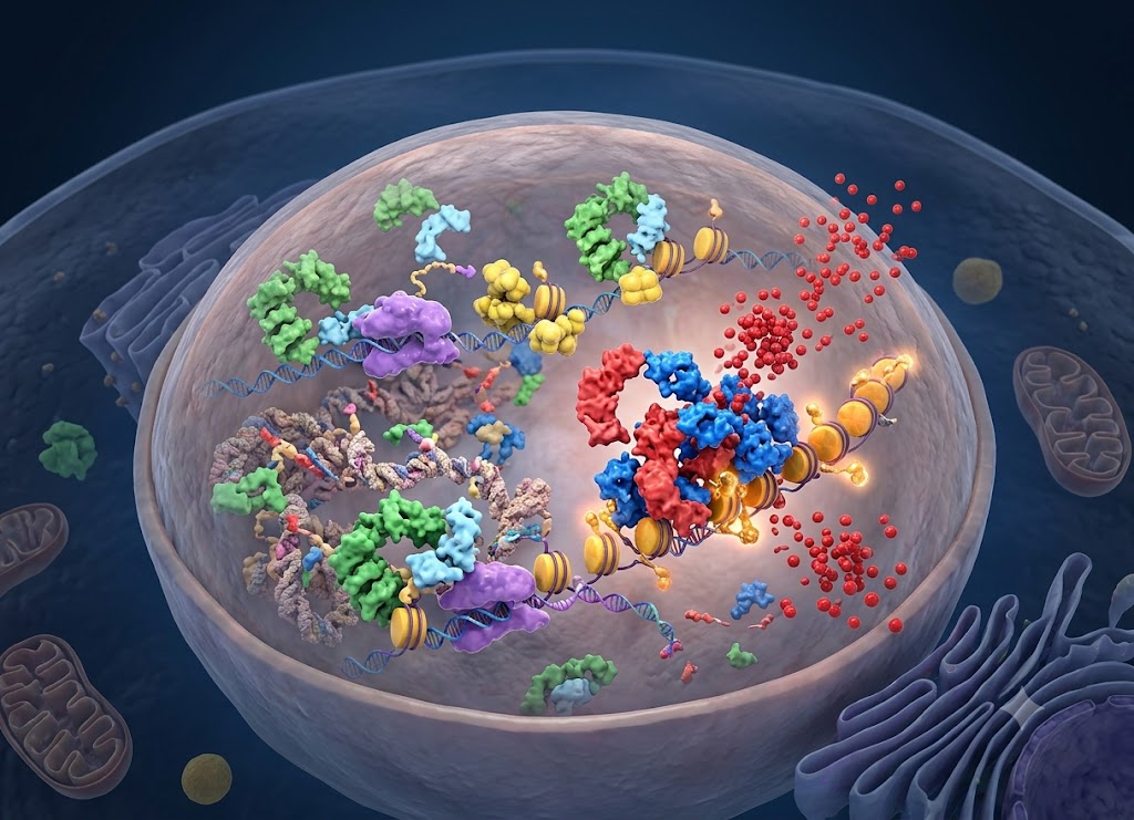
NUT carcinoma is a rare and highly aggressive cancer caused by genetic changes involving the NUTM1 gene. These changes interfere with normal cell development and promote uncontrolled cell growth. The disease most often develops in midline areas of the body, particularly the chest, head, and neck, although it can occur in other locations.
Because NUT carcinoma can grow very quickly and may look similar to other poorly differentiated cancers, identifying it can be challenging. Specialized testing is often necessary to confirm the diagnosis. Recognizing the disease early and obtaining an accurate diagnosis can be important when determining the most appropriate treatment approach.
The symptoms of NUT carcinoma vary depending on where the tumor begins and whether it has spread. Possible symptoms include a growing mass, chest pain, difficulty swallowing, breathing problems, persistent coughing, unexplained weight loss, and fatigue.
Since these symptoms can overlap with other illnesses and cancers, diagnosis usually involves a biopsy followed by specialized molecular testing to look for changes involving the NUTM1 gene. This guide explains the symptoms, causes, diagnostic process, treatment options, prognosis, and other important information about this uncommon cancer.
Symptoms of NUT Carcinoma
NUT carcinoma, previously called NUT-midline carcinoma, is an uncommon and exceptionally aggressive form of poorly differentiated squamous cell carcinoma. There is no single symptom that immediately identifies the disease, so its initial presentation can vary considerably.
The symptoms largely depend on where the primary tumor develops. NUT carcinoma most commonly affects structures along the body’s midline, particularly the head, neck, sinonasal passages, lungs, and mediastinum.
One of the defining characteristics of this cancer is its rapid progression. Symptoms that initially seem mild or similar to common illnesses can become much more severe within only a few weeks.
Common Signs by Tumor Location
As the tumor grows, it can press against, invade, or obstruct nearby tissues and organs. The resulting symptoms depend heavily on the tumor’s location.
Head and Neck (Including Sinonasal Tract)
When NUT carcinoma develops in the head, neck, or sinus region, the early symptoms can look very similar to a stubborn sinus infection or upper respiratory illness.
- Nasal Obstruction: Persistent or worsening blockage in one or both nostrils that does not improve with typical treatments.
- Facial Pain and Pressure: Pressure, swelling, or a feeling of fullness around the cheeks, palate, and eyes.
- Sensory and Cranial Changes: Ongoing headaches, reduced or complete loss of smell, and repeated nosebleeds may occur.
- Advanced Nerve/Eye Involvement: As the tumor grows into nearby tissue or bone, it may cause facial numbness, weakness, bulging of the eye, or vision problems.
- Palpable Mass: A lump in the neck may develop when the cancer spreads to nearby lymph nodes.
Thoracic and Chest (Mediastinum or Lungs)
When NUT carcinoma begins in the chest, symptoms are often respiratory and may initially be mistaken for pneumonia, bronchitis, or another type of lung cancer.
- Chronic Cough: A persistent cough, often without mucus, is a common early symptom.
- Breathing Difficulties: Shortness of breath or wheezing may gradually become worse, particularly with physical activity or while lying down, as the tumor affects the airways.
- Chest Discomfort: Patients may experience dull or aching chest pain, or sharper pain that becomes worse with deep breathing.
- Compression Symptoms: Pressure on the esophagus can make swallowing difficult, while involvement of the recurrent laryngeal nerve may cause ongoing hoarseness.
- Superior Vena Cava (SVC) Syndrome: If the tumor compresses the large vein that carries blood from the upper body back to the heart, swelling can develop in the face, neck, and arms. This is a serious complication that requires medical attention.
General Systemic Symptoms
As the cancer advances, symptoms can become more generalized. These may include severe fatigue, a general feeling of illness or weakness, and rapid, unexplained weight loss.
Why Misdiagnosis and Delayed Diagnoses are Common
NUT carcinoma is frequently diagnosed later than other cancers because its symptoms and microscopic appearance can resemble more common diseases.
The “Master of Disguise” Presentation: Early symptoms often look like ordinary illnesses, so doctors may initially investigate more common explanations. For example, a young person with nasal congestion and sinus pressure may first receive treatment for sinusitis. If symptoms continue despite treatment and the person’s condition worsens quickly, additional imaging and testing may eventually be required.
Pathological Mimicry: Even after a biopsy, the cancer cells may appear very primitive and poorly differentiated under a microscope. Without specialized testing, the tumor may be classified as poorly differentiated squamous cell carcinoma (PDSCC) or sinonasal undifferentiated carcinoma (SNUC).
Because NUT carcinoma is rare, specialized testing may not automatically be performed. Doctors may need to consider the possibility when a younger patient develops an unusually aggressive tumor in a midline location. Testing can include immunohistochemistry for the NUT protein and molecular analysis to identify a NUTM1 gene fusion.
How is NUT Carcinoma Diagnosed?
Diagnosing NUT carcinoma generally requires several steps, beginning with imaging and tissue sampling and progressing to specialized molecular testing.
Because the cancer can develop quickly and may not have distinctive characteristics under routine microscopic examination, molecular confirmation is especially important.
Step 1: Initial Structural and Metabolic Imaging
When symptoms persist despite standard treatment, doctors may use advanced imaging to locate a possible tumor, determine how far it has invaded nearby structures, and look for signs of disease elsewhere in the body.
Computed Tomography (CT): A contrast-enhanced CT scan can provide detailed images of bones and soft tissues. It can help identify tumors in the chest, lungs, head, or neck and reveal involvement of nearby lymph nodes.
Magnetic Resonance Imaging (MRI): MRI is particularly useful when a tumor is located in the sinonasal area or near the skull base. Its detailed soft-tissue images can help doctors distinguish the tumor from nearby brain tissue, nerves, blood vessels, and other structures.
Positron Emission Tomography (PET/CT): PET/CT can be used to assess metabolic activity throughout the body. It can help identify distant areas of disease and provide an overall picture of the cancer’s extent.
Step 2: Tissue Biopsy and Early Pathology Clues
Once imaging identifies a suspicious mass, a biopsy is generally required to obtain tissue for laboratory examination. The method depends on the tumor’s location and may range from a needle biopsy to a surgical procedure.
Under a microscope, NUT carcinoma can show several unusual characteristics.
Undifferentiated Appearance: The cancer cells often appear primitive and relatively uniform, with large nuclei, rapid cell division, and a loss of normal tissue structure.
Abrupt Squamous Differentiation: Some tumors contain areas where primitive cells suddenly develop into flattened, keratin-producing squamous cells. This abrupt change can provide an important clue, although microscopic examination alone is usually not enough to establish the diagnosis.
Step 3: Definitive Molecular Confirmation (The Gold Standard)
Because NUT carcinoma can closely resemble other poorly differentiated cancers, specialized molecular and protein testing is essential for confirming the diagnosis.
1. Immunohistochemistry (IHC) — The Frontline Screen
NUT immunohistochemistry is one of the primary tests used to screen for NUT carcinoma. It uses a specific antibody to detect the NUT protein in tumor cells.
Abnormal NUT protein expression outside its normal tissue distribution can provide a strong diagnostic clue. A characteristic nuclear staining pattern can support the diagnosis, although results should be interpreted by specialists and confirmed in the appropriate clinical and molecular context.
2. Advanced Molecular Genetic Assays
Additional molecular tests can confirm the diagnosis and determine the genetic structure of the tumor.
Fluorescence In Situ Hybridization (FISH): FISH uses fluorescent DNA probes to examine the NUTM1 gene region. When a rearrangement involving NUTM1 is present, the test can provide visual evidence of the genetic change.
RNA-Based Next-Generation Sequencing (NGS): RNA sequencing can be particularly valuable because some NUTM1 gene fusions may not be detected reliably with standard DNA-based testing. RNA-based analysis can identify active fusion transcripts and determine which partner gene is involved. Common partners include BRD4, BRD3, and NSD3. Identifying the specific fusion can also help determine eligibility for certain targeted clinical trials.
Modern Treatments for NUT Carcinoma
Treatment for NUT carcinoma generally requires an aggressive and coordinated approach. Because this cancer can progress rapidly, treatment often combines therapies aimed at controlling the tumor locally with treatments designed to address disease throughout the body.
The most appropriate strategy depends on factors such as the tumor’s location, stage, genetic characteristics, overall health, and whether the disease can be surgically removed.
Conventional Multimodal Therapy: The Structural Backbone
When NUT carcinoma is diagnosed before it has spread extensively, aggressive treatment of the primary tumor can be an important part of care.
1. Radical Surgical Resection
When a tumor is accessible and can be safely removed, complete surgical removal with clear margins may be an important part of treatment.
Head and Neck Primaries: Surgery may involve complex procedures affecting the sinonasal cavities, skull base, or other nearby midline structures. Removal of affected neck lymph nodes may also be necessary.
Thoracic Primaries: Tumors located in the lungs or central chest may require removal of part or all of a lung or affected mediastinal structures. Complete removal can be challenging because NUT carcinoma often grows deeply into surrounding tissues.
2. High-Dose Radiation Therapy
Radiation therapy can help strengthen local tumor control. It may be used after surgery to target microscopic cancer cells that could remain in the treatment area.
For tumors that cannot be removed surgically, high-dose radiation may be combined with systemic chemotherapy as part of a chemoradiation approach.
3. Intensive Systemic Chemotherapy
Chemotherapy may be used before surgery to reduce tumor size, after surgery to address microscopic disease, or as treatment for advanced cancer that cannot be surgically removed.
Because NUT carcinoma behaves differently from many conventional lung or head and neck cancers, oncology teams may use intensive combinations of drugs commonly used for other aggressive cancers.
- Common Multi-Drug Combinations: Treatment regimens may include drugs such as ifosfamide, etoposide, cisplatin, vincristine, and doxorubicin.
- The Durability Challenge: Chemotherapy can sometimes cause substantial initial tumor shrinkage, but NUT carcinoma has a high risk of developing resistance and returning after an initial response.
Targeted Breakthroughs: Epigenetic Therapy with BET Inhibitors
The discovery of the BRD4-NUT oncoprotein has helped researchers develop treatments that directly target the biological processes driving NUT carcinoma.
The BRD4-NUT fusion protein acts as an abnormal regulator of gene activity. It attaches to chromatin and promotes continued cell growth while preventing cancer cells from developing into mature, normal cells.
BET inhibitors are designed to interfere with the bromodomain activity of proteins involved in this process. By blocking these interactions, they may interrupt the molecular signals that allow the cancer to continue growing.
When this pathway is disrupted, cancer cells may stop multiplying, begin to differentiate into more mature cells, and eventually undergo programmed cell death.
BET inhibitors that have been studied in NUT carcinoma include birabresib (MK-8628), molibresib (GSK525762), and ZEN-3694. Because NUT carcinoma is extremely rare, access to these therapies is often through clinical trials.
Current Horizons: Emerging Synergies and Immunotherapy
As of 2026, research is increasingly focused on combining treatments that target different biological pathways. The goal is to improve responses and reduce the likelihood that the cancer develops resistance.
BET Inhibitor Combination Regimens: Clinical trials are examining whether BET inhibitors can work more effectively when combined with other treatments. For example, ZEN-3694 has been investigated alongside the CDK4/6 inhibitor abemaciclib and with chemotherapy combinations such as cisplatin/etoposide.
The Evolving Role of Immunotherapy: NUT carcinoma has traditionally been viewed as an immunologically “cold” tumor. However, some clinical reports suggest that immune checkpoint inhibitors such as pembrolizumab or cemiplimab may provide meaningful disease control in selected patients, particularly when used alongside chemotherapy or other treatments.
Because the disease can progress rapidly and some standard genetic tests may fail to identify the relevant fusion, referral to a specialized cancer center with experience in NUT carcinoma, advanced molecular testing, and clinical trials can be especially important.
Prognosis and Long-Term Outlook for NUT Carcinoma
NUT carcinoma remains a serious and difficult cancer to treat because of its aggressive behavior. However, outcomes are not identical for every patient. Information from international registries, including the International NUT Carcinoma Registry at the Dana-Farber Cancer Institute, has helped researchers identify factors that may influence prognosis.
Historically, the median overall survival for the general NUT carcinoma population has been reported at approximately 6.5 to 10 months. However, this statistic does not determine the outcome for an individual patient. Factors such as tumor location, stage, genetic fusion partner, treatment response, and whether the tumor can be completely removed can all influence survival.
Risk Stratification: What Drives the Outlook?
Clinical registry data indicates that prognosis can vary significantly according to several major factors.
Anatomical Location (The Thoracic Penalty): The location of the primary tumor is one of the strongest factors associated with outcome.
- Thoracic Primary: Tumors that begin in the lungs, mediastinum, or other parts of the chest are associated with a particularly difficult prognosis. Reported data show a median overall survival of approximately 4.4 months and a 2-year survival rate of around 5%.
- Non-Thoracic Primary: Tumors that begin in the head, neck, or sinonasal region may follow a somewhat less aggressive course than thoracic tumors.
The Genetic Fusion Partner: The gene that fuses with NUTM1 can also influence tumor behavior and prognosis.
- Non-BRD4 Fusions: Patients with non-thoracic tumors driven by non-BRD4 partners, such as BRD3 or NSD3, have been associated with more favorable outcomes, with reported median overall survival reaching approximately 36.5 months in some registry analyses.
- BRD4 Fusions: Non-thoracic tumors driven by the classic BRD4-NUTM1 fusion have been associated with a median overall survival closer to 10 months.
The Surgical Margin Factor: Long-term survival remains uncommon, but the patients most likely to achieve extended survival are generally those with localized, non-metastatic disease that can be completely removed through radical surgery.
NUT Carcinoma vs. Poorly Differentiated Squamous Cell Carcinoma (PDSCC)
NUT carcinoma and poorly differentiated squamous cell carcinoma can look very similar under a standard microscope. This similarity can lead to an initial misdiagnosis. However, they are different diseases with distinct genetic features, patient populations, and treatment considerations.
| Feature | NUT Carcinoma | Poorly Differentiated Squamous Cell Carcinoma (PDSCC) |
|---|---|---|
| Primary Genetic Driver | A chromosomal rearrangement involving the NUTM1 gene, often with a partner such as BRD4. | A combination of genetic mutations that generally does not involve a NUTM1 rearrangement. |
| Patient Demographics | More often identified in children, adolescents, and young adults, with a median age around 23 to 24. | More commonly diagnosed in older adults, particularly those over age 50. |
| Environmental Links | No established connection to lifestyle, environmental exposures, or inherited risk factors. | Strongly associated with factors such as tobacco, alcohol, and certain HPV-related cancers. |
| Clinical Velocity | Extremely rapid progression, with many patients presenting with local or distant metastatic disease. | Can be aggressive but generally follows a more typical progression pattern. |
| Treatment Success | Often difficult to control with standard chemotherapy, while targeted BET inhibitors may produce mechanism-based responses. | Often treated with established chemotherapy and radiation approaches, depending on the cancer’s location and stage. |
Shifting the Outlook: Modern Clinical Trials
Because conventional chemotherapy may not provide durable control, researchers are increasingly focusing on targeted treatments and combination strategies.
Next-Generation BET Inhibitors: Early BET inhibitors such as birabresib and molibresib have sometimes produced rapid tumor shrinkage, but resistance can develop. Newer studies are evaluating compounds designed to more effectively target the BRD4-NUT pathway while improving tolerability.
Targeted Combinations: Research is moving beyond single-drug treatment. Clinical studies are investigating combinations of BET inhibitors such as ZEN-3694 with cell-cycle inhibitors such as abemaciclib, as well as combinations with conventional chemotherapy.
Primed Immunotherapy: NUT carcinoma is often considered an immunologically “cold” tumor. Nevertheless, some reports suggest that adding immune checkpoint inhibitors such as pembrolizumab to chemotherapy or radiation may produce partial responses or periods of disease stabilization in certain patients.
Connecting with Specialized Support
Because NUT carcinoma is extremely rare, patients and families may have difficulty finding people with similar experiences through general cancer support networks.
Specialized organizations and research centers can provide important resources.
The International NUT Carcinoma Registry: Based at the Dana-Farber Cancer Institute, this international registry collects information about patients with NUT carcinoma and supports research into diagnosis and treatment. It can also help connect patients with specialists and information about clinical trials.
- Global Caregiver Support: Private patient and caregiver communities can provide opportunities to share experiences, discuss treatment challenges, and find emotional support.
- Rare Disease Advocacy: Organizations such as the National Organization for Rare Disorders (NORD) provide educational resources, patient navigation, and other forms of support for people affected by rare diseases.
Conclusion
NUT carcinoma is a rare and highly aggressive cancer that can be difficult to recognize because its symptoms may resemble other illnesses and its cells can look similar to other poorly differentiated cancers.
Because the disease can progress rapidly, persistent or unusual symptoms should receive appropriate medical evaluation. Confirming the diagnosis often requires a biopsy combined with specialized protein and molecular testing.
Treatment depends on the tumor’s location, stage, genetic characteristics, and overall health. Options may include surgery, radiation therapy, chemotherapy, targeted therapies, and participation in clinical trials. Although NUT carcinoma remains challenging to treat, continued research is improving our understanding of the disease and creating new opportunities for targeted treatment.
Frequently Asked Questions
1. What is NUT carcinoma?
NUT carcinoma is a rare and aggressive cancer associated with genetic changes involving the NUTM1 gene. These changes interfere with normal cell development and allow abnormal cells to grow rapidly.
The cancer is classified as poorly differentiated because its cells often do not resemble mature normal tissue when examined under a microscope. NUT carcinoma can occur in different parts of the body but commonly develops in the chest, head, and neck. Specialized testing is often necessary to confirm the diagnosis.
2. What are the symptoms of NUT carcinoma?
Symptoms depend on where the tumor develops and how far the disease has progressed.
Possible symptoms include a rapidly growing lump, persistent cough, chest pain, difficulty breathing, difficulty swallowing, hoarseness, unexplained weight loss, and severe fatigue.
Because these symptoms can occur with many other illnesses, NUT carcinoma may not be recognized immediately. Symptoms that persist, worsen quickly, or do not respond to standard treatment should be evaluated by a healthcare professional.
3. What causes NUT carcinoma?
NUT carcinoma develops because of genetic changes involving the NUTM1 gene. These changes commonly occur through a chromosome rearrangement that creates a gene fusion.
The genetic alteration is generally acquired during a person’s lifetime rather than inherited from their parents. Researchers are continuing to investigate why these changes occur and whether additional biological factors contribute to the development of the disease.
4. How is NUT carcinoma diagnosed?
Diagnosis usually begins with a biopsy, in which a sample of the tumor is examined by a pathologist.
Because NUT carcinoma can resemble other aggressive cancers under a microscope, specialized tests may be needed. These can include NUT protein immunohistochemistry and molecular testing to identify a NUTM1 gene rearrangement.
CT, MRI, and PET scans may also be used to identify the tumor, evaluate nearby structures, and determine whether the cancer has spread.
Accurate diagnosis is especially important because NUT carcinoma requires a different treatment strategy from many cancers that may look similar under routine pathology.
5. How is NUT carcinoma treated?
Treatment usually involves more than one approach because NUT carcinoma can grow and spread rapidly.
Depending on the tumor’s location and stage, treatment may include surgery, radiation therapy, chemotherapy, targeted therapies, and clinical trial participation.
Newer treatments are being developed to target the genetic and molecular mechanisms involved in NUT carcinoma. Because the disease is rare, treatment at a specialized cancer center with experience in NUT carcinoma may be beneficial.
6. What is the outlook for people with NUT carcinoma?
NUT carcinoma generally has a challenging prognosis because it tends to grow rapidly and is often diagnosed after the disease has already spread.
However, individual outcomes can differ considerably. Factors such as the tumor’s location, stage, genetic fusion partner, response to treatment, ability to completely remove the tumor, and overall health can all affect prognosis.
Advances in molecular diagnosis, targeted treatment, and clinical research are helping doctors better understand the disease and develop new treatment options. Ongoing specialist care and regular follow-up are important for people living with NUT carcinoma.