Pulmonary Embolism (PE): Causes, Symptoms, Diagnosis, and Treatment
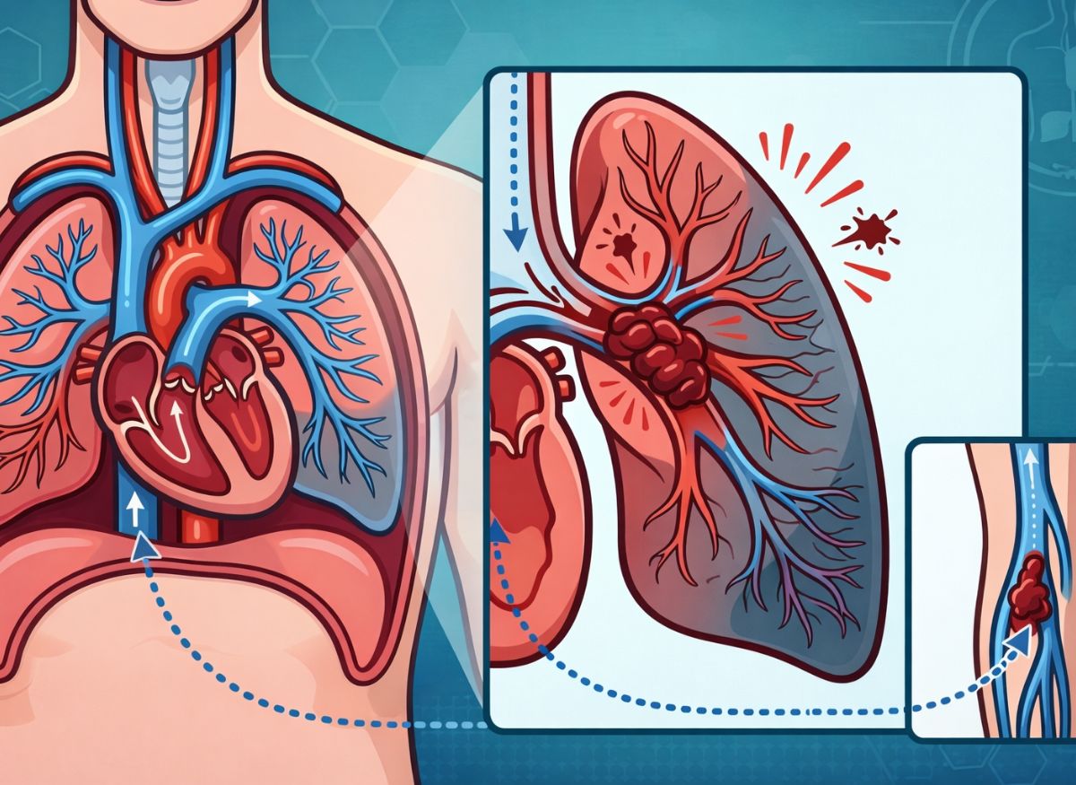
Pulmonary embolism (PE) is a medical emergency that frequently strikes with little warning and calls for immediate action. Typically, a small clot forms in a leg vein, breaks free, travels through the circulation, and lodges in an artery of the lung. The result is interrupted oxygen delivery and serious stress on the body.
PE is more widespread and deadly than most people assume. It affects hundreds of thousands of people around the world every year. In the U.S. alone, an estimated 900,000 cases of venous thromboembolism (PE plus deep vein thrombosis, or DVT) occur annually, and between 60,000 and 100,000 of them end in death. These figures show both how common the condition is and how lethal it can be when symptoms go unnoticed or care is delayed.
Part of the danger is that PE is easy to miss. Breathlessness, chest discomfort, or a racing pulse can seem minor or be blamed on something less serious, even as a dangerous blockage develops.
Knowing about PE is about more than medical facts; it is about noticing warning signs early enough to respond. The gap between prompt awareness and a slow reaction can decide whether someone lives. The sections below cover the causes, symptoms, diagnosis, and treatment of PE so you can spot it and understand why speed matters.
What Is Pulmonary Embolism?
A pulmonary embolism happens when a blood clot obstructs one or more arteries in the lungs and interferes with normal circulation. In most cases the clot begins in the deep veins of the leg (a condition called deep vein thrombosis, or DVT) and is carried by the blood to the lungs. Once it becomes stuck in a pulmonary artery, it can block blood flow partially or entirely, lowering blood oxygen and straining the heart.
How serious a PE is depends on how large the clot is and how many there are. One small clot might cause only mild symptoms or none at all, whereas a big clot or several clots can become life-threatening quickly. When blood cannot reach part of the lung, that tissue is starved of oxygen and may be damaged. Because PE can appear abruptly and deteriorate fast, it is treated as an emergency. Left untreated, it can cause lasting lung injury, heart problems, or death.
Types of Pulmonary Embolism
PE is generally grouped into three categories according to clot size and how much it affects the heart and lungs:
- Massive PE: A large clot blocks a major lung artery, causing blood pressure to plunge and putting severe strain on the heart. This is life-threatening and needs emergency care right away.
- Submassive PE: Blood pressure stays reasonably stable, but the heart is under strain and oxygen levels drop. The patient may look fine at first, yet complications can emerge without prompt treatment.
- Low-risk (non-massive) PE: The clots are smaller and have little effect on blood pressure or heart function. Symptoms are mild to moderate, but treatment is still needed so the condition doesn’t worsen.
Massive vs. Submassive PE
What separates the two is their immediate effect on the circulatory system, specifically blood pressure and heart function. This distinction matters because it determines how urgently and how aggressively doctors must treat. The deciding factor is hemodynamic instability, meaning the body can no longer maintain adequate blood pressure and circulation.
A massive PE is the most dangerous category. It is defined by sustained low blood pressure: a systolic reading under 90 mmHg lasting more than 15 minutes, or a fall of at least 40 mmHg. Such a drop means the clot is obstructing lung blood flow so badly that the heart cannot push enough blood to the rest of the body. This condition, called obstructive shock, demands immediate, forceful treatment, often clot-dissolving drugs (thrombolytics) or surgical removal of the clot (embolectomy).
In a submassive PE, sometimes called intermediate-risk PE, blood pressure remains normal, but the heart shows signs of strain from pumping against the obstruction. This strain is known as right ventricular dysfunction. It can be identified on imaging such as an echocardiogram, which may reveal an enlarged right ventricle that isn’t contracting well, or through raised blood markers such as troponin or brain natriuretic peptide (BNP), which are released when the heart muscle is stressed. A submassive PE is less immediately dangerous than a massive one, but it can still worsen. It calls for close observation and standard blood-thinning treatment, with thrombolytics considered if the patient’s condition declines.
Causes of Pulmonary Embolism
A PE develops when a fragment of a clot, most often from a leg DVT, detaches, passes through the heart, and becomes wedged in a pulmonary artery. In effect, a problem confined to a vein turns into a dangerous blockage in the lungs’ blood vessels. This chain of events, from a vein in the limb to the arteries of the lung, is the basic mechanism behind the disease.
Understanding Deep Vein Thrombosis (DVT)
DVT is the formation of a clot (thrombus) inside a deep vein, most often in the calf, thigh, or pelvis. These veins lie deep within the muscles beside the arteries and carry a large share of the body’s blood back to the heart.
DVT is the source of roughly 90% of acute PEs, which is why preventing and detecting it is central to managing PE. Clots in these veins are especially hazardous because of their size and their direct route to the heart and lungs. When a piece breaks off the vein wall, it becomes an embolus. Blood flow carries it up through increasingly large leg veins, into the inferior vena cava (the body’s largest vein), through the right atrium and right ventricle, and out into the pulmonary arteries.
Because the pulmonary arteries get narrower as they branch through the lungs, the embolus eventually gets trapped. This blocks blood from reaching the lung region that artery supplies, leaving the body short of oxygenated blood and raising pressure on the right side of the heart.
Main Risk Factors for Blood Clots
The major risk factors fall into three groups described by Virchow’s Triad: slow blood flow (venous stasis), damage to the vein lining (endothelial injury), and blood that clots too readily (hypercoagulability). Together these cover many conditions, behaviors, and inherited traits that raise the chance of DVT and, in turn, PE.
- Prolonged immobility and venous stasis: When you stay inactive for long stretches, your calf muscles stop squeezing blood back toward the heart. Blood moves sluggishly, clotting factors build up, and a clot becomes more likely. Examples include flights or car trips longer than four hours, hospital stays or bed rest after illness or surgery, and paralysis from spinal cord injury.
- Surgery and vein injury: Major operations, especially hip or knee surgery, carry substantial risk. Surgery can injure the inner lining of veins, exposing tissue underneath that triggers clotting, and the immobility of recovery adds to the risk. Serious trauma such as fractures can also directly damage vessels.
- Medical conditions and hypercoagulability: Some illnesses make blood clot more easily. Cancer is a major one, since certain tumors release substances that promote clotting. Heart failure and chronic obstructive pulmonary disease (COPD) can cause blood to pool. Inflammatory diseases such as Crohn’s disease and rheumatoid arthritis raise risk too. Inherited clotting disorders (thrombophilias), such as Factor V Leiden, create a genetic tendency toward excess clotting.
- Lifestyle and hormonal factors: Obesity raises vein pressure and promotes inflammation; smoking harms vessel linings and alters clotting; and risk climbs notably after age 40. Estrogen-containing medications, including birth control pills and hormone replacement therapy, increase clotting factors in the blood. Pregnancy also raises risk because of hormonal shifts and pressure from the growing uterus on pelvic veins.
Symptoms of Pulmonary Embolism
The key warning signs are abrupt, unexplained severe shortness of breath, sharp chest pain that intensifies when breathing in deeply, and a lingering cough that may bring up blood. Symptoms often come on without notice, and their intensity varies with clot size, how much of the lung is affected, and the person’s general health. Knowing these classic signs is vital, because PE requires prompt evaluation and treatment to avoid a fatal outcome.
Common Symptoms
The most typical symptoms are sudden breathlessness (dyspnea), pain with breathing (pleuritic chest pain), and cough. Together they form a classic triad, though not everyone has all three. They arise directly from the arterial blockage and the body’s reaction to it.
- Sudden, unexplained shortness of breath: This is the most commonly reported symptom. The blocked artery keeps part of the lung from exchanging gases, so blood is poorly oxygenated and the body detects the shortfall. The brain speeds up breathing, creating a feeling of not getting enough air even at rest. The onset is typically abrupt.
- Sharp, stabbing chest pain: Another hallmark, this pain occurs when a clot settles in an artery near the lung’s outer edge and inflames the pleura, the double-layered membrane around the lungs. Friction between the inflamed layers causes sharp, localized pain that is clearly worse with deep breaths, coughing, or sneezing.
- Cough and coughing up blood: A persistent, often dry cough may accompany PE. Sometimes inflammation and high pressure in the lung vessels cause slight bleeding into the airways, producing blood-streaked sputum (hemoptysis). It is less common than breathlessness and chest pain, but it is a serious red flag.
- Other signs: Beyond the classic triad, people may have a fast or irregular heartbeat as the heart labors against the blockage. Lightheadedness, dizziness, or fainting can occur when the obstruction is large enough to seriously reduce cardiac output and blood pressure. Swelling, pain, or redness in one leg, usually the calf, may point to the DVT that started it all.
Pulmonary Embolism Without Symptoms
A PE can occur with no noticeable symptoms at all, a situation often called a silent PE. This is risky because without warning signs like chest pain or breathlessness, the person doesn’t seek care, and the problem goes undiagnosed and untreated.
Silent PEs are more likely when clots are small. Small emboli can lodge in tiny, outlying branches of the pulmonary arteries, blocking only a small portion of lung blood supply. The disruption may be too slight to cause obvious symptoms, and the body’s reserve capacity can mask it.
Still, “silent” does not mean harmless. Any embolic event, whatever its size, causes some lung tissue damage (infarction) and strains the cardiovascular system. A silent PE also often indicates an existing DVT that may keep shedding clots, and a small, symptom-free PE can foreshadow a much larger, life-threatening one.
These cases are often found by accident on imaging, such as a CT scan done for another reason. Finding one calls for a full workup to locate the clot’s source and start treatment, usually anticoagulants, to prevent more serious events. This underlines why people at high risk should have their DVT and PE risk factors assessed.
Diagnosis and Management of Pulmonary Embolism
Handling PE well involves a fast, accurate diagnostic process, preventive measures for high-risk people, an understanding of the long-term outlook, and classification of severity to guide treatment. These pieces work together, beginning before a PE ever occurs and continuing well into recovery. Management is tailored to each patient’s overall health, the cause of the clot, and how the embolism is affecting the heart and lungs.
How PE Is Diagnosed
Diagnosis is a multi-step process. It starts with assessing symptoms and risk factors, but definitive testing is needed to confirm a clot. Clinicians first use scoring tools such as the Wells score to estimate the likelihood of PE. Depending on that estimate, a D-dimer blood test may follow; it measures a substance released when clots break down. A negative D-dimer in a low-risk patient effectively rules out PE. A positive result, however, isn’t specific to PE, since many other conditions raise D-dimer, so imaging is needed for confirmation.
The gold standard is CT pulmonary angiography (CTPA). In this noninvasive test, contrast dye is injected into a vein and a CT scanner produces detailed cross-sectional pictures of the pulmonary arteries, letting radiologists see blockages clearly. For patients who can’t have contrast dye because of kidney problems or allergy, a ventilation/perfusion (V/Q) scan is the alternative.
This nuclear medicine test compares airflow (ventilation) with blood flow (perfusion) in the lungs. An area with good airflow but poor blood flow strongly suggests PE. To find where the clot came from, doctors often perform a compression ultrasound of the legs to look for DVT, since more than 90% of PEs start in leg clots.
How to Prevent PE
Prevention focuses on reducing the factors that promote clotting: immobility, surgery, and certain illnesses. Hospitalized patients, particularly those having major surgery such as orthopedic procedures or those on prolonged bed rest, routinely receive preventive care combining mechanical and medication-based approaches to keep blood moving smoothly through the veins.
These measures are essential for stopping the initial DVT that often leads to PE, and they are adjusted to each person’s risk.
- Medication-based prevention: For many high-risk patients, such as those recovering from surgery or staying long in hospital, doctors prescribe low-dose anticoagulants like heparin or low-molecular-weight heparin, given by injection. They thin the blood just enough to discourage clots without causing major bleeding.
- Mechanical prevention: Graduated compression stockings are widely used; they gently squeeze the legs to push blood back toward the heart. Intermittent pneumatic compression (IPC) devices, inflatable leg sleeves that repeatedly inflate and deflate, mimic the muscle action of walking.
- Lifestyle and behavior: For the general public, particularly during long periods of sitting such as air or car travel, simple habits help: getting up to walk every one to two hours, doing seated leg exercises such as ankle circles and foot pumps, drinking enough fluids, and avoiding tight clothing.
Treatment for Pulmonary Embolism
Treatment relies mainly on anticoagulants to stop new clots from forming, with thrombolytics to dissolve large existing clots and surgical or catheter-based procedures for severe cases. The immediate aims are to stabilize the patient, stop the current clot from growing, prevent further clots from the source DVT, and restore blood flow through the pulmonary arteries to relieve the heart.
Medications Used to Treat PE
Two main drug categories are used. Anticoagulants are the foundation of treatment for almost every patient, while thrombolytics are kept for severe, life-threatening cases. They work in fundamentally different ways to handle the acute event and prevent recurrence.
- Anticoagulants (blood thinners): These are the most common and essential PE medications. Their job isn’t to dissolve the existing clot but to keep it from enlarging and to block new clots. By stopping the clotting cascade, they give the body’s own clot-dissolving system (fibrinolysis) time to break the embolus down gradually. Treatment usually starts in the hospital with a fast-acting injectable such as heparin or low-molecular-weight heparin (LMWH), then moves to an oral anticoagulant for at least three to six months. Oral choices include warfarin (Coumadin), which needs regular blood monitoring, and the newer direct oral anticoagulants (DOACs) such as apixaban (Eliquis), rivaroxaban (Xarelto), and dabigatran (Pradaxa), which are often favored for their fixed dosing and fewer interactions.
- Thrombolytics (clot busters): These strong drugs actively and rapidly dissolve existing clots. The most common is tissue plasminogen activator (tPA), which activates plasminogen to form plasmin, an enzyme that breaks down the clot’s fibrin mesh and quickly restores blood flow. Because they are so potent, they carry a much higher risk of serious bleeding, including bleeding in the brain. They are therefore reserved for patients with massive PE who are hemodynamically unstable, with dangerously low blood pressure or signs of shock, where the risk of dying from the clot outweighs the bleeding risk.
Anticoagulants vs. Thrombolytics
The two differ in how they work, what they aim to achieve, and when they are used: anticoagulants prevent and stabilize, while thrombolytics actively dissolve life-threatening clots. Understanding this difference is key to choosing the right strategy. Despite the nickname, anticoagulants neither thin the blood nor break up existing clots. They interfere with the chain of chemical reactions that builds a fibrin clot.
By blocking particular clotting factors, drugs such as heparin and warfarin essentially pause clot formation. That keeps the lung embolus from growing and, importantly, prevents new clots in the legs or elsewhere. The goal is to stabilize the patient while the body’s own fibrinolytic system slowly clears the clot over days to weeks. They are the standard of care for the great majority of hemodynamically stable PE patients.
Thrombolytics, by contrast, are meant for fast, aggressive intervention. They directly dissolve the clot causing the dangerous blockage. Drugs like tPA convert plasminogen to plasmin, the body’s main clot-dissolving enzyme, rapidly breaking down the clot’s fibrin structure and reopening the blocked artery.
Their goal is immediate restoration of blood flow to reverse shock and prevent death. Because they are so powerful, they carry a high risk of major bleeding anywhere in the body, so they are limited to patients with severe PE showing hemodynamic instability, such as persistent low blood pressure or right heart strain. In short, anticoagulants play defense by preventing worsening, while thrombolytics play offense by attacking the clot itself.
Long-Term Outlook After a PE
For people who survive a PE, the long-term outlook is generally good as long as it was diagnosed and treated promptly and they keep up with follow-up care. Anticoagulant therapy is the mainstay of long-term management, and most patients take it for at least three to six months. The exact length depends on circumstances: if a temporary factor such as surgery or immobility provoked the PE, a shorter course may do. If the PE had no clear trigger, or the patient has an ongoing risk such as an inherited clotting disorder, lifelong anticoagulation may be advised to prevent recurrence.
Most patients recover fully, but some develop lingering symptoms known as post-PE syndrome, including ongoing breathlessness, chest pain, and reduced exercise tolerance, which can affect quality of life. A rarer and more serious complication is chronic thromboembolic pulmonary hypertension (CTEPH), which affects roughly 1–4% of PE survivors when clots in the pulmonary arteries fail to dissolve completely.
Instead, the clots turn into scar-like tissue that permanently narrows or blocks the arteries. This raises pressure in the pulmonary arteries and forces the right side of the heart to work harder, and over time it can lead to right-sided heart failure. CTEPH needs specialized treatment, which may involve surgery to remove the scar tissue (pulmonary thromboendarterectomy) or medications to control the high lung blood pressure.
Frequently Asked Questions
1. How can you tell if you have a pulmonary embolism?
PE usually shows up with sudden, noticeable symptoms, though intensity varies. The most common are shortness of breath, sharp chest pain that worsens with deep breaths, a fast heartbeat, and an unexplained cough, sometimes with blood. Some people also feel dizzy, sweat heavily, or faint. Since these signs overlap with other conditions, PE is hard to identify without medical tests. If these symptoms appear suddenly, especially together, seek medical help immediately.
2. Can you recover completely from a pulmonary embolism?
Yes, many people do, particularly when it is diagnosed early and treated properly. Blood thinners and other therapies stop new clots from forming while the body gradually dissolves existing ones. Recovery time depends on clot size and general health. Some people have lingering symptoms, but with follow-up care and lifestyle changes, long-term results are often good.
3. What is the best treatment for pulmonary embolism?
The main treatment is anticoagulant medication, which keeps clots from growing and lowers the chance of new ones. In more severe cases, doctors may use clot-dissolving drugs or perform procedures to remove the clot. Oxygen and supportive care are given as needed. The best option depends on the embolism’s severity and the patient’s stability at diagnosis.
4. How long can someone have a PE without knowing it?
A small PE can go unnoticed for days or even weeks, especially when symptoms are mild or blamed on something else. Even so, the situation can deteriorate quickly. That unpredictability is exactly why PE is so dangerous and why early evaluation is essential.
5. How painful is a pulmonary embolism?
Pain varies, but it is commonly described as sharp and stabbing, particularly with deep breathing or coughing. Some people feel only slight discomfort, while others have intense, frightening pain. Severity depends on the clot’s size and location.
Conclusion
Pulmonary embolism gives little advance notice, and its effects can be swift and severe. A small clot can rapidly become a life-threatening blockage, so awareness is one of your best defenses. Knowing the causes, recognizing the symptoms, and understanding when to act can make a critical difference. Many cases can be treated and recovery is possible, but timing matters: the sooner PE is identified, the better the odds of avoiding serious complications.
Your body often signals trouble before things become critical. Taking sudden breathlessness, chest pain, or unusual discomfort seriously is not an overreaction; it is a way of protecting your life. With pulmonary embolism, acting fast can save it.