9 Key Spina Bifida Signs to Watch For
For parents and caregivers, noticing unusual physical changes or developmental differences in a child can naturally lead to questions about their health. Spina bifida is one condition where recognizing possible signs can help families seek medical advice and understand what may be happening. This birth defect affects the development of the spine and spinal cord during pregnancy.
Spina bifida develops when the neural tube, which eventually forms the brain and spinal cord, does not close completely during early pregnancy. The condition can range from very mild forms that cause few or no symptoms to more complex forms that affect movement, sensation, bladder and bowel function, and other areas of neurological health.
The signs of spina bifida can vary considerably depending on the type and location of the spinal defect. Some signs are noticeable at birth, such as a visible sac or unusual skin markings on the lower back. Others may become apparent later through muscle weakness, walking difficulties, changes in sensation, or bladder and bowel problems.
Because spina bifida affects the spinal cord and the nerves that branch from it, its effects can be different from one person to another. Early evaluation can help healthcare professionals determine whether additional testing or treatment is needed.
In this guide, we will explore 9 key spina bifida signs, explain how they may be related to the condition, and discuss when medical evaluation may be appropriate.
9 Key Symptoms of Spina Bifida to Watch For?
Sac-Like Protrusion
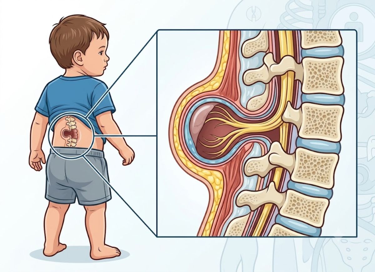
A visible, fluid-filled sac on a baby’s back can be a sign of one of the more significant forms of spina bifida, including myelomeningocele or meningocele.
In myelomeningocele, the sac can contain the membranes surrounding the spinal cord, cerebrospinal fluid, and part of the spinal cord or nerves. The area may be covered by very thin tissue or may not have normal skin coverage.
Meningocele is different because the sac generally contains the protective membranes and fluid, while the spinal cord remains within the spinal canal.
A visible spinal sac requires prompt medical evaluation after birth. Babies with open spina bifida usually receive specialized care soon after delivery to protect the affected area and determine whether surgical treatment is needed.
Abnormal Tuft of Hair, Dimple, or Birthmark
Certain skin changes on the lower back can sometimes be associated with spina bifida, particularly hidden forms such as spina bifida occulta.
Possible skin markers include an unusual patch of hair, a deep or atypical dimple, a birthmark, or a small fatty lump. These findings do not automatically mean that a child has spina bifida, as many skin markings are harmless.
However, when a healthcare provider believes a skin finding may be associated with an underlying spinal abnormality, imaging such as an ultrasound or MRI may be recommended.
In some cases, an underlying problem such as tethered cord syndrome can occur alongside these findings and may cause neurological symptoms as a child grows.
Paralysis and Muscle Weakness
Weakness or paralysis can occur when the spinal nerves are affected by spina bifida. The extent of movement difficulties generally depends on the location and severity of the spinal defect.
A defect higher on the spine may affect more of the nerves responsible for movement in the lower body. Lower spinal defects may result in less severe weakness, although the effects can still vary considerably.
Some people may walk independently, while others may use braces, crutches, walkers, or wheelchairs for mobility.
Differences in muscle strength can also affect posture, balance, joint development, and movement patterns. Ongoing assessment by medical and rehabilitation specialists can help determine appropriate support.
Lack of Sensation
Spina bifida can affect the nerves responsible for transmitting sensations such as touch, pressure, pain, and temperature.
When sensation is reduced, a person may not immediately notice an injury, blister, burn, or pressure area on the legs or feet. This can make regular skin checks particularly important for people with reduced sensation.
Reduced sensation can also affect awareness of body position, which may influence balance and coordination.
The extent of sensory changes depends on the location and severity of the nerve involvement.
Neurogenic Bladder
The nerves that control the bladder may be affected by spina bifida. When this happens, the bladder may not store or empty urine normally, a condition commonly referred to as neurogenic bladder.
Some people may have difficulty emptying the bladder completely, while others may experience urinary leakage. Incomplete emptying can increase the risk of urinary tract problems and, in some cases, can affect kidney health.
Management depends on the individual’s bladder function. Some people may need a structured bladder program, which can include clean intermittent catheterization when recommended by a healthcare professional.
Regular medical monitoring can help protect urinary tract and kidney health.
Neurogenic Bowel
Nerve involvement can also affect the muscles and nerves responsible for bowel function. This may contribute to constipation, difficulty emptying the bowel, or problems controlling bowel movements.
A structured bowel management plan can help many individuals manage these symptoms. Depending on the person’s needs, treatment may include dietary changes, adequate fluids, medications, stool-softening products, or other techniques recommended by a healthcare provider.
The goal is to establish predictable and comfortable bowel function while reducing complications associated with constipation or incontinence.
Clubfoot (Talipes Equinovarus)
Clubfoot is a foot deformity that can occur in some babies with spina bifida. The foot may be turned inward and downward because of differences in muscle strength and nerve function.
The severity varies, and treatment depends on the individual child’s condition. Orthopedic specialists may use approaches such as stretching, casting, bracing, or, in some cases, surgery to improve foot position and function.
Early evaluation can help determine the most appropriate treatment and support future mobility.
Hip Dysplasia and Dislocation
Weakness or imbalance in the muscles surrounding the hips can affect hip development in children with spina bifida.
Hip dysplasia occurs when the hip joint does not develop normally, while hip dislocation happens when the head of the thigh bone moves out of the hip socket.
Children with spina bifida may have their hips monitored regularly through physical examinations and, when appropriate, imaging studies.
Treatment depends on the child’s age, severity of the problem, mobility, and overall condition. The aim is to support comfortable movement, sitting, standing, and mobility whenever possible.
Scoliosis and Kyphosis
Spinal curvature can occur in people with spina bifida. Scoliosis refers to an abnormal sideways curvature of the spine, while kyphosis involves excessive forward rounding.
These conditions may be related to differences in the vertebrae, muscle imbalance, or other changes associated with spina bifida. Spinal curves may become more noticeable during periods of growth.
Mild curvature may simply be monitored. More significant or progressive curves may require treatment such as bracing or surgery, depending on their severity and effect on posture, comfort, and function.
What is Spina Bifida and What are its Main Types?
Spina bifida is a neural tube defect that develops when the spinal column and related structures do not form or close completely during early pregnancy.
The condition has three main forms: myelomeningocele, meningocele, and spina bifida occulta. These forms differ in how much the spinal cord and surrounding structures are involved.
What is Myelomeningocele (Open Spina Bifida)?
Myelomeningocele is generally the most severe form of spina bifida. In this condition, part of the spinal cord and its surrounding membranes can extend through an opening in the spine.
Because the spinal cord and nerves are affected, myelomeningocele can cause weakness or paralysis, reduced sensation, and bladder or bowel problems. The effects depend largely on the location and extent of the spinal defect.
Myelomeningocele is also frequently associated with other neurological conditions, including hydrocephalus and Chiari II malformation.
Hydrocephalus occurs when cerebrospinal fluid builds up in or around the brain, potentially increasing pressure. Some children require treatment to help control the buildup of fluid.
Chiari II malformation involves changes in the position of structures at the lower part of the brain and can affect the normal flow of cerebrospinal fluid. Some individuals may experience neurological symptoms related to this condition.
Babies born with myelomeningocele require specialized medical care shortly after birth. Surgery is often performed early to close the spinal opening, protect the affected tissues, and reduce the risk of complications. However, surgery cannot reverse nerve damage that occurred during development.
Meningocele vs. Myelomeningocele
The main difference between meningocele and myelomeningocele is what is contained within the protruding sac.
With meningocele, the sac generally contains the protective membranes around the spinal cord and cerebrospinal fluid, while the spinal cord itself remains inside the spinal canal.
With myelomeningocele, the spinal cord and nerves are involved in the defect. As a result, myelomeningocele is more likely to cause significant neurological problems.
People with meningocele may have few or no neurological symptoms, although the condition can sometimes be associated with other spinal abnormalities.
Treatment depends on the size and location of the defect and whether neurological problems are present. Surgery may be recommended to close the defect and protect the affected structures.
Because meningocele is less likely to involve the spinal cord directly, its long-term effects can be less severe than those associated with myelomeningocele. However, each case requires individual assessment.
What is Spina Bifida Occulta (“Hidden” Spina Bifida)?
Spina bifida occulta is generally the mildest form of spina bifida. It occurs when one or more vertebrae do not completely form around the spinal cord, but the skin usually covers the area and there is no external sac.
Many people with spina bifida occulta have no symptoms and may never know they have it. The condition is sometimes discovered accidentally during an imaging test performed for another reason.
In some cases, however, a person may have symptoms related to an associated spinal cord abnormality.
Skin changes over the lower back can sometimes provide a clue. These may include an unusual hair patch, a deep dimple, a birthmark, or a small fatty area.
These findings do not necessarily indicate a serious problem, but certain combinations of symptoms and skin markers may lead a healthcare provider to recommend further evaluation.
One possible associated condition is tethered cord syndrome, in which the spinal cord is abnormally attached to surrounding tissues. As a child grows, this can sometimes lead to neurological or bladder and bowel symptoms.
Treatment is not usually necessary for uncomplicated spina bifida occulta that causes no symptoms. If associated problems are identified, treatment is based on the specific condition.
Spina Bifida Diagnosis
Spina bifida can sometimes be identified during pregnancy through prenatal screening and ultrasound examinations.
One screening method is a maternal blood test that measures alpha-fetoprotein (AFP). This test is often performed during the second trimester. Higher-than-expected AFP levels can be associated with an open neural tube defect, although an abnormal result does not by itself confirm spina bifida.
When screening suggests a possible neural tube defect, a detailed fetal ultrasound may be performed. Ultrasound can provide images of the baby’s spine and may identify changes associated with spina bifida.
In certain situations, additional testing may be considered. Amniocentesis can measure substances such as AFP and acetylcholinesterase in amniotic fluid and may provide additional information about a suspected open neural tube defect.
After birth, physical examination and imaging tests can help determine the type and severity of spina bifida. Depending on the child’s symptoms, doctors may use ultrasound, MRI, or other tests to evaluate the spine and nervous system.
Long-term Health Complications of Spina Bifida
The long-term effects of spina bifida vary significantly. People with mild forms may have few or no health problems, while those with myelomeningocele may require ongoing care from several medical specialists.
Hydrocephalus is one of the conditions commonly associated with myelomeningocele. When cerebrospinal fluid builds up and causes pressure, treatment may be necessary to manage the fluid and protect brain function.
Chiari II malformation can also occur and may contribute to neurological symptoms or problems with cerebrospinal fluid flow.
Another possible complication is tethered cord syndrome. This occurs when the spinal cord becomes abnormally attached to surrounding tissues. As the child grows, stretching of the spinal cord can contribute to symptoms such as weakness, changes in sensation, pain, or bladder and bowel problems.
Mobility difficulties are also common in more severe forms of spina bifida. Depending on their abilities, individuals may use braces, crutches, walkers, or wheelchairs.
Bladder and bowel problems may require long-term management and regular medical monitoring. Some individuals may also experience difficulties with attention, learning, or executive functioning.
Latex sensitivity is another concern that can occur more frequently among people with spina bifida. Healthcare providers may recommend appropriate precautions when latex exposure is a concern.
How to Prevent Spina Bifida
Not every case of spina bifida can be prevented because its development involves multiple genetic and environmental factors. However, adequate folic acid intake before and during early pregnancy can substantially reduce the risk of neural tube defects.
Folic acid is a B vitamin that plays an important role in cell growth and neural tube development. Because the neural tube closes very early in pregnancy, often before someone knows they are pregnant, getting enough folic acid before conception is important.
The CDC recommends that women who could become pregnant consume 400 micrograms of folic acid each day. This can be obtained through supplements, fortified foods, or foods containing folate, depending on individual circumstances.
People who have a higher risk of having a baby with a neural tube defect may be advised to take a higher dose under medical supervision. Anyone with a previous pregnancy affected by a neural tube defect or another relevant risk factor should discuss the appropriate dose with a healthcare professional before pregnancy.
Food fortification with folic acid has also helped reduce the occurrence of neural tube defects in many countries.
The Differences Between Spina Bifida and Other Neural Tube Defects
Spina bifida and anencephaly are both neural tube defects, meaning they develop when the neural tube does not close normally during early development. However, they affect different parts of the developing nervous system.
Spina bifida primarily involves the lower portion of the neural tube that develops into the spinal cord and spine. Its effects can range from very mild vertebral changes to significant neurological impairment.
Anencephaly affects the upper portion of the developing neural tube and results in severe abnormalities involving the brain and skull. It is a life-limiting condition and is generally not compatible with prolonged survival after birth.
The major difference between the two conditions is therefore the part of the neural tube affected and the resulting impact on development.
Spina bifida has a wide range of outcomes, and many individuals live into adulthood with appropriate medical care and support. Anencephaly, by contrast, has a very different prognosis because of the extensive effect on brain development.
FAQs
1. Can a person live with spina bifida?
Yes. Many people with spina bifida live into adulthood and lead fulfilling lives. The effects depend on the type of spina bifida, the location of the spinal defect, and the degree of nerve involvement. Medical care, rehabilitation, assistive devices, and other forms of support can help manage symptoms and improve independence.
2. Are you able to walk with spina bifida?
Walking ability varies widely. Some people with spina bifida walk independently, while others use braces, crutches, walkers, or wheelchairs. Mobility is influenced by the location of the spinal defect and how strongly the nerves controlling the legs are affected.
3. What is the average life span of a person with spina bifida?
Life expectancy varies depending on the type and severity of spina bifida and the presence of related health problems. Advances in medical treatment have helped many people with spina bifida live into adulthood and beyond. People with more complex forms may require lifelong monitoring and specialized care.
4. What’s the worst case of spina bifida?
Myelomeningocele is generally considered the most severe form of spina bifida. It can involve significant nerve damage and may lead to problems such as weakness or paralysis, reduced sensation, bladder and bowel dysfunction, hydrocephalus, and other neurological complications.
5. Is spina bifida painful?
Spina bifida does not cause pain in every person. However, some individuals may develop discomfort related to nerve problems, muscle imbalance, orthopedic conditions, tethered cord syndrome, or other complications. Pain can vary considerably from one person to another.
6. Do all people with spina bifida need a wheelchair?
No. Wheelchair use is not required for everyone with spina bifida. Some people walk independently, while others use mobility devices only for certain activities or distances. The need for a wheelchair depends on the person’s strength, sensation, balance, and overall neurological function.
7. Which celebrity has spina bifida?
Some public figures have spoken about living with spina bifida. John Cougar Mellencamp, for example, has publicly discussed having the condition. Public discussions from people living with spina bifida can help increase awareness and show the wide range of experiences associated with the condition.
8. Is spina bifida linked to autism?
Spina bifida is not considered a direct cause of autism. However, some individuals with spina bifida may experience learning, attention, or developmental differences. Researchers continue to study the relationship between neurological development, spina bifida, and other developmental conditions.
Conclusion
Spina bifida is a neural tube defect that can affect the spine, spinal cord, nerves, movement, sensation, and bladder or bowel function. Its signs and severity can vary greatly depending on the type and location of the spinal defect.
Recognizing possible signs, such as a visible spinal sac, unusual lower-back skin markings, muscle weakness, reduced sensation, mobility problems, or bladder and bowel difficulties, can help families seek appropriate medical evaluation.
Although some forms of spina bifida can cause significant lifelong challenges, many people with the condition are able to live active and fulfilling lives with appropriate medical care and support. Treatment plans are individualized according to each person’s symptoms and needs.
Anyone concerned about possible signs of spina bifida should discuss them with a qualified healthcare professional. Early evaluation, ongoing monitoring, and appropriate support can help manage complications and promote better long-term health and quality of life.