9 Signs of Corneal Edema and What They Mean for Your Vision
The cornea is the clear, curved surface at the front of the eye, and it depends on a very specific balance of fluid to stay transparent. Corneal edema happens when that balance breaks down and extra fluid collects inside the tissue. The result is vision that turns blurry, cloudy, or distorted. It can strike one eye or both, and it can come on fast or creep in slowly, depending on what’s driving it.
Catching the warning signs early matters because it gives doctors a chance to step in before the damage becomes permanent. Corneal edema can be triggered by many things surgery (cataract operations are a common culprit), an injury to the eye, infection, inflammation, glaucoma, or a breakdown of the endothelial cells that normally keep the cornea drained. Below are nine warning signs to watch for, along with what tends to cause them, how doctors diagnose the condition, and how it’s typically treated.
What Is Corneal Edema?
Corneal edema is swelling of the cornea the transparent, dome-like window at the front of the eye brought on by a buildup of excess fluid. A healthy cornea stays clear and holds a very consistent thickness, which is what lets light travel cleanly through to the retina for sharp sight. Once fluid starts pooling in the tissue, that clarity and smoothness break down, producing blurred or foggy vision and sometimes glare or halos around lights.
The condition can show up in a single eye or both, and its onset ranges from sudden to gradual depending on the root cause. Frequent triggers include intraocular surgery (cataract or glaucoma procedures, for example), physical trauma, infection, inflammation, a spike in eye pressure, or injury to the endothelial cells the layer responsible for pumping fluid out of the cornea.
If it’s allowed to persist, corneal edema can bring ongoing discomfort, worsening visual distortion, and, in the worst cases, permanent loss of sight. Catching it early and treating it promptly are key to restoring clear vision and protecting the eye long-term.
The 9 Visual and Physical Warning Signs
Swelling inside the eye’s structure produces nine recognizable warning signs:
- Blurry or Cloudy Vision a hazy, indistinct clouding of sight.
- Halos Around Lights rings or starburst patterns scattered around bright points of light.
- Eye Pain or Discomfort sharp physical distress that appears once tissue damage advances.
- Light Sensitivity (Photophobia) discomfort or pain set off by bright surroundings.
- Watery Eyes (Epiphora) the eye’s reflex response of heavy tearing in reaction to surface irritation.
- Foreign Body Sensation a gritty, scratchy feeling caused by an irregular corneal surface.
- Eye Redness ongoing discoloration linked to continuous tissue inflammation.
- Decreased Contrast Sensitivity a shift in vision where objects look faded or washed out.
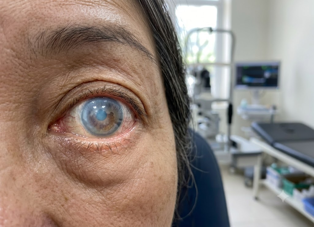
- Visibly Hazy or Blue-Tinted Corneacloudiness in the eye that becomes noticeable to other people in later stages.
The Specific Nature of Blurry or Cloudy Vision
Blurred vision is usually the first sign to appear, and it has a distinctive quality. People describe it as looking through thick fog, rising steam, or a smudged window sight becomes hazy, unclear, or smeared. Reading, spotting fine details, and recognizing faces all become difficult. Unlike ordinary blurriness, this kind doesn’t go away with blinking and can’t be fixed with regular glasses, which sets it apart from a standard vision problem.
There’s a notable pattern to when it strikes: it’s worst right after waking up. While the eyes are shut during sleep, tears can’t evaporate off the corneal surface.
With the eyes closed for hours, fluid has nowhere to go and builds up inside the cornea overnight there’s no evaporation to draw it out, and a weakened endothelial pump can’t keep pace with the incoming fluid. Once the eyes open and are exposed to air again, evaporation resumes, gradually pulling the extra fluid back out and sharpening vision as the day continues.
In the early phases of conditions like Fuchs’ Dystrophy, this pattern blurry in the morning, clearer later is a telltale diagnostic sign. But as more endothelial cells are lost and pump function keeps declining, the fluid buildup stops clearing on its own. Blurriness then becomes constant throughout the day, interfering with everyday activities no matter the hour.
Why Halos and Glare Form Around Light Sources
Seeing halos around lights comes from an optical effect: excess fluid trapped in the swollen cornea bends and scatters incoming light before it can focus properly on the retina.
People notice this as rainbow-colored rings, starburst patterns, or harsh glare surrounding bright points lamps, streetlights, or oncoming headlights. It’s especially disruptive and hard to ignore at night, when the contrast between a bright light and dark surroundings is greatest.
A healthy, well-packed cornea is built from tightly organized collagen fibers arranged so light can pass through directly with almost no scattering, producing sharp images. When edema sets in, the tissue becomes waterlogged, and the excess fluid pushes the collagen fibers apart, forming tiny fluid pockets within the stroma the cornea’s main structural layer.
As light enters the eye, it hits these fluid pockets, which act like small, uneven prisms. Rather than passing straight through, the light bends and splits off in many directions.
That scattering sends light rays landing around rather than exactly on the correct focal point on the retina, which is what produces the sensation of halos and glare. Beyond being visually distracting, this internal scatter also lowers overall image quality and contrast sensitivity, making something like night driving considerably riskier.
Advanced Structural Swelling, Severe Pain, and Discomfort
Pain and physical discomfort in the eye signal that the swelling has reached a more serious, advanced stage a condition doctors call bullous keratopathy. Mild-to-moderate swelling is typically painless, but once fluid buildup becomes severe and chronic, it forces small, fluid-filled blisters bullae to form on the epithelium, the cornea’s outer layer.
The epithelium is packed with nerve endings, making it one of the body’s most sensitive tissues. As pressure builds beneath it from the swollen stroma, the epithelial layers are pushed apart, forming these painful blisters. They can burst on their own or simply from the friction of blinking.
Once a blister bursts, it leaves an exposed, raw patch on the eye, putting sensitive corneal nerves directly in contact with air and the movement of the eyelid. That exposure causes sharp, intense pain often described as feeling like sand, glass, or an eyelash caught in the eye. This kind of pain typically comes along with heavy tearing, noticeable redness, and strong light sensitivity.
If this blister-and-rupture cycle keeps repeating, it can turn into a chronic problem, bringing ongoing discomfort, repeated corneal erosions, and a greater chance of secondary infection making it one of the more difficult complications of advanced corneal swelling.
The Underlying Mechanics of Light Sensitivity (Photophobia)
Photophobia heightened sensitivity to light stems from light scattering inside the hazy, fluid-soaked layers of a swollen cornea. That scattering overstimulates the retina, turning ordinary bright light into something uncomfortable or even painful. Normally, a clear cornea channels light along a tight, focused path straight to the retina. But once the tissue swells, that structural precision is lost.
Trapped fluid throws off the cornea’s uniform structure, effectively turning it into a hazy filter. Light especially strong sunlight or harsh artificial lighting can no longer pass through cleanly. Instead, it scatters in countless directions as it hits the fluid pockets inside the stroma. This scattering effect, called straylight, floods the retina with unfocused, inconsistent light that overwhelms the rods and cones responsible for vision.
The brain reads this flood of disorganized stimulation as glare, discomfort, and pain, prompting a reflex to limit light exposure squinting hard, shutting the eyes, or retreating to a dimmer space. In short, light sensitivity from a swollen cornea reflects the eye’s inability to properly focus incoming light, which is why some people end up wearing sunglasses indoors and avoiding brightly lit places altogether.
Identifying the Underlying Triggers
Looking at what causes corneal edema and fluid buildup on the cornea, the root issue usually traces back to something disrupting the fluid balance normally maintained by the endothelium. The main processes behind corneal edema include:
- Endothelial Dysfunction: inherited disorders such as Fuchs’ Dystrophy that gradually destroy the endothelial pump cells.
- Surgical Trauma: physical damage or cell loss sustained during procedures inside the eye, like cataract removal or glaucoma surgery.
- Inflammatory and Infectious Processes: deep infections, corneal ulcers, or internal inflammation (uveitis) that impair cell performance.
- Elevated Intraocular Pressure: sharp increases in eye pressure, as seen in acute glaucoma, that force fluid into the cornea faster than the pump cells can remove it.
Recognizing these symptoms and understanding what triggers them lets people get evaluated sooner, before chronic swelling causes permanent scarring.
Causes and Treatments of Corneal Edema
Nearly every case of corneal edema traces back to the health of the corneal endothelium. This single layer of specialized cells lines the inside of the cornea and constantly pumps fluid out to keep the tissue clear.
When these cells are damaged, reduced in number, or stop functioning properly, they lose the ability to carry out that pumping job and fluid starts collecting in the tissue. Identifying the exact cause is the critical first step in building an effective treatment plan.
The routes that lead to corneal swelling differ widely, from unintentional damage during routine eye procedures to the slow, progressive cell loss seen in inherited disorders.
Because of that variation, treatment has to be tailored based on how severe the swelling is, how much vision has been affected, and how much natural healing capacity the cornea still has. Non-surgical approaches aim to ease symptoms temporarily, while surgery offers a lasting fix by replacing the damaged tissue outright.
The Link Between Eye Surgery and Corneal Swelling
The connection between intraocular surgery and subsequent swelling comes down to the risk of damaging the endothelial cell layer during the procedure, which can compromise the eye’s ability to stay clear afterward. The endothelium is a single layer of cells that cannot regenerate once they’re gone, they’re gone.
During procedures like cataract surgery, the surgical tools, irrigation fluid, and the ultrasonic energy used to break apart the cataract (phacoemulsification) can unintentionally harm or destroy some of these cells. Losing a small number of endothelial cells is considered a normal, expected part of most eye surgeries.
In a healthy eye that starts with a high cell count, this modest loss isn’t a big deal nearby healthy cells stretch to cover the gaps, leading to only mild, short-lived swelling that resolves within days or weeks. But if a patient already has a low cell count going into surgery (due to age or a condition like Fuchs’ Dystrophy), or if the surgery itself is unusually long or complicated, the cell loss can be much more significant.
Once endothelial cell density drops below a critical point generally around 500 cells/mm² the remaining cells can no longer drain fluid effectively, and the swelling becomes chronic. When this happens after cataract surgery, it’s known clinically as pseudophakic bullous keratopathy (or aphakic bullous keratopathy when there’s no lens implant), and it often calls for a corneal transplant to restore clear sight.
How Fuchs’ Dystrophy Triggers Fluid Accumulation
Fuchs’ Dystrophy causes swelling by triggering a gradual, genetically driven decline in the corneal endothelial cells. This inherited condition is marked by premature, slow deterioration of that inner cell layer. As it progresses, two key changes take place:
- Accelerated Cell Death and Polymegathism: Endothelial cells die off faster than normal. Since they can’t be replaced, the remaining cells stretch and enlarge to cover the gaps on the cornea’s back surface. This stretching (polymegathism) temporarily maintains coverage, but the oversized, irregular cells are less efficient at pumping fluid out of the stroma.
- Guttae Formation: The disease also causes small, abnormal bumps called guttae to form on Descemet’s membrane, the thin layer supporting the endothelial cells. These bumps disturb the normal cell arrangement and further weaken pump function.
Early on, this cellular decline is mild, producing the familiar pattern of hazy vision after waking that clears through the day. But as more cells are lost and the pumping system fails more completely, the cornea can no longer regulate its own fluid levels.
That leads to swelling that persists all day, significant vision loss, and eventually the painful epithelial blisters of bullous keratopathy the disease’s final stage.
Non-Surgical Solutions for Mild Cases
For mild swelling, treatment usually starts with hypertonic saline eye drops and ointments. These work through osmosis, drawing extra fluid out of the swollen tissue. They’re the go-to first option for managing symptoms and improving vision in early-stage Fuchs’ Dystrophy or during short-term recovery after surgery.
Hypertonic saline solutions carry a much higher salt concentration (usually 2% or 5% sodium chloride) than the fluid naturally found in the cornea. Putting these on the eye’s surface sets up a concentration gradient that pulls trapped water out of the swollen epithelium and stroma and onto the tear film, where it evaporates away.
Hypertonic Saline (2% or 5% Salt) → Applied to Eye Surface → Creates Osmotic Gradient → Draws Water Out of Stroma/Epithelium → Evaporates via Tear Film
- Topical Drops (e.g., Muro 128 5%): Usually used several times during the day to help keep the cornea clear while a person is up and active.
- Topical Ointment: A thicker formula that works longer, best applied at bedtime. It counters the fluid buildup that happens overnight while the eyes are shut, cutting down on morning blurriness.
- Bandage Contact Lenses: Used in more advanced cases where bullae have already burst. This doesn’t treat the underlying swelling it’s strictly for comfort. The soft lens covers the exposed nerve endings, cutting down on friction from blinking and offering real pain relief while the epithelium heals.
When Surgical Intervention is Required
Doctors typically recommend a corneal transplant once swelling has become permanent and advanced enough to interfere with everyday life reading, driving, or recognizing people’s faces.
Surgery is only considered after non-surgical treatments have failed to give enough relief, which usually signals that the endothelial pump has failed for good common in late-stage Fuchs’ Dystrophy or after significant surgical trauma.
The standard modern approach is endothelial keratoplasty (EK), a partial-thickness transplant that swaps out only the damaged inner layer of the cornea while leaving the healthy outer tissue in place. There are two main versions:
| Transplant Type | Acronym | Tissue Layers Replaced | Recovery Characteristics |
|---|---|---|---|
| Descemet’s Membrane Endothelial Keratoplasty | DMEK | Replaces only the damaged endothelial cells and their extremely thin basement membrane (Descemet’s membrane). | A refined technique that can offer faster visual recovery and better final vision. |
| Descemet’s Stripping Endothelial Keratoplasty | DSEK | Replaces the endothelial cells, Descemet’s membrane, and a thin supporting layer of donor stromal tissue. | Gives a slightly thicker graft for extra structural support during placement. |
In both procedures, the surgeon removes the patient’s failing endothelial layer and replaces it with a thin disc of healthy donor tissue carrying fresh, functioning endothelial cells. This restores the eye’s ability to pump fluid out, letting the waterlogged tissue clear and vision return.
In rare cases where chronic swelling has already caused widespread scarring throughout the cornea, a full-thickness transplant penetrating keratoplasty (PKP) may be needed to replace the entire cornea.
Broader Aspects of Diagnosing and Managing Corneal Conditions
Diagnosing and managing corneal conditions goes well beyond spotting symptoms it involves advanced imaging, careful differentiation from similar diseases, and treatment plans tailored to the condition’s cause and the patient’s age.
It also means paying attention to preventable secondary causes and recognizing how congenital cases differ from those that develop in adulthood. Taking this wider view supports more accurate diagnosis, better treatment, and improved outcomes.
Ophthalmic Diagnosis of Corneal Edema
Eye doctors diagnose corneal edema using a set of specialized exams designed to assess the cornea’s structure, measure its thickness, and evaluate the condition of its cell layers.
Slit-Lamp Examination
This high-powered microscope uses a focused light beam and serves as the backbone of eye examinations. It lets the doctor examine each corneal layer closely, working from the outer epithelium down to the inner endothelium. During the exam, the doctor looks for classic signs of fluid retention, such as:
- Loss of tissue clarity (haziness).
- Microcysts or larger fluid blisters (bullae) on the surface.
- An overall increase in corneal thickness.
Pachymetry
To get an exact measurement of the swelling, doctors use pachymetry, which measures corneal thickness precisely. It can be done with an ultrasound probe touching the eye or through non-contact optical scanning.
A cornea thicker than expected confirms edema. Doctors often take repeated pachymetry readings over time to track how the condition is progressing or how well it’s responding to treatment.
Specular Microscopy
To pinpoint the exact cause of fluid buildup especially when Fuchs’ dystrophy or similar pump failure is suspected doctors use specular microscopy, which provides a detailed look at the endothelial cell layer.
This lets specialists measure the exact cell density and check for changes in cell size and shape. A low cell count or irregular cell patterns known as polymegathism and pleomorphism confirm that the endothelial pump isn’t working properly.
Differentiating Corneal Edema from Keratitis
Although both conditions affect the same part of the eye and impair vision, they’re distinct diseases with different causes and presentations.
Corneal Edema → Endothelial Pump Dysfunction → Passive Fluid Buildup in Stroma (Non-Inflammatory) Keratitis → Infection, Injury, or Autoimmunity → Active Inflammatory Response (Cellular Infiltration)
| Clinical Parameter | Corneal Edema | Keratitis |
|---|---|---|
| Primary Mechanism | Tissue swelling from passive fluid buildup in the stroma when the endothelium fails to pump fluid out. | An active inflammatory reaction in the cornea, usually from external agents, infection, or a systemic issue. |
| Root Drivers | Damage, dysfunction, or progressive loss of endothelial cells. | Bacterial, viral, fungal, or parasitic infection, physical trauma, or autoimmune conditions. |
| Key Symptoms | Foggy or hazy vision, halos around lights, and pain only if surface blisters develop. | Severe pain, a foreign body sensation, strong redness, light sensitivity, and watery or pus-like discharge. |
| Nature of Condition | A physiological fluid imbalance rather than active inflammation. | An active inflammatory process that can cause secondary edema if severe, though the inflammation itself needs direct treatment. |
Preventability and Risk Mitigation Strategies
Whether corneal edema can be prevented depends largely on its origin. Cases fall into two categories primary and secondary each with a different outlook for prevention.
Primary Cases (Non-Preventable)
Primary edema linked to inherited genetic conditions can’t be prevented. Fuchs’ Endothelial Corneal Dystrophy is the classic example a progressive, DNA-driven disorder that causes premature death of endothelial cells. No lifestyle change or medication can stop it from developing. People with a family history can only get regular monitoring from an eye specialist to catch and manage symptoms as they appear.
Secondary Cases (Preventable)
On the other hand, many cases caused by external factors or other eye conditions can be prevented or reduced through targeted steps:
- Preventing Traumatic Injury: Wearing safety goggles or protective eyewear during sports, construction, or yard work protects the eye from injuries that can destroy the endothelial layer.
- Controlling Intraocular Pressure: High eye pressure from glaucoma can slowly damage endothelial cells. Sticking to prescribed medications and regular pressure checks helps protect corneal health long-term.
- Refining Surgical Protocols: During procedures like cataract surgery, surgeons can lower the risk of post-operative swelling by using careful technique, high-quality protective solutions for the endothelium, and keeping surgery time as short as possible.
- Practicing Strict Lens Hygiene: Careful cleaning, handling, and timely replacement of contact lenses helps prevent the serious infections that can lead to secondary swelling.
Congenital Versus Adult-Onset Pathologies
Congenital and adult-onset forms of corneal edema differ in their causes, how they present, when they appear, and how urgently they need to be treated.
Congenital Edema → Present at Birth → Widespread Cloudiness (CHED) → High Risk of Amblyopia → Urgent Full-Thickness Transplant (PKP) Adult-Onset → Develops Later in Life → Slow, Degenerative Progression → Clears During the Day → Selective Layer Transplant (DMEK/DSEK)
Congenital Corneal Edema
This form appears at birth or shortly after, caused by genetic mutations that interfere with normal eye development before birth. Congenital Hereditary Endothelial Dystrophy (CHED) is a key example, where the endothelial pump doesn’t work properly from birth. This produces a cloudy, blue-gray appearance in one or both of an infant’s eyes.
It shows up immediately and is often paired with light sensitivity and involuntary eye movements (nystagmus). Treating it quickly is critical because clear vision is essential for the visual pathways in the brain to develop correctly in early childhood. Without treatment, the child risks permanent vision loss from amblyopia (lazy eye). Because of this, a full-thickness corneal transplant penetrating keratoplasty is often needed within the first few months of life.
Adult-Onset Corneal Edema
By contrast, adult-onset cases like those caused by progressive Fuchs’ dystrophy or complications after surgery such as pseudophakic bullous keratopathy develop slowly over years or decades. These patients start out with healthy corneas, but the endothelial cells break down gradually later in life due to aging, genetics, or surgical damage.
Symptoms like morning blurriness that clears during the day appear gradually and worsen slowly over time. Treatment usually starts conservatively with hypertonic saline drops or ointments to draw fluid out osmotically. Surgery is held off until vision becomes consistently impaired and starts interfering with daily life.
The preferred surgical option is endothelial keratoplasty (DMEK or DSEK), a less invasive procedure that replaces only the damaged inner layer, allowing for a much quicker recovery than the full-thickness transplants needed in congenital cases.
By understanding these causes through this framework, doctors can determine the right diagnostic and treatment path for lasting visual stability.
Conclusion
Corneal edema can seriously affect both vision and comfort if it isn’t treated. Symptoms like blurred or cloudy vision, halos around lights, eye pain, light sensitivity, or visible corneal swelling shouldn’t be brushed off. Treatment depends on the underlying cause and can range from eye drops and medication to contact lenses or, in more severe cases, surgery such as a corneal transplant. Catching it early and managing it promptly goes a long way toward protecting vision and quality of life.
Frequently Asked Questions
1. What is corneal edema?
Corneal edema is swelling of the cornea caused by a buildup of fluid. It disrupts the cornea’s normal clarity and shape, leading to blurred vision, light sensitivity, and sometimes discomfort. It can be sudden or long-term and may affect one or both eyes depending on the cause.
2. What are the common causes of corneal edema?
It can result from trauma, eye surgery (like cataract or glaucoma procedures), infection, inflammation, elevated eye pressure, or damage to the endothelial cells. Hereditary conditions, overuse of contact lenses, or toxin exposure can also play a role. Finding the underlying cause is essential for effective treatment.
3. How does corneal edema affect vision?
It disrupts the smooth, clear surface of the cornea, which can cause blurred or hazy vision, halos around lights, glare, and trouble seeing in bright conditions. In severe cases, vision can fluctuate or become significantly impaired, making everyday tasks harder.
4. What are the warning signs of corneal edema?
Warning signs include blurry or cloudy vision, halos around lights, eye pain or pressure, light sensitivity, watery eyes, a gritty feeling, redness, visible corneal swelling, and vision that’s worse right after waking. Catching these early and getting checked out can help prevent complications.
5. How is corneal edema diagnosed?
Doctors diagnose it through a full eye exam, which may include slit-lamp microscopy, corneal thickness measurement, and a visual acuity check. Additional tests like endothelial cell counts or imaging can help determine severity and cause, guiding the right treatment approach.
6. How is corneal edema treated?
Treatment depends on the cause. Mild cases may respond well to hypertonic saline drops or ointments that draw out excess fluid. Treating underlying issues, like high eye pressure or infection, is also important. Severe or ongoing cases may need surgery, such as endothelial keratoplasty or a corneal transplant, to restore clarity and vision.
7. Can corneal edema be prevented?
Not every case can be prevented, but certain steps can lower the risk good contact lens hygiene, regular eye exams, prompt treatment of eye infections, careful monitoring after surgery, and managing conditions like glaucoma. Protecting the eyes from injury and following post-surgical care instructions also help reduce risk.