10 Signs of a Hematoma and When to Be Concerned
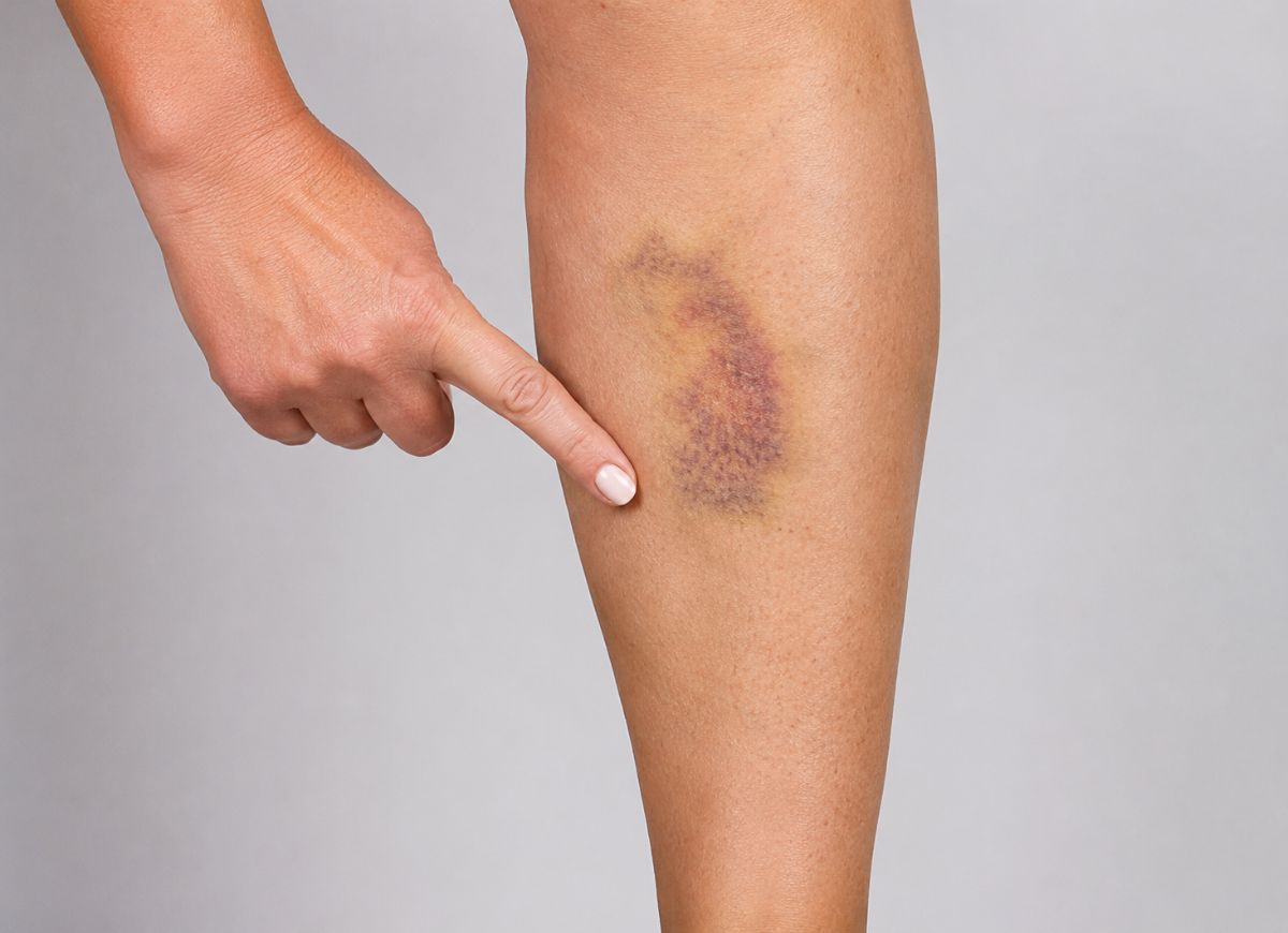
Have you ever looked at a bruise and wondered why it felt harder, bigger, or more painful than it should? Most bruises are harmless and fade on their own, but sometimes blood collects deeper under the skin or inside the body, forming what’s called a hematoma. At first, it may look like an ordinary bruise. Then the area may swell, feel firm, become tender, or change color in a way that makes you pause.
A hematoma can happen after a fall, bump, sports injury, surgery, injection, or even a minor accident that seemed forgettable at the time. For many people, it shows up as a dark purple or blue patch with swelling underneath. For others, especially those on blood thinners or living with bleeding disorders, a hematoma can form more easily and grow faster than expected.
That’s what makes it worth understanding. A small hematoma near the skin may only need rest, ice, compression, and time. A larger one can press on nearby tissue, limit movement, cause significant pain, or signal bleeding that needs medical attention. Some hematomas aren’t visible at all, forming deeper in muscles, organs, or around the brain after a head injury.
Below are 10 signs of a hematoma and what separates one that just needs time from one that needs urgent care.
What is a Hematoma?
A hematoma is a localized collection of blood, often clotted, that has escaped a blood vessel and pooled within body tissue or a cavity, essentially an internal blood blister. This can happen just under the skin (subcutaneous), deep within a muscle (intramuscular), or in more critical spots like the space around the brain (intracranial). The defining feature is that the blood collects in a specific pocket, creating a palpable mass.
This is different from an ordinary bruise (ecchymosis), where blood from damaged capillaries spreads out thinly through the skin’s surface layers, making it flat and diffuse. A hematoma is a more significant bleed, forming a three-dimensional collection that often feels firm, rubbery, or lumpy. The concentrated volume of blood is what leads to more pronounced pain and swelling, since it presses harder on surrounding nerves and tissue than the spread-out blood of a typical bruise.
The 10 Warning Signs
1. Deep, dark skin discoloration. Pooled blood under the skin appears more intense than a standard bruise, dark blue, deep purple, or even black. As the body breaks down hemoglobin in the collected blood, the color gradually shifts to green and then yellow over days or weeks.
2. A firm, rubbery, or lumpy feeling. This is a hallmark sign. The pooled blood, liquid or clotted, forms a distinct mass that feels denser and more defined than the surrounding tissue.
3. Significant swelling and inflammation. The body’s response to injury, combined with the physical volume of pooled blood, causes noticeable swelling that contributes to pain and tightness.
4. Localized pain and tenderness. Pressure from the hematoma on nearby nerve endings causes pain directly over the area, ranging from a dull ache to severe, throbbing pain, especially when touched or moved.
5. Warmth at the site. As the body directs more blood flow to the injury to begin healing, the skin over the hematoma may feel warm, a classic sign of inflammation.
6. Limited range of motion. A hematoma forming in or near a muscle or joint can physically restrict movement. A large hematoma in the thigh, for example, can make it difficult to bend the knee or walk.
7. A visible, raised area. Unlike a flat bruise, a significant hematoma often creates a visible bulge or dome-shaped swelling due to the volume of blood collected beneath the skin.
8. Skin that feels tight. Swelling and the underlying mass of blood can stretch the skin over it, creating a sensation of tightness or tautness.
9. Headache or confusion. This applies specifically to hematomas inside the head (subdural or epidural). Blood collecting inside the rigid skull increases pressure on the brain, causing severe headache, dizziness, confusion, vomiting, or loss of consciousness.
10. Numbness or tingling. A hematoma large enough to compress nearby nerves can interfere with nerve signaling, causing numbness, tingling, or a “pins and needles” sensation in the affected area or beyond it.
Can a Hematoma Form Without an Obvious Injury?
Yes. While direct trauma, like a fall or a blow, is the most common cause, several medical factors can lead to spontaneous bleeding, where even minor, unnoticed stress on a vessel results in a significant bleed.
Medications are a common cause. Blood thinners like warfarin, apixaban, or rivaroxaban, and antiplatelet drugs like aspirin or clopidogrel, all work by making it harder for blood to clot, meaning even a minor bump someone doesn’t remember can produce a substantial hematoma.
Medical conditions also raise risk. Blood clotting disorders like hemophilia or Von Willebrand disease directly impair the body’s ability to stop bleeding. Liver disease, particularly cirrhosis, can reduce production of the proteins needed for clotting. Thrombocytopenia (low platelet count) means there aren’t enough platelets to plug damaged vessels. Structural issues like an aneurysm can also rupture spontaneously and cause a hematoma with no external trauma at all.
Epidural vs. Subdural Hematoma
Both are bleeds within the skull, but they differ in location, the vessel involved, and how symptoms progress.
Epidural hematoma occurs when blood collects between the skull and the dura mater (the tough membrane covering the brain), usually from a skull fracture that tears an artery, most often the middle meningeal artery. Because arterial blood is under high pressure, these can expand rapidly. A classic pattern is a brief loss of consciousness at the time of injury, followed by a “lucid interval” where the person seems fine, then a rapid decline as the hematoma grows. It’s most associated with head trauma in younger people, from sports or car accidents, with symptoms typically appearing within minutes to hours.
Subdural hematoma forms between the dura mater and the next layer covering the brain (the arachnoid mater), typically from torn “bridging veins.” It’s more common in older adults, since age-related brain shrinkage stretches these veins, making them vulnerable to tearing from even minor head trauma. Symptoms can appear quickly (within 72 hours), more gradually (3 to 21 days), or slowly over weeks to months, making it harder to diagnose, especially in older adults.
On a CT scan, an epidural hematoma typically looks lens-shaped, while a subdural hematoma appears crescent-shaped, following the brain’s curve.
When to Be Concerned
Certain signs mean a hematoma needs immediate medical evaluation rather than home care.
Rapid growth. A hematoma visibly expanding over minutes to hours means the vessel is still actively bleeding and hasn’t stabilized, risking significant blood loss and pressure buildup.
Severe or worsening pain. Pain that’s excruciating, out of proportion to the visible size, or steadily intensifying can indicate rising pressure on nerves or the onset of compartment syndrome, where pressure inside a muscle compartment cuts off blood supply.
Neurological symptoms. Weakness, inability to move a joint, numbness, or tingling in the area or further down a limb means the hematoma may be compressing nerves or blood flow, and needs immediate attention to prevent permanent nerve damage.
Any head hematoma. Even a small one deserves close monitoring. A severe or persistent headache, nausea or vomiting, confusion, dizziness, blurred vision, unequal pupil sizes, slurred speech, or loss of consciousness after a head injury are signs of a possible intracranial hematoma, a life-threatening emergency.
Location in the neck or abdomen. A hematoma in the neck can compress the airway and interfere with breathing. One in the abdomen could signal bleeding from an internal organ like the spleen or liver. Both need urgent evaluation.
What Happens If It’s Left Untreated
An untreated, severe hematoma can lead to serious, sometimes permanent, complications, mostly from the pressure it exerts on surrounding structures and the risk of infection.
Nerve damage. An expanding hematoma pressing on nerves can cause chronic pain, persistent numbness, muscle weakness, or, in severe cases, paralysis if not relieved in time.
Compartment syndrome. This urgent complication, typically in the lower leg or forearm, happens when pressure from a hematoma inside a muscle compartment rises high enough to cut off blood flow. Without immediate surgery (a fasciotomy) to relieve pressure, it can cause permanent muscle death, nerve damage, and potentially require amputation.
Infection. A large pool of stagnant blood is a good environment for bacteria. If the skin over it breaks or bacteria enter the bloodstream another way, the hematoma can become infected, potentially forming an abscess and leading to sepsis if untreated.
Pressure on vital organs. This is the most critical risk with intracranial hematomas, since the rigid skull can’t expand and any blood collection directly compresses brain tissue, potentially causing irreversible brain damage or death. A large hematoma in the chest can similarly collapse a lung or press on the heart.
A permanent hardened lump. Sometimes the body can’t fully reabsorb a large hematoma, and over time it can calcify into a permanent hard lump that causes chronic pain and may need surgical removal.
Types That Need Immediate Medical Attention
Any suspected intracranial hematoma. Anyone with a head injury followed by severe headache, vomiting, confusion, seizure, or loss of consciousness needs emergency care immediately, given the risk of brain compression.
Limb hematomas with the “5 P’s.” Severe pain, pale skin, numbness or tingling, a faint or absent pulse, and inability to move the digits together signal an emergency requiring urgent assessment to prevent limb loss.
Rapidly growing, tense hematomas causing functional impairment, such as making it impossible to bear weight on a leg, need a doctor’s evaluation, since this suggests ongoing bleeding that may need a procedure to locate and stop the source.
How It’s Diagnosed
Diagnosis starts with a physical exam checking for swelling, discoloration, tenderness, and warmth, along with questions about how the injury happened and when symptoms began. For superficial hematomas, this is often enough.
For deeper or more concerning hematomas, imaging becomes important. An ultrasound can identify hematomas in muscles or the abdominal cavity. For suspected head injuries, a CT scan is the preferred first step, fast and effective at detecting acute bleeding and pressure on the brain. An MRI may follow for a more detailed look, helping distinguish between an acute, subacute, or chronic hematoma. This imaging determines whether a hematoma can be managed with observation and home care, or needs urgent medical or surgical intervention.
How Hematomas Are Treated
For minor, superficial hematomas, the standard approach is R.I.C.E.: Rest (avoiding strain on the area), Ice (15-20 minutes several times a day to limit swelling and numb pain), Compression (an elastic bandage, not tight enough to restrict circulation), and Elevation (raising the area above heart level to help fluid drain).
For large, painful, or deep hematomas, medical intervention may be needed. Aspiration, using a needle and syringe to drain the blood, provides immediate relief and is common for large muscular hematomas or ones under a nail.
For the most severe cases, surgery may be necessary. A craniotomy, temporarily removing part of the skull, is used to drain an epidural or subdural hematoma causing neurological symptoms. A fasciotomy, cutting open the tissue enclosing a muscle, relieves dangerous pressure in cases of compartment syndrome.
Who’s at Higher Risk
- People on blood thinners or antiplatelet medications (warfarin, apixaban, rivaroxaban, clopidogrel, even daily aspirin) face a higher risk, since these drugs intentionally impair clotting, meaning even minor injuries can cause significant, prolonged bleeding.
- Older adults face elevated risk for several reasons: a higher likelihood of falls, age-related brain changes that stretch and weaken bridging veins (raising subdural hematoma risk), and a greater likelihood of being on blood thinners.
- People with bleeding disorders, like hemophilia or von Willebrand disease, have a compromised ability to clot, so even trivial injuries can cause deep, extensive hematomas requiring specialized treatment.
- People with severe liver disease, such as cirrhosis, often have impaired clotting factor production, making them prone to easy bruising and serious hematomas from minor trauma.
- Heavy alcohol use compounds risk by damaging the liver, having a mild blood-thinning effect, and increasing the likelihood of falls and injuries.
Frequently Asked Questions
How serious is a hematoma?
It depends on size, location, cause, and how quickly it’s growing. Small ones after a bump often resolve with rest, ice, and elevation. It becomes more concerning when it’s large, expanding, extremely painful, warm, infected-looking, or causing numbness or weakness, especially after a head, chest, or abdominal injury, or in someone on blood thinners.
Do hematomas go away on their own?
Yes, in most cases. The body gradually reabsorbs the trapped blood, with smaller ones improving within days to weeks and larger ones taking longer. Color typically shifts from dark purple or blue to green, yellow, and brown as it heals. Growing pain, heat, redness, or lack of improvement warrant a checkup.
Is it okay to massage a hematoma?
Generally not early on. Pressing or rubbing a fresh hematoma, especially in the first 24 to 48 hours, can irritate damaged vessels and worsen swelling or bleeding. Gentle movement may help later, but deep massage should wait until pain and swelling have clearly improved.
Which type is most common?
Hematomas under the skin or in soft tissue after a bump, fall, injection, or minor trauma are the most common. Deeper types, in muscles, under a nail, or internally after trauma, and head-related hematomas like subdural bleeds are less common but far more serious.
Can I fly with a leg hematoma?
It depends on severity. A small, improving one from a known minor injury usually isn’t a problem, though long flights can worsen leg swelling. Avoid flying without medical advice if the leg is very swollen, painful, warm, red, numb, or worsening, since these can overlap with a blood clot.
Can I exercise with a leg hematoma?
Light movement may be fine once pain is mild and swelling is improving, but intense exercise, running, heavy lifting, or contact sports too soon can make it worse. Rest and elevation are usually more helpful early on.
What can make a hematoma worse?
Repeated bumping, massaging too early, applying heat too soon, straining, intense exercise, alcohol use, and blood-thinning medications can all worsen bruising or bleeding.
Why does a hematoma hurt so much?
Blood collecting under the skin or deeper in tissue creates pressure that stretches surrounding tissue and irritates nerves. Pain can feel worse near a joint, inside a muscle, under a nail, or in a tight space with little room for swelling.
Is a hot bath good for a hematoma?
Not in the first 24 to 48 hours, since heat can increase blood flow and worsen swelling early on. Ice is preferred initially; heat may help later once swelling has settled and stopped expanding.
How do I know a hematoma is healing?
It should become progressively less painful, less swollen, and softer, with color shifting from dark blue or purple toward green, yellow, and brown, and the lump gradually shrinking. Growth, increased heat or redness, drainage, fever, or worsening pain suggest it isn’t healing normally.
What vitamin speeds up bruise healing?
No vitamin makes a hematoma disappear overnight, but good nutrition supports healing generally: vitamin C for collagen production and vessel repair, vitamin K for normal clotting, and adequate protein, zinc, iron, and fluids for tissue recovery. Food sources are generally preferred over supplements, and anyone on blood thinners should be cautious with vitamin K in particular.
The Bottom Line
A hematoma can look like a simple bruise, but the swelling, firmness, and pain may tell a deeper story. Many small ones heal on their own with rest, ice, compression, and elevation. Others need medical attention, especially when they grow quickly, cause severe pain, follow a head injury, or appear in someone on blood thinners. The key is watching the pattern: a healing hematoma should slowly shrink, soften, fade in color, and hurt less over time. One that’s growing, getting hotter, more painful, numb, or harder to move around deserves a closer look rather than being dismissed.