12 Early Warning Signs of MALT Lymphoma You Shouldn’t Ignore
Can a slow-moving cancer stay hidden behind symptoms that feel completely routine? MALT lymphoma short for mucosa-associated lymphoid tissue lymphoma is a form of non-Hodgkin lymphoma that typically takes root in the tissue lining an organ rather than in a lymph node itself. Most cases are described as indolent, meaning they grow slowly, but “slow” doesn’t mean “safe to overlook.”
MALT lymphoma is rare, making up roughly 5% to 8% of all non-Hodgkin lymphomas, with the stomach being the site where it shows up most often. In many stomach cases, a long-standing infection with Helicobacter pylori (H. pylori) plays a central role which is also why some early gastric cases can improve simply by clearing that infection with antibiotics.
What makes this disease tricky is that it doesn’t look the same in every patient. In the stomach it might show up as indigestion, cramping, nausea, appetite loss, weight loss, or bleeding. In the eyes, lungs, salivary glands, thyroid, skin, or intestines, the picture changes entirely. Some people live with mild complaints for months before anything is found; others stumble onto it while being tested for something unrelated.
Below are 12 warning signs worth knowing. None of them guarantee cancer on their own most have far more common explanations but swelling that won’t go away, weight loss you can’t explain, night sweats, ongoing tiredness, persistent stomach trouble, or symptoms that keep coming back are worth having checked out. Recognizing these signs early can mean earlier answers, better questions for your doctor, and treatment that starts sooner rather than later.
What is MALT Lymphoma?
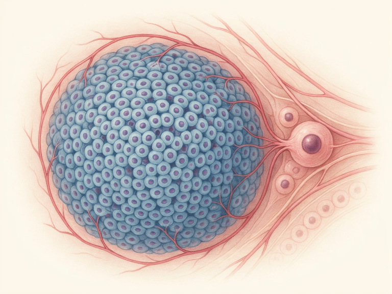
MALT lymphoma is a slow-progressing form of non-Hodgkin lymphoma that develops from B-lymphocytes living within mucosa-associated lymphoid tissue a loosely distributed network of immune cells found along the mucosal linings of the body. Understanding it means breaking down what that tissue is and where it typically shows up.
What sets this cancer apart is that it starts outside the classic lymphatic organs like lymph nodes, which is why it’s labeled an extranodal lymphoma. It tends to develop out of long-running inflammation usually from a chronic infection or an autoimmune condition that keeps a specific part of the immune system switched on for an extended stretch of time.
Over time, that constant stimulation can trigger genetic changes in the local B-cells, pushing them to multiply out of control and eventually form a tumor. Because the process is gradual, MALT lymphoma often takes years to progress, and its early symptoms can be so mild they get mistaken for far more ordinary problems which is exactly why catching it early is so difficult.
How is MALT Lymphoma Medically Defined?
In clinical terms, MALT lymphoma falls under extranodal marginal zone B-cell lymphoma a distinct subtype of non-Hodgkin lymphoma marked by the runaway growth of B-lymphocytes that originate in the marginal zone of MALT tissue. That definition packs in a few important details.
“Extranodal” simply means the disease starts somewhere other than a lymph node, setting it apart from most other lymphomas. “Marginal zone” points to a particular region inside secondary lymphoid follicles where these B-cells normally live and work cells that are part of the adaptive immune system, tasked with responding to threats encountered at mucosal surfaces.
The path to MALT lymphoma unfolds in stages. It starts with a chronic inflammatory trigger commonly a persistent bacterial infection such as H. pylori in the stomach. That ongoing stimulation draws B-cells and T-cells to the site and causes them to multiply, gradually building organized lymphoid tissue in a place where it wouldn’t normally exist in any real quantity.
In that environment of constant immune activity, B-cells become more prone to picking up genetic mutations. A chromosomal translocation known as t(11;18)(q21;q21) shows up frequently in MALT lymphoma and is thought to be a key turning point the moment a reactive immune response tips over into a true malignancy.
These genetic changes switch on signaling pathways that let the cells survive and multiply on their own, no longer dependent on the original inflammatory trigger. Because the disease grows slowly, patients often go months or years with gradually worsening symptoms rather than a sudden onset.
Where in the Body Does MALT Lymphoma Commonly Develop?
The stomach is by far the most frequent site, responsible for roughly 50–70% of all cases, though MALT lymphoma can technically arise almost anywhere there’s mucosal tissue. That range in location is exactly why the symptoms can look so different from one patient to the next.
Cases that start in the stomach are called gastric MALT lymphoma, and they’re almost always tied to chronic H. pylori infection. The ongoing irritation this bacterium causes builds the perfect conditions for the lymphoma to take hold, and patients frequently mistake the early symptoms for gastritis or an ulcer.
Cases outside the stomach fall under non-gastric MALT lymphoma and can turn up in a wide variety of places. The salivary glands especially the parotid gland are a common non-gastric site, often connected to Sjögren’s syndrome, an autoimmune disorder that inflames the glands responsible for tears and saliva.
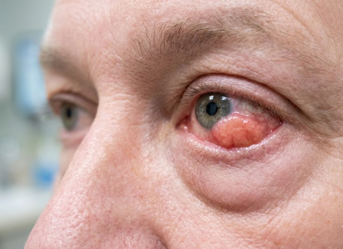
The ocular adnexa (the eye area) covers the conjunctiva, lacrimal gland, and orbit. Lymphoma here often shows up as a painless, salmon-colored patch on the eye’s surface, or as swelling and redness that won’t clear. It has been linked to infection with Chlamydia psittaci.
Pulmonary MALT lymphoma is rare and usually silent, often spotted by chance on a chest X-ray or CT scan. When it does cause symptoms, they tend to be vague a lingering cough or breathlessness.
Cutaneous MALT lymphoma shows up as one or several red-to-purple bumps, patches, or plaques on the skin. In certain parts of the world, it has been tied to Borrelia burgdorferi, the bacterium behind Lyme disease.
Thyroid MALT lymphoma is almost exclusively found in people with a history of Hashimoto’s thyroiditis, an autoimmune disease that inflames the thyroid over time. Intestinal MALT lymphoma is less common still and has occasionally been connected to infections such as Campylobacter jejuni.
12 Warning Signs of MALT Lymphoma
Persistent Indigestion or Heartburn
This tends to be one of the first complaints people notice a burning discomfort in the upper abdomen, often worse after eating. What separates it from ordinary heartburn is that it lingers and doesn’t really respond to over-the-counter antacids.
Upper Abdominal Pain or Discomfort
Some describe it as a dull ache, others as a constant pressure sitting high in the stomach. It can come and go or stay steady, and it happens because the tumor itself is pressing on the stomach wall and nearby nerves.
Nausea and Vomiting
As the lymphoma grows, it can get in the way of the stomach’s normal digestion and emptying, leaving a person feeling queasy much of the time. If the tumor partially blocks the stomach, this can progress into actual vomiting.
Loss of Appetite
Between the pain, discomfort, and nausea, eating often stops feeling worth it. Many patients notice food simply stops appealing to them, even meals they’d normally enjoy.
Feeling Full After Eating a Small Amount (Early Satiety)
A stomach wall that’s thickened by the tumor can’t expand the way it normally would, so even a modest meal can trigger an uncomfortable feeling of fullness. Over time, this often adds up to unplanned weight loss.
Lungs (Pulmonary MALT Lymphoma)
Often found without any symptoms at all, but when signs do appear, they usually involve the respiratory system a dry, ongoing cough or shortness of breath that’s more noticeable during activity, caused by tumor tissue spreading through the lung’s airways or tissue. Less often, this can bring chest pain or small amounts of blood when coughing.
Eyes (Ocular Adnexal MALT Lymphoma)
Most often affecting the conjunctiva or the lacrimal glands, the telltale sign is a painless, raised, salmon-colored patch on the eye. Other clues include ongoing redness, irritation, blurry vision, watery eyes, or a visible swelling of the eyelid frequently mistaken for chronic pink eye.
Skin (Cutaneous MALT Lymphoma)
This form appears as skin lesions one or several reddish-purple rashes, patches, or raised nodules that build up gradually. They’re usually painless, sometimes itchy, and most likely to appear on the trunk or arms.
Salivary Glands
When it develops in the salivary glands, most often the parotid gland in the cheek, it usually shows up as a painless, firm swelling or lump along the jaw. Dry mouth or dry eyes can accompany it, especially when linked to Sjögren’s syndrome.
Unexplained Weight Loss
This refers to losing more than 10% of body weight over roughly six months without dieting or new exercise habits. It happens because the cancer cells burn through the body’s energy reserves and can throw off metabolism and appetite.
Drenching Night Sweats
These go well beyond a light sweat they’re heavy enough to soak through nightclothes and bedding, sometimes forcing a change of sheets overnight. The cause isn’t fully understood, but it’s believed to involve cytokines released by lymphoma cells that interfere with the brain’s temperature control.
Unexplained Fever
A fever above 100.4°F (38°C) that keeps recurring without any sign of infection, often worse in the evenings. Like night sweats, this is thought to come from substances released by the cancer cells that trigger a body-wide inflammatory response. Any of these so-called “B symptoms” call for prompt medical attention, since they can affect both prognosis and how urgently treatment is needed.
Causes and Risk Factors for MALT Lymphoma
Chronic inflammation is the main driver behind MALT lymphoma, usually set off by a lingering infection or an autoimmune condition. It’s this prolonged immune activity at one specific site in the body that ultimately fuels the disease.
Unlike cancers tied to inherited genetic conditions, MALT lymphoma is mostly something the body develops over time, from a drawn-out standoff between the immune system and whatever is irritating it. That ongoing immune response causes lymphoid tissue to build up in places it normally wouldn’t, such as the stomach lining.
In that setting of constant cell activity and turnover, B-cells face a higher chance of picking up mutations that push them toward becoming cancerous. This is also why MALT lymphoma can appear in so many different organs anywhere there’s mucosal tissue and the potential for ongoing inflammation.
The Connection Between H. pylori Infection and MALT Lymphoma
Few relationships in oncology are as well documented as the one between H. pylori infection and gastric MALT lymphoma. H. pylori is a bacterium that settles into the stomach lining, and long-term infection is already a known cause of gastritis, ulcers, and stomach cancer. In MALT lymphoma, it acts as a constant trigger for the immune system.
The body keeps fighting the bacterium, pulling in large numbers of T-cells and B-cells to the stomach lining, which over time builds up acquired MALT tissue there.
The sequence tends to follow a predictable path. T-cells responding to H. pylori signal local B-cells to multiply. Some of those B-cells eventually pick up mutations the t(11;18) translocation being a well-known one which produces a fusion protein called API2-MALT1. That protein switches on the NF-κB pathway, giving the B-cells a survival edge and letting them grow without needing further T-cell input.
At that point, what started as a normal immune reaction has become a true lymphoma. What’s notable is what this means for treatment: in more than 75% of patients with early, localized gastric MALT lymphoma, simply eliminating the H. pylori infection with antibiotics is enough to shrink and often eliminate the lymphoma entirely, without chemotherapy or radiation.
Other Infections or Autoimmune Diseases Considered Risk Factors
Outside of H. pylori, several other chronic infections and autoimmune diseases are known to raise the risk of non-gastric MALT lymphoma. The pattern is always the same ongoing immune stimulation in a particular organ eventually leads to lymphoma there. Each non-gastric site tends to have its own associated trigger, underscoring just how localized this disease is.
Sjögren’s syndrome is an autoimmune condition in which the immune system attacks the glands that make tears and saliva, causing long-term inflammation. People with Sjögren’s face a risk of salivary gland MALT lymphoma that’s up to 44 times higher than the general population.
Hashimoto’s thyroiditis involves chronic immune-driven damage to the thyroid gland, and it’s the leading cause of underactive thyroid in developed countries. It’s also strongly tied to primary thyroid MALT lymphoma.
Chlamydia psittaci, usually carried by birds, is recognized as a risk factor for ocular adnexal (eye) MALT lymphoma. As with H. pylori in the stomach, treating this infection with antibiotics can sometimes cause the lymphoma to regress.
Borrelia burgdorferi, the bacterium behind Lyme disease, has been connected to cutaneous MALT lymphoma, particularly in parts of Europe where the infection is common. The ongoing skin infection is thought to be the trigger.
Campylobacter jejuni typically causes food poisoning, but in rare cases chronic infection has been linked to a specific intestinal MALT lymphoma known as immunoproliferative small intestinal disease (IPSID).
MALT Lymphoma Diagnosis
Diagnosing MALT lymphoma involves a physical exam, a look at medical history, bloodwork, imaging, and most importantly a biopsy of the affected tissue for lab analysis. The process usually starts once a patient shows up with persistent symptoms like the ones described earlier.
A doctor’s first step is usually digging into those symptoms and checking for risk factors, such as a past H. pylori infection or an autoimmune disorder. Because the symptoms overlap with so many common conditions, the workup tends to be methodical, ruling out more likely explanations first.
Imaging CT scans or ultrasounds can help visualize the affected organ and gauge how far the disease has spread, while bloodwork gives a broader picture of overall health and can flag issues like anemia. None of these tests alone can confirm MALT lymphoma, though; that requires an actual tissue sample.
Is a Biopsy Always Required For a Definitive Diagnosis?
Yes a biopsy is essential and remains the gold standard for confirming MALT lymphoma. Imaging and bloodwork can raise suspicion, but they can’t provide certainty.
Only by studying a tissue sample under a microscope can a pathologist spot the specific features of the cancer and rule out other explanations, including infections, benign inflammation, or different cancer types. How the biopsy is taken depends entirely on where the suspected lymphoma is located.
For the most common form, gastric MALT lymphoma, the biopsy is usually taken during an upper endoscopy (EGD). A doctor guides a thin, flexible, camera-equipped tube through the mouth to examine the esophagus, stomach, and duodenum, then uses small forceps passed through the scope to collect samples from any areas that look abnormal.
Once collected, the tissue goes to a pathology lab, where the cells’ structure is examined and specialized tests such as immunohistochemistry and flow cytometry are run. Immunohistochemistry relies on antibodies to identify specific markers on the cell surface.
In MALT lymphoma, cells typically test positive for B-cell markers CD20 and CD79a, while testing negative for CD5, CD10, and Cyclin D1 a pattern that helps distinguish it from other B-cell lymphomas. Genetic testing may also look for translocations like t(11;18), adding further confirmation and useful prognostic detail.
Imaging and Blood Tests In the Diagnostic Process
Imaging and bloodwork play a supporting but essential role, mainly used to determine how far the disease has spread and to check on a patient’s general health. While the biopsy is what confirms the diagnosis, these tools round out the picture needed for treatment planning.
For suspected gastric MALT lymphoma, upper endoscopy remains the main tool for both viewing the area and collecting a biopsy. Endoscopic ultrasound (EUS) is sometimes done at the same time, using a probe on the endoscope tip to produce detailed images of the stomach wall and nearby lymph nodes, showing how deep the lymphoma has grown.
CT scans of the chest, abdomen, and pelvis are the standard for staging, used to check whether the disease has reached other organs, distant MALT sites, or enlarged lymph nodes helping determine whether it’s localized (Stage I/II) or more widespread (Stage III/IV).
Ultrasound works particularly well for non-gastric sites close to the skin’s surface, such as the salivary glands, thyroid, or superficial lymph nodes.
Routine bloodwork also plays a role. A complete blood count (CBC) checks red cells, white cells, and platelets anemia can develop from ongoing bleeding tied to a gastric tumor. A comprehensive metabolic panel (CMP) checks kidney and liver function ahead of treatment. Lactate dehydrogenase (LDH), an enzyme that can rise with lymphoma, may signal a larger tumor burden when elevated.
Depending on where the lymphoma is located, additional tests a urea breath test, stool antigen test, or blood test for H. pylori may be used to check for an underlying infection.
Broader Medical Considerations for MALT Lymphoma
Beyond spotting the warning signs, understanding MALT lymphoma also means understanding how it’s staged, how it differs by location, what the outlook typically looks like, and why it responds so well to treatments that don’t always involve chemotherapy.
These details matter because they set MALT lymphoma apart from more aggressive cancers, giving patients and families useful context as they work through diagnosis and treatment decisions.
This bigger picture covers how doctors track the disease’s spread, the real differences between its common locations, its generally encouraging prognosis compared with other lymphomas, and its notable responsiveness to non-chemo treatment options in certain cases. Each piece feeds into building a treatment plan suited to the individual patient.
How is MALT Lymphoma Staged?
Like most lymphomas, MALT lymphoma is staged using the Ann Arbor system, which tracks how far the cancer has spread through the body. Because it’s an extranodal lymphoma starting outside the lymph nodes an “E” is often added to the stage label (for example, Stage IE).
Since MALT lymphoma usually begins in a single organ, like the stomach, staging plays a big role in shaping both treatment and outlook. Stage IE, where the disease is confined to one site, is the most typical presentation at diagnosis.
That tendency to stay localized is a big part of why the outlook is generally favorable. As the disease advances, it can spread to nearby lymph nodes or more distant sites, raising the stage and often calling for more systemic treatment beyond localized options like antibiotics or radiation.
Stage I (IE): Confined to one extranodal site the stomach, a salivary gland, or a patch of skin, for example. This is both the earliest and the most commonly diagnosed stage.
Stage II (IIE): Involves an extranodal site along with nearby lymph nodes on the same side of the diaphragm a gastric MALT lymphoma that has spread to nearby abdominal lymph nodes, for instance.
Stage III: Involves lymph node regions on both sides of the diaphragm, or an extranodal site plus lymph nodes on the opposite side a sign of more widespread disease.
Stage IV: The lymphoma has spread broadly, reaching one or more extranodal organs (such as the liver or bone marrow) in a scattered pattern, with or without lymph node involvement.
The Difference Between Gastric and Non-gastric MALT Lymphoma
The key difference between gastric and non-gastric MALT lymphoma comes down to where it starts, what typically causes it, and how it’s treated. Gastric MALT lymphoma forms in the stomach lining and makes up the bulk of all MALT lymphoma cases. It’s closely and specifically tied to chronic H. pylori infection, which drives long-term inflammation that can eventually push B-cells toward becoming cancerous.
Non-gastric MALT lymphoma, on the other hand, develops outside the stomach commonly in the salivary glands, thyroid, lungs, skin, or the tissue around the eyes. What triggers it varies more widely and tends to involve other chronic inflammatory or autoimmune conditions rather than one specific bacterial infection.
The Prognosis for MALT Lymphoma Compared to Other Lymphomas
The outlook for MALT lymphoma is generally quite good notably better than many other non-Hodgkin lymphomas. That’s largely because of its indolent nature: it grows slowly over years and tends to stay confined to where it started for a long stretch of time.
When caught early, it can often be managed with minimal or non-invasive treatment, leading to strong long-term survival. The five-year survival rate for localized cases frequently tops 90%.
Compare that with aggressive lymphomas like diffuse large B-cell lymphoma (DLBCL), which grow fast, cause serious symptoms quickly, and demand immediate, intensive chemotherapy. DLBCL can be cured, but its speed makes it a far more urgent threat.
Early-stage gastric MALT lymphoma tied to H. pylori tends to respond remarkably well to antibiotics alone, with remission rates between 70% and 80%. For cases that don’t respond, or that occur outside the stomach, localized radiation therapy also delivers strong long-term control with few side effects. Even in rare cases where it transforms into a more aggressive lymphoma, having started as an indolent disease generally works in the patient’s favor.
FAQs
1. How quickly do MALT lymphoma symptoms develop?
Usually very gradually over months or even years. Early on, symptoms tend to be mild and easy to dismiss, such as indigestion, fatigue, swelling, or general discomfort. Because it’s typically slow-growing, it’s sometimes found while testing for something else entirely.
2. What does stage 1 lymphoma feel like?
Stage 1 may cause few noticeable symptoms, or none at all. When something does show up, it usually depends on the location a lump, swelling, stomach discomfort, fatigue, or unexplained appetite or weight changes. Some cases are caught before symptoms even appear.
3. What is the 10-year survival rate for MALT lymphoma?
Outlook is generally favorable, especially with early detection. Many studies put 10-year survival rates in the 80% to 90% range, though this shifts based on age, stage, location, overall health, and whether the disease transforms into something more aggressive.
4. Which bacteria causes MALT lymphoma?
H. pylori is the bacterium most strongly linked to gastric MALT lymphoma. Long-term infection triggers chronic stomach inflammation, which can contribute to the lymphoma developing in some people.
5. Where does MALT lymphoma start?
It starts in mucosa-associated lymphoid tissue immune tissue found in places like the stomach, salivary glands, thyroid, lungs, eyes, skin, and intestines. The stomach is the most common starting point.
6. What is the new name for MALT lymphoma?
It’s formally classified as extranodal marginal zone lymphoma of mucosa-associated lymphoid tissue, though most people still use the shorter, more familiar name, MALT lymphoma.
7. Can MALT lymphoma turn into a different lymphoma?
Yes in some cases it can transform into a faster-growing lymphoma, most commonly diffuse large B-cell lymphoma. This doesn’t happen often, but it’s a key reason regular follow-up matters, even when the disease seems to be moving slowly.
Conclusion
MALT lymphoma tends to move slowly, but its warning signs still deserve attention. Ongoing stomach trouble, unexplained weight loss, night sweats, persistent tiredness, swelling, bleeding, or unusual symptoms in the eyes, lungs, skin, thyroid, or salivary glands are all worth having checked.
Because it can start in so many different places, no two cases look exactly alike. Some people notice clear symptoms early on; others only find out after imaging, an endoscopy, a biopsy, or bloodwork done for a completely different reason. Catching it early gives doctors more room to choose the right approach whether that’s antibiotics, radiation, immunotherapy, chemotherapy, or simply close monitoring.
The bottom line is simple: don’t ignore symptoms that stick around. When something unusual doesn’t improve, or keeps coming back, getting it checked can bring clarity, peace of mind, and earlier treatment if it’s ever needed.