7 Symptoms of Mediastinal Cancer That Can Be Easy to Overlook
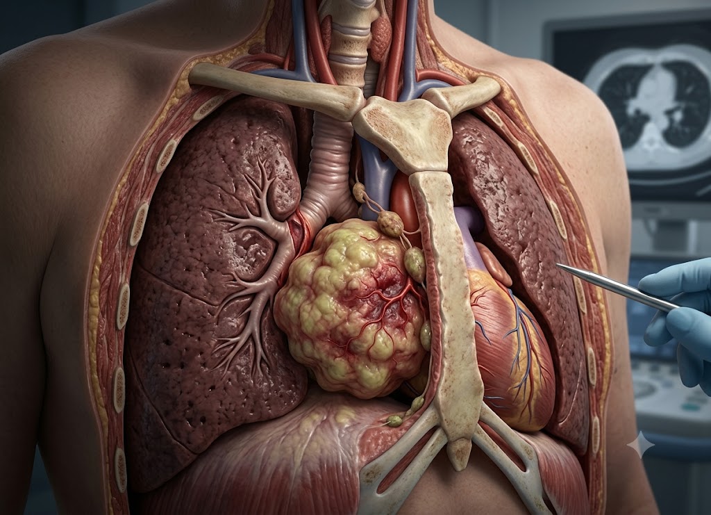
Mediastinal cancer is a malignant tumor that develops in the mediastinum, the central part of the chest located between the lungs. This area contains several important structures, including the heart, major blood vessels, trachea, esophagus, thymus, and lymph nodes. Because a tumor can gradually press on these structures before producing obvious problems, its early symptoms may be vague and can easily be mistaken for common respiratory, digestive, or fatigue-related conditions.
Symptoms of mediastinal cancer can vary depending on the tumor’s size, location, and specific type. Some people may not notice symptoms initially, while others can develop coughing, chest discomfort, shortness of breath, hoarseness, difficulty swallowing, night sweats, swelling, or unexplained weight loss. These symptoms may seem mild at first, but they become more concerning when they persist, worsen, or occur together. This article covers seven symptoms of mediastinal cancer that can be overlooked and explains why ongoing or unexplained symptoms should receive medical attention.
What is Mediastinal Cancer and Why Are Its Symptoms Often Subtle?
Mediastinal cancer describes malignant tumors that develop inside the mediastinum, the central chest compartment positioned between the lungs. This region is relatively crowded and contains many important organs, blood vessels, airways, and nerves. As a result, a growing mediastinal mass may gradually push against surrounding structures before producing obvious symptoms.
                    [Structural Displacement in the Confined Chest]
                                           │
         ┌─────────────────────────────────┼─────────────────────────────────┐
         ▼                                 ▼                                 ▼
 [Pliable Tissue Buffer]         [Limited Early Pain Signals]       [Slow Spatial Shift]
 - Fat and connective tissue      - Deep chest structures may       - Mass can enlarge gradually
   allow gradual compression        not cause immediate pain          over an extended period
 - Nearby organs can adjust       - Discomfort may appear once      - Early symptoms may resemble
   without sudden blockage         nerves or other tissues are affected  common conditions
One reason diagnosis can be challenging is that the mediastinum contains fat and flexible connective tissue that can temporarily accommodate an enlarging mass. A tumor may therefore grow for an extended period while gently displacing nearby structures instead of causing an immediate obstruction. When noticeable symptoms eventually develop, the mass may already be relatively large, making awareness of the possible warning signs important.
Anatomical Boundaries of the Mediastinum
To determine where a tumor is located and what type it may be, healthcare professionals divide the mediastinum into different anatomical regions. The overall space is bordered by the sternum at the front, the spine at the back, the lungs on both sides, the thoracic inlet above, and the diaphragm below.
Anterior Compartment: This region lies behind the breastbone and in front of the heart. It contains the thymus, fat, and lymphatic tissue. Tumors in this area can include thymomas, lymphomas, and germ cell tumors and may be described as an anterior superior mediastinal mass when located toward the upper front part of the mediastinum.
Middle Compartment: The middle section contains the heart, trachea, main bronchi, and major blood vessels, including the aorta and superior vena cava. Masses in this region can involve lymph nodes or structures such as bronchial cysts.
Posterior Compartment: This area is located behind the heart and follows the spinal column. It contains the esophagus, descending thoracic aorta, and several important nerve structures. Tumors in this compartment are often neurogenic, meaning they develop from tissues associated with nerves.
Why Mediastinal Tumors Produce Vague Initial Symptoms
The early symptoms of a mediastinal mass can be difficult to recognize because the chest cavity can accommodate gradual changes for some time.
Incremental Spatial Compression: Unlike a sudden heart problem or acute respiratory infection, a tumor usually applies pressure gradually. Airways, blood vessels, and nerves may slowly shift as the mass grows rather than becoming blocked immediately. This can produce mild symptoms, such as an occasional cough or breathlessness during exercise, that are easy to overlook.
Absence of Early Pain Receptors: Many deep structures in the center of the chest are not highly sensitive to pain. Significant discomfort may not develop until the tumor affects nearby nerves, bones, chest wall structures, ribs, or the spine.
Subconscious Physiological Adaptation: When breathing capacity or airway space changes gradually, people may unconsciously adjust their routines. For example, they may walk more slowly, avoid stairs, or reduce physical activity without recognizing why. These changes can delay medical evaluation.
Understanding Mediastinal Cancer and Its Structural Impact
Mediastinal cancer involves malignant growth within the mediastinum, the central chest compartment separating the lungs and containing several vital structures. This region is bordered by the sternum in front, the spine behind, the lungs on both sides, the thoracic inlet above, and the diaphragm below.
                       [The Mediastinal Compartments]
                                     │
         ┌───────────────────────────┼───────────────────────────┐
         ▼                           ▼                           ▼
[Anterior Compartment]      [Middle Compartment]        [Posterior Compartment]
- Behind the sternum         - Central chest area        - Along the spine
- Thymus and lymph tissue    - Heart, trachea, bronchi   - Nerve-related structures
- Fat and other tissues      - Major blood vessels      - Descending thoracic aorta
Because the mediastinum is surrounded by the chest’s relatively rigid structures, even a growing mediastinal mass can eventually place pressure on nearby organs, nerves, airways, and blood vessels.
A tumor located toward the upper front part of the mediastinum may be identified as an anterior superior mediastinal mass. Because the area is enclosed, such tumors may grow considerably before causing severe symptoms. Early complaints can therefore be vague and may be mistaken for common respiratory, digestive, or other non-cancerous problems.
Respiratory and Voice Changes
As a mediastinal tumor grows, it may affect the main airways and nerves involved in breathing and voice production.
Chronic Non-Productive Cough: A growing mass may press against the trachea or major bronchial tubes. This can narrow or irritate the airway and trigger a persistent dry cough. Because the underlying cause is external pressure rather than a simple airway infection, ordinary cough remedies may not resolve the symptom.
Vocal Hoarseness: The recurrent laryngeal nerve controls movement of the vocal cords and travels through the upper chest near important structures. An anterior superior mediastinal mass may place pressure on this nerve and interfere with vocal cord movement. This can result in a weak, breathy, or hoarse voice that does not improve with normal rest or hydration.
Shortness of Breath (Dyspnea): As a tumor becomes larger, it may limit how well the lungs expand or place pressure on the airways. Fluid can also collect around the lungs, producing a pleural effusion. In more advanced situations, pressure on the superior vena cava can lead to superior vena cava syndrome, which may cause swelling of the face and upper body, facial flushing, and significant breathing difficulty.
Esophageal and Neurological Indicators
A tumor that grows toward the back or upper part of the mediastinum may affect the esophagus or nearby nerve pathways.
Difficulty Swallowing (Dysphagia): The esophagus travels through the posterior chest. Pressure from a tumor can narrow this muscular passage and make swallowing more difficult. Early on, patients may feel as though solid or dry foods are sticking behind the breastbone. Over time, swallowing liquids may also become harder. Some people may unconsciously take smaller bites or drink more during meals to compensate.
Horner’s Syndrome: A tumor located near the top of the chest or superior mediastinum can sometimes affect the sympathetic nerve chain. Damage to this nerve pathway can produce Horner’s syndrome, which may include a drooping upper eyelid (ptosis), a smaller pupil (miosis), and reduced or absent sweating on one side of the face (anhidrosis).
Non-Specific Chest Discomfort and Localized Pain
Chest discomfort caused by a mediastinal tumor may differ from the sudden, intense pain associated with certain acute heart conditions. It may instead feel vague, dull, or difficult to identify precisely.
Vague Chest Pressure or Fullness: As a tumor enlarges, it can stretch or press against nearby tissues and nerves. This may produce a sensation of pressure, fullness, or an unusual feeling in the chest that is difficult to describe.
Progressive, Constant Pain: When pain develops, it may gradually become more persistent. Some people may notice greater discomfort when taking deep breaths, coughing, or changing position.
Skeletal Invasion: In advanced disease, a tumor may extend into nearby bones such as the sternum, ribs, or vertebrae. When this occurs, pain may become more localized and intense and can sometimes be worse at night.
Systemic Manifestations and Lymphatic Spread
Some types of mediastinal cancer, including Hodgkin and non-Hodgkin lymphomas, can produce systemic symptoms because cancer cells and the immune response may release inflammatory substances.
Constitutional “B Symptoms”: These systemic effects can alter normal body processes and produce a group of symptoms known as B symptoms. They may include unexplained weight loss, significant night sweats that soak clothing or bedding, and persistent fever without an obvious infection. Unexplained weight loss is often defined clinically as losing more than 10% of body weight over six months.
Supraclavicular Lymphadenopathy: Lymphatic drainage from the chest can carry cancer cells toward lymph nodes above the collarbone or in the neck. As a result, a firm, painless, or persistent lump in the supraclavicular area can sometimes be an early visible sign of an underlying chest tumor.
Primary Risk Factors for Mediastinal Tumors
Risk factors for a mediastinal mass vary significantly depending on the specific type of tumor. The mediastinum contains many different tissues, including thymic tissue, lymphatic structures, nerves, and other cell types, so tumors can develop through different biological pathways.
                       [Etiological Pathways of Mediastinal Masses]
                                             │
         ┌───────────────────────────────────┴───────────────────────────────────┐
         ▼                                                                       ▼
[Hereditary & Genetic Syndromes]                                [Autoimmune & Systemic Disorders]
- MEN1 mutations -> Certain thymic tumors                       - Myasthenia Gravis -> Thymic tumors
- NF1 mutations -> Neurogenic tumors                             - HIV/AIDS -> Certain lymphomas
- Carney Complex -> Schwannoma risk                              - Immune disorders -> Thymic associations
Many people diagnosed with mediastinal cancers, including primary mediastinal B-cell lymphoma and certain germ cell tumors, may not have an obvious preventable lifestyle risk factor. These tumors can occur in otherwise healthy people, including younger adults. However, some inherited genetic conditions and autoimmune disorders have recognized associations with particular mediastinal tumors.
Hereditary and Genetic Syndromes
Certain inherited conditions caused by germline genetic changes can increase the likelihood of developing tumors in specific areas of the chest.
Multiple Endocrine Neoplasia Type 1 (MEN1): This inherited condition results from changes in the MEN1 tumor suppressor gene. Although MEN1 is best known for tumors involving the parathyroid, pituitary, and pancreatic glands, people with the condition can also have an increased risk of certain neuroendocrine tumors and thymic tumors within the mediastinum.
Neurofibromatosis Type 1 (NF1): NF1 is caused by changes in the NF1 gene and can lead to abnormal growth along nerve pathways. Since the posterior mediastinum contains important nerve structures, people with NF1 have an increased risk of developing neurogenic tumors, including schwannomas, neurofibromas, and malignant peripheral nerve sheath tumors.
Carney Complex: This uncommon inherited disorder can increase the risk of several types of tumors. Some affected individuals develop schwannomas involving nerve-sheath tissue, including tumors that may occur in the posterior mediastinal region.
Pre-Existing Medical and Autoimmune Conditions
The connection between the immune system and thymic tissue means that some autoimmune conditions have important associations with thymic tumors.
Myasthenia Gravis (MG): This autoimmune neuromuscular condition causes fluctuating muscle weakness and has a well-known association with thymomas. A proportion of people with Myasthenia Gravis are diagnosed with a thymoma, while some people with thymoma also develop Myasthenia Gravis. Changes involving the thymus may contribute to abnormal immune responses and the development of antibodies that interfere with neuromuscular communication.
Secondary Autoimmune Conditions: Other immune-related disorders have also been associated with thymoma. These include pure red cell aplasia, systemic lupus erythematosus, and polymyositis. These relationships reflect the complex interaction between thymic tissue and immune-system regulation.
HIV/AIDS and Immunosuppression: Severe immune suppression can increase the risk of certain cancers, including some lymphomas. People with advanced HIV or AIDS have an increased risk of particular hematologic malignancies, including primary mediastinal B-cell lymphoma and other forms of non-Hodgkin lymphoma.
Clinical Diagnostic Framework for Mediastinal Cancer
Diagnosing mediastinal cancer generally involves several organized steps. Because the mediastinum contains many critical structures, doctors first determine the location and characteristics of a mass before obtaining tissue for a definitive diagnosis.
                          [The Mediastinal Diagnostic Pathway]
                                           │
         ┌─────────────────────────────────┴─────────────────────────────────┐
         ▼                                                                   ▼
 [Advanced Diagnostic Imaging]                                      [Tissue Acquisition / Biopsy]
 - CT: Shows size, shape & invasion                              - CT-Guided Needle: Tissue sample
 - MRI: Evaluates nerves and spine                                - Mediastinoscopy: Samples nodes
 - PET: Measures metabolic activity                               - VATS: Larger sample or removal
A complete evaluation generally combines detailed imaging with tissue sampling when appropriate. Imaging helps map the mediastinal mass, while pathology determines the exact cell type and whether the growth is malignant.
Advanced Diagnostic Imaging Modalities
Imaging studies provide a detailed view of the chest and help identify the location, size, and relationship of a mass to surrounding structures.
Chest X-Ray Screening: A mediastinal mass may sometimes be discovered unexpectedly on a chest X-ray performed for another reason. It can appear as widening of the central chest silhouette or an unusual bulge. However, an X-ray does not provide enough detail to determine the exact type of tissue involved.
Computed Tomography (CT) Scans: A chest CT is one of the main imaging tests used to assess a mediastinal mass. When contrast is used, CT can show the size, shape, density, and location of the growth and help determine whether it is affecting nearby structures such as the trachea, pericardium, blood vessels, or chest wall.
Magnetic Resonance Imaging (MRI): MRI can be particularly useful when a mass is located in the posterior mediastinum or close to the spine and nerves. It provides detailed soft-tissue images and can help determine whether a tumor extends toward the spinal canal or affects nearby nerve roots. MRI can also be useful when detailed vascular imaging is required in certain circumstances.
Positron Emission Tomography (PET) Scans: PET imaging evaluates metabolic activity using a radioactive tracer. Some cancers absorb more of the tracer than surrounding tissues, making them appear more active on the scan. PET can help doctors assess disease activity, identify possible lymph-node involvement, and determine whether cancer has spread to other areas.
Definitive Tissue Acquisition and Biopsy Approaches
Although imaging can provide important clues, a tissue biopsy is often needed to confirm mediastinal cancer and determine the exact type. A pathologist examines the tissue to distinguish among tumors such as thymoma, lymphoma, germ cell tumors, or other growths and to help guide treatment.
CT-Guided Needle Biopsy: If the tumor can be safely reached, particularly an anterior mediastinal mass near the chest wall, a radiologist may use CT imaging to guide a needle into the tumor and collect a core tissue sample. This is minimally invasive, although certain conditions, including some lymphomas, may require a larger tissue sample for accurate classification.
Mediastinoscopy: Mediastinoscopy is a surgical procedure that can be used to sample lymph nodes in selected areas of the mediastinum. A small incision is made near the base of the neck, allowing a thin illuminated instrument to enter the central chest area. The surgeon can then examine and collect tissue from accessible lymph nodes.
Video-Assisted Thoracoscopic Surgery (VATS): When a mass cannot be safely reached with a needle or a larger sample is required, VATS may be used. The surgeon makes small incisions between the ribs and inserts a camera and specialized instruments. This approach can provide a substantial tissue sample and, in selected cases, may allow the mass to be removed during the same operation.
Systemic and Multidisciplinary Management of Mediastinal Cancer
Treatment for mediastinal cancer is highly individualized and may involve thoracic surgeons, medical oncologists, radiation oncologists, pulmonologists, and other specialists. Since the mediastinum contains critical structures such as the heart, trachea, and major blood vessels, treatment must be carefully planned according to the tumor’s type, location, and extent.
                          [Multidisciplinary Treatment Framework]
                                             │
         ┌───────────────────────────────────┼───────────────────────────────────┐
         ▼                                   ▼                                   ▼
 [Surgical Resection]               [Systemic Therapies]                [Radiation Oncology]
 - Used for selected solid masses   - Common for lymphomas              - Treats selected localized
 - Thymomas & some neurogenic       - Chemotherapy                         or unresectable disease
   tumors                            - Targeted/immunotherapy             - May follow surgery
 - VATS or robotic approaches
Even a non-cancerous mediastinal growth may require treatment or monitoring if it presses on important organs. Correctly identifying the mass is therefore an important first step. Long-term care may include follow-up imaging, monitoring for treatment effects, and management of symptoms and overall quality of life.
Comprehensive Classification of Mediastinal Tumors
A mediastinal mass is classified according to both its exact cell type and the compartment where it develops.
Anterior Compartment Tumors: The front portion of the mediastinum is a common location for an anterior superior mediastinal mass. Tumors may originate from thymic tissue, including thymomas and thymic carcinomas. Lymphomas and germ cell tumors, such as teratomas and seminomas, can also occur in this area.
Middle Compartment Growths: The middle mediastinum contains the heart, major airways, and blood vessels. Masses found here may include enlarged lymph nodes, lymphomas, or metastatic disease that has spread from another primary cancer.
Posterior Compartment Lesions: The posterior mediastinum follows the spinal column and contains many nerve-related structures. Neurogenic tumors, including schwannomas and neurofibromas, are common in this compartment. Although many are benign, they may require monitoring or treatment if they grow or place pressure on the spinal cord or nearby nerves.
Key Differences: Mediastinal Cancer vs. Lung Cancer
Although both conditions occur within the chest and can cause symptoms such as coughing, chest discomfort, and shortness of breath, mediastinal cancer and lung cancer originate in different anatomical locations.
Anatomical Origin: Lung cancer begins within the lungs, either in lung tissue or cells lining the respiratory airways. Mediastinal tumors develop outside the lungs, within the central chest structures separating the two lungs.
Cellular Profiles: Because the tumors develop from different tissues, their cell types also differ. Lung cancer most often consists of epithelial cancers such as adenocarcinoma or squamous cell carcinoma. Mediastinal tumors can arise from lymphatic tissue, thymic tissue, nerve-related structures, or germ cells.
Diagnostic and Staging Procedures: Both conditions may initially be evaluated with a CT scan, but further testing can differ. Lung cancer may require bronchoscopy and staging using the TNM system. Mediastinal tumors may require techniques such as mediastinoscopy or surgical biopsy, while lymphomas are evaluated and staged using criteria specific to blood and lymphatic cancers.
Standard Treatment Modalities
After doctors determine the tumor’s location, cellular type, and stage, a multidisciplinary team develops an individualized treatment plan.
Surgical Resection: Surgery may be the main treatment for localized solid tumors that can be safely removed. Depending on the tumor, this can include thymomas, selected teratomas, and some neurogenic tumors. Surgeons may use open surgery or minimally invasive techniques such as VATS or robotic-assisted thoracic surgery.
Systemic Therapies: Chemotherapy is commonly used for blood-related cancers such as Hodgkin and non-Hodgkin lymphomas because the medication can circulate throughout the body. Depending on the cancer type, targeted therapies or immunotherapies may also be considered to act on specific molecular features of the tumor.
Radiation Therapy: Radiation uses high-energy beams to damage and destroy cancer cells. Depending on the tumor, it may be used before surgery to shrink a mass, after surgery to treat remaining microscopic disease, or as part of treatment for tumors that cannot be safely removed.
Primary Factors Influencing Patient Prognosis
The outlook for mediastinal cancer depends on several tumor-related and patient-related factors.
Histological Type and Cellular Grade: The specific type of tumor has a major influence on prognosis. A benign neurogenic tumor that is completely removed may have an excellent outlook, while malignant tumors vary considerably in their behavior. The cellular grade can also provide information about how aggressively a tumor may grow.
Stage at Diagnosis and Resectability: The extent of disease at diagnosis is another important factor. Smaller tumors that remain localized and have not invaded nearby structures may be easier to treat. When a surgeon can completely remove a tumor with clear margins, this can also influence long-term outcomes.
Overall Health and Treatment Tolerance: A patient’s general health, age, physical condition, and other medical conditions can affect treatment options and recovery. Conditions involving the heart, lungs, immune system, or other organs may influence whether someone can safely undergo major surgery, chemotherapy, radiation, or combined treatment.
Conclusion
Mediastinal cancer can be challenging to identify early because its symptoms may resemble ordinary respiratory, digestive, or chest-related problems. Persistent coughing, chest pressure, shortness of breath, hoarseness, difficulty swallowing, unexplained weight loss, night sweats, or swelling around the face and neck can warrant medical evaluation when they continue without an obvious explanation. Since the mediastinum contains many vital structures, a growing tumor can cause problems by placing pressure on nearby organs, nerves, airways, or blood vessels. When symptoms persist or become worse, a healthcare professional can determine whether imaging, biopsy, or specialist assessment is needed.
Frequently Asked Questions
1. What is mediastinal cancer?
Mediastinal cancer is a malignant growth that develops in the mediastinum, the central area of the chest located between the lungs. This compartment contains structures such as the heart, major blood vessels, trachea, esophagus, thymus, and lymph nodes. Tumors can originate from several types of tissue, including lymphatic, thymic, nerve-related, and germ-cell tissue. Some masses in the mediastinum are benign, while others are cancerous and require further evaluation.
2. What symptoms can mediastinal cancer cause?
Possible symptoms include persistent coughing, chest discomfort, shortness of breath, wheezing, hoarseness, difficulty swallowing, fever, night sweats, and unexplained weight loss. Some patients may also develop swelling in the face, neck, or upper chest if a tumor affects blood flow. Symptoms can occur when a mass places pressure on nearby airways, nerves, organs, or blood vessels. In some cases, however, a mediastinal tumor may cause no noticeable symptoms and may only be discovered through imaging.
3. Why can mediastinal cancer be overlooked?
Mediastinal cancer can initially be overlooked because its symptoms may look similar to common conditions such as respiratory infections, asthma, acid reflux, or other everyday health problems. A mild cough, hoarse voice, or occasional chest discomfort may not immediately appear serious. Some tumors also grow gradually, allowing the body to adapt to the changes. Persistent, unexplained, or worsening symptoms should therefore be discussed with a healthcare professional.
4. How is mediastinal cancer diagnosed?
Doctors may begin with a medical history and physical examination followed by imaging such as a chest X-ray, CT scan, MRI, or PET scan. These tests can show where a mediastinal mass is located, how large it is, and whether it affects nearby structures. A biopsy is often needed to determine whether the mass is cancerous and identify its exact type. Treatment decisions are then based on the tumor type, location, stage, and the patient’s overall health.
5. Is mediastinal cancer treatable?
Treatment depends on the exact type of mediastinal tumor, its stage, location, and whether it can be surgically removed. Depending on the diagnosis, treatment may include surgery, chemotherapy, radiation therapy, targeted treatment, immunotherapy, or a combination of approaches. Some tumors respond well to treatment, while others require more intensive or specialized care. Evaluation by a multidisciplinary medical team helps determine the most appropriate treatment plan.