Echo Stress Test: 10 Reasons Your Doctor May Recommend It
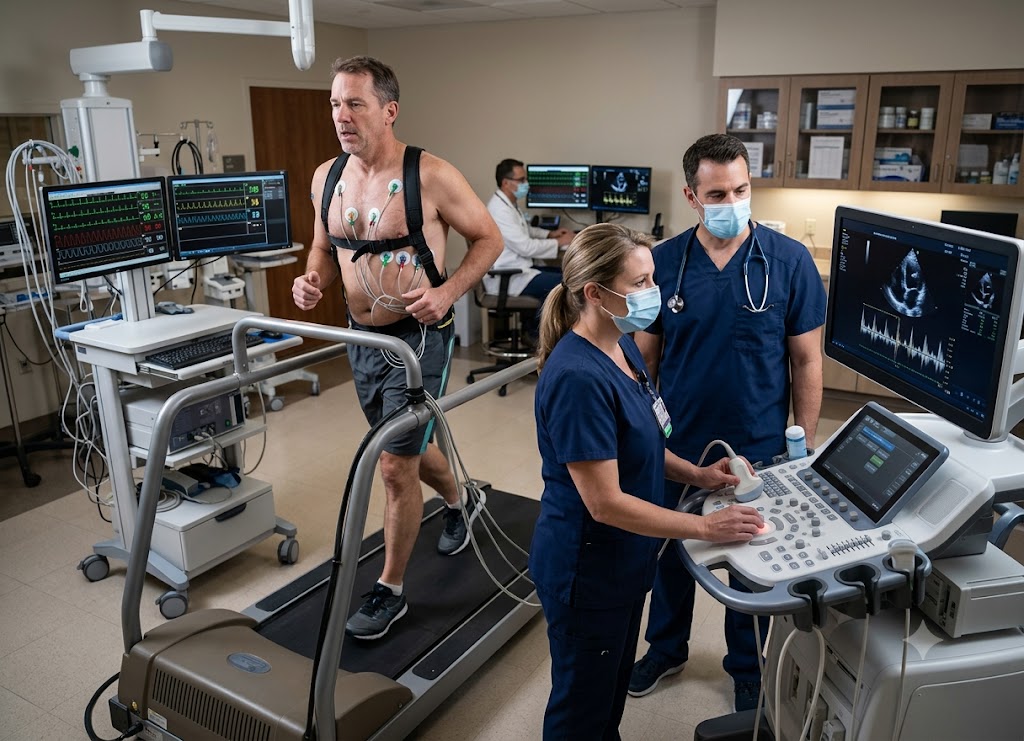
An echo stress test pairs a heart ultrasound with physical exertion (or a medication that mimics it) so doctors can see how your heart behaves under pressure, not just at rest. Watching the heart pump faster lets clinicians spot problems that a resting scan alone would miss things like restricted blood flow, weakened heart muscle, or valves that don’t hold up once the workload increases.
There are many reasons a doctor might order this test chest pain, breathlessness, dizziness, or fatigue that shows up only during activity are common triggers. It’s also used to check the impact of coronary artery disease, track an existing heart condition, confirm a treatment worked, or clear someone for a more demanding exercise routine. Below are 10 of the most common reasons doctors order an echo stress test, along with what the results can tell you.
What Defines an Echo Stress Test and How Does It Work?
An echo stress test is a specialized diagnostic exam that pairs a cardiac ultrasound (echocardiogram) with a classic exercise stress test, producing live images of your heart muscle and valves while they’re under physical load.
Its purpose is to reveal how the heart responds when the body suddenly needs more oxygen-rich blood something exercise reliably triggers. To fully understand the exam, it helps to see how it differs from a plain stress test and to walk through what actually happens during each stage.
Comparing Core Diagnostics: Echo Stress Test vs. Standard Stress Test
[Standard Stress Test] ──► EKG tracing only ──────► Indirect hints of a blockage
[Echo Stress Test]     ──► EKG + live ultrasound ──► Direct view of the muscle moving
To understand what a stress echo actually looks for, it helps to line it up against the more basic version of the test:
The standard exercise stress test only tracks the heart’s electrical activity. You walk on a treadmill while hooked up to an EKG, and doctors watch for telltale electrical changes such as ST-segment depression. It’s useful, but it only offers circumstantial evidence it can’t actually show the heart muscle itself.
The exercise stress echocardiogram goes further by adding an ultrasound scan right before and immediately after exercise. Using high-frequency sound waves, it captures detailed moving images of the heart, much like a standard echocardiogram would at rest.
Comparing the pre-exercise images with the peak-exertion images lets a cardiologist watch the heart walls contract in real time.
                       [What the images reveal]
                                  │
      ┌────────────────────────────┴────────────────────────────┐
      ▼                                                          ▼
[A healthy response]                                 [A restricted-flow response]
 ├── Every wall segment squeezes harder               ├── The area past a blockage is starved
 ├── Pumping stays even and strong                    ├── That wall barely moves, or stops moving
 └── Confirms blood is reaching the whole heart        └── Flags a "wall motion abnormality" (CAD)
Beyond blood flow, this test also lets cardiologists check the heart valves under stress revealing whether narrowing (stenosis) or leaking (regurgitation) gets worse under exertion, something a resting scan can easily miss.
Phase-by-Phase Stress Test Procedure
A stress echo runs through a set protocol designed to safely push the heart while still capturing sharp images before the heart rate has a chance to settle back down.
[Phase 1: Resting Baseline] ──► [Phase 2: Exercise Load] ──► [Phase 3: Peak-Exertion Scan]
Phase 1: Resting baseline. The test starts with setup: a technician attaches EKG electrodes to your chest to track your heart’s rhythm and wraps a blood pressure cuff around your arm. You’ll then lie on your left side while a sonographer applies gel and moves a transducer across your chest, capturing baseline images of your heart’s chambers and valves at rest.
Phase 2: The exercise workload. Once the resting images are saved, you’ll walk on a treadmill or pedal a stationary bike following a set protocol (the Bruce protocol is a common one), with speed and incline stepping up every few minutes. Throughout this phase, the team keeps an eye on your EKG rhythm, blood pressure, and how you’re feeling, working toward a target heart rate usually around 85% of your predicted maximum for your age.
Phase 3: Immediate post-exercise imaging. This is the most time-critical part of the whole test. The instant you hit peak exertion, you stop, step off, and quickly lie back down on your left side. The sonographer has roughly 60 to 90 seconds to capture images from the same angles as the resting scan any longer and your heart rate drops enough that exercise-induced weak spots can disappear. That side-by-side comparison is what lets the cardiologist check for coronary artery disease.
Diagnostic Protocol Summary
| Stage | Goal | Equipment |
|---|---|---|
| 1. Prep & rest | Capture baseline structure, resting wall movement, and valve function | Ultrasound transducer, EKG electrodes, blood pressure cuff |
| 2. Active exercise | Raise cardiac workload and oxygen demand to surface hidden flow issues | Treadmill or stationary bike, continuous EKG |
| 3. Post-stress scan | Capture peak contractions within 90 seconds to catch wall motion abnormalities | Ultrasound transducer, synced imaging workstation |
This two-part imaging approach before and immediately after exertion is what makes the test so useful: it gives your care team a direct visual read on how your heart handles stress, informing decisions about your cardiovascular care.
Top 10 Reasons for an Echo Stress Test
The most common reasons doctors order this test include: diagnosing coronary artery disease behind chest pain, checking how well a heart valve holds up under load, measuring overall pumping strength in conditions like heart failure, setting safe exercise limits for cardiac patients, mapping damage after a heart attack, clearing patients for surgery, helping diagnose pulmonary hypertension, confirming that treatments like stents or bypass surgery worked, tracking congenital heart conditions over time, and tracking down the cause of dizziness or palpitations that only appear during activity.
Each use leans on the same core strength of the test showing exactly how the heart behaves under physical stress. Here’s a closer look at each one.
Investigating Ischemia: Chest Pain and Shortness of Breath
An echo stress test is often the first choice for investigating chest pain (angina) or breathlessness that only shows up during activity. At rest, narrowed coronary arteries can still deliver just enough blood to keep symptoms quiet, so a resting echo often looks normal. Exercise changes that: as the heart’s oxygen demand rises, a narrowed artery can’t keep up, and that mismatch causes ischemia.
[Physical exertion] ──► Oxygen demand outpaces supply ──► Ischemia ──► A wall segment moves poorly
To see what’s happening, cardiologists compare the before-and-after ultrasound images:
- A healthy response: every part of the heart wall contracts more forcefully under stress.
- An ischemic response: a segment starved of blood weakens it may contract feebly (hypokinesis) or stop moving altogether (akinesis).
This lets physicians pinpoint which artery is likely blocked and gauge how serious the problem is.
Valvular Assessment Under Hemodynamic Load
A resting echocardiogram can already flag structural valve problems like aortic stenosis (narrowing) or mitral regurgitation (leaking) but it can’t show how those valves cope with real exertion. An exercise stress echo recreates that load to show how the valve actually behaves under high blood flow.
                          [How valves are tested under stress]
                                      │
      ┌────────────────────────────────┴────────────────────────────────┐
      ▼                                                                  ▼
[Aortic stenosis]                                    [Mitral regurgitation]
 ├── Tracks pressure gradients as they climb          ├── Measures how much blood leaks backward
 ├── Flags critical flow restrictions                 ├── Tracks rising pressure in the lungs
 └── Helps time surgical valve replacement             └── Helps explain unexplained breathlessness
Using Doppler ultrasound, a cardiologist can watch pressure gradients shift across a narrowed valve, or see leakage worsen during exercise giving objective evidence for whether surgery should happen now or can wait.
Measuring Pumping Strength and Ejection Fraction
A full stress echo also measures your heart’s ejection fraction (EF) the percentage of blood the left ventricle pushes out with each beat. A normal resting EF usually falls between 50% and 70%.
- Normal response: ejection fraction climbs during exercise as the muscle contracts more powerfully.
- Abnormal response: if EF drops or stays flat under stress, it can point to muscle weakness, a past unnoticed heart attack, or significant blockages.
This segment-by-segment picture helps specialists plan treatment for people living with chronic heart failure.
Crafting Safe Exercise Prescriptions
For anyone recovering from a major cardiac event, this test helps set a safe path forward by finding their ischemic threshold the exact heart rate and workload where wall motion problems or rhythm issues start showing up.
Continuous EKG and blood pressure monitoring throughout the test lets specialists identify:
- Arrhythmias triggered by exercise
- Dangerous blood pressure spikes or drops during exertion
- A safe upper heart-rate limit for independent workouts or cardiac rehab
Post-Heart Attack Damage Mapping
After a heart attack, an echo stress test helps track recovery. A heart attack leaves permanent scar tissue, but nearby muscle may have survived and could still be at risk from other partially blocked arteries.
[Stress test after a heart attack] ──► Map permanent scar tissue ──► Find muscle still at risk ──► Assess next steps
The post-exercise imaging is what separates unrecoverable scar tissue from muscle that’s alive but vulnerable crucial for deciding whether a stent or bypass surgery is needed to prevent another attack.
Pre-Operative Cardiac Risk Assessment
Major surgery puts real strain on the cardiovascular system, thanks to anesthesia, fluid shifts, and the body’s inflammatory response. An echo stress test is often used beforehand to check whether the heart can handle that strain.
- Strong functional capacity: completing the exercise protocol without symptoms, EKG changes, or wall abnormalities suggests good cardiac reserve and lower surgical risk.
- Unmasking hidden disease: for less active patients, the test can reveal coronary artery disease that wasn’t previously known letting the surgical team adjust their approach or treat the heart issue first.
Identifying Exercise-Induced Pulmonary Hypertension
Pulmonary hypertension means abnormally high blood pressure in the lungs’ arteries. A resting echo can estimate these pressures, but some people have normal or borderline readings at rest and only show elevated pressure during activity.
Doppler measurements taken during a stress echo let technicians track how fast blood leaks backward through the tricuspid valve under load. Using the modified Bernoulli equation, that speed converts into an estimate of the pulmonary artery systolic pressure:
Delta P = 4v^2
Watching how these pressures shift under stress helps doctors figure out whether lung-vessel disease is behind someone’s breathing trouble especially when standard heart and lung tests come back normal.
Evaluating the Success of Cardiac Treatments
An exercise stress echo gives objective, visual confirmation that a recent cardiac treatment worked. For patients who’ve had angioplasty, a stent, or bypass surgery, it verifies that blood flow to the heart muscle has actually improved.
[Before treatment: weak wall movement] ──► [Procedure] ──► [After treatment: normal wall movement]
If a wall segment that used to weaken under stress now contracts normally, that’s a good sign the stent or graft is working. If the weakness persists, it can be an early warning of re-narrowing (restenosis), prompting the team to adjust medication or plan further treatment.
Long-Term Monitoring of Congenital Heart Defects
Adults born with congenital heart defects often need lifelong monitoring, even after successful childhood surgery. The long-term effects of those early structural issues can create hidden strain that only surfaces during physical activity.
                        [What's tracked in congenital monitoring]
                                       │
      ┌─────────────────────────────────┴─────────────────────────────────┐
      ▼                                                                    ▼
[Blood shunting]                                        [Right ventricle reserve]
 ├── Tracks shifting pressure overloads                  ├── Measures overall structural limits
 ├── Watches blood pass through unresolved wall openings ├── Catches early signs of right-side strain
 └── Flags dynamic blockages as they appear               └── Monitors for exercise-triggered arrhythmias
A stress echo lets clinicians watch blood shunting through small unresolved wall openings, gauge how well the right ventricle is holding up, and check for developing blockages or scar-related arrhythmias before symptoms appear.
Diagnosing Atypical Exertional Symptoms
Symptoms like sudden dizziness, lightheadedness, a racing heart, or extreme fatigue often show up only during activity and vanish by the time you’re sitting in a doctor’s office which makes them hard to catch on a standard visit or a resting echo.
A stress echo gets around this by safely recreating those symptoms in a controlled setting. Tracking EKG rhythm and ultrasound images at the exact moment symptoms appear can help identify:
- Dynamic outflow obstructions (as seen in hypertrophic cardiomyopathy, where thickened heart muscle blocks blood flow during exertion)
- Arrhythmias triggered by exercise that reduce pumping efficiency
- Sudden blood pressure drops during physical strain
Summary of Diagnostic Triggers
| Symptom / clinical need | What’s measured | Main diagnostic goal |
|---|---|---|
| Exertional angina / chest pain | Regional wall motion abnormalities | Confirm or rule out coronary artery disease |
| Moderate structural valve issues | Pressure gradients across the valve | Time valve repair or replacement |
| Prior heart attack | Post-stress ejection fraction | Separate viable muscle from scar tissue |
| Clearance for major surgery | Functional capacity | Estimate cardiac reserve, reduce surgical risk |
| Unexplained exertional dizziness | Left ventricular outflow | Check for exercise-triggered blood flow drops |
Pairing an EKG with live ultrasound is what makes this dual approach so valuable it gives your cardiologist a direct, real-time look at how your heart is really doing, supporting a more accurate diagnosis and treatment plan.
What Practical Information Should You Know About Your Test?
Knowing how to prepare, what the results mean, what risks exist, and what alternatives are available all go a long way toward a smoother, less stressful test experience. It also puts you in a better position to ask informed questions and work alongside your care team on next steps.
This section breaks down what to expect, before and after the procedure.
Preparing for the Procedure: Fasting and Restrictions
Certain foods, drinks, and medications can shift your heart rate and blur the results, so a few precautions matter:
[12–24 hours before] ──► Cut out all caffeine (coffee, tea, soda, chocolate)
[3–4 hours before]   ──► Full fast (nothing but water)
[Day of the test]    ──► No smoking / review heart medications with your doctor
Caffeine: Avoid all caffeine for 12 to 24 hours beforehand coffee, tea, energy drinks, colas, even decaf and chocolate. Caffeine artificially shifts your resting heart rate and can throw off the results.
Fasting: Skip food, gum, and anything besides plain water for at least 3 to 4 hours before the test. An empty stomach helps prevent nausea and keeps breathing comfortable during exercise. Skip smoking that day too, since nicotine narrows blood vessels.
Medications: Go over your current prescriptions with your cardiologist beforehand. Beta-blockers (like metoprolol or atenolol) and some calcium channel blockers slow the heart rate taking them the morning of the test can stop your heart from reaching its target zone, making the results inconclusive. Never stop a prescribed medication without your doctor’s direct instruction.
Day-of-Apparel Checklist
Clothing: Wear loose, comfortable two-piece athletic wear. Skip one-piece outfits or dresses the sonographer needs quick, easy access to your bare chest for the EKG electrodes and ultrasound transducer.
Footwear: Stick to supportive sneakers. Heels, dress shoes, boots, sandals, or loose slippers are a slipping hazard on a moving treadmill.
Navigating Abnormal Findings and Next Steps
An abnormal result means the test found a mismatch between how much oxygen your heart needs during exercise and how much blood it’s actually getting.
[Post-exercise scan] ──► Detects weak wall movement ──► Points to ischemia (a blocked artery)
Deciphering the results. The most common abnormal finding is a regional wall motion abnormality the resting scan might look perfectly normal, but the post-exercise images show a specific segment contracting weakly (hypokinesis) or not moving at all (akinesis). Since different coronary arteries feed different zones of the heart, pinpointing exactly where that weakness is tells the cardiologist which vessel is likely narrowed. A drop in ejection fraction or ischemic EKG changes (like ST-segment depression) are other common findings.
The diagnostic pipeline. An abnormal result isn’t a final diagnosis it’s one piece of evidence your care team uses to plan next steps:
                       [What happens after an abnormal result]
                                     │
      ┌────────────────────────────────┼────────────────────────────────┐
      ▼                                ▼                                ▼
[Putting it in context]        [Further testing]                [Planning treatment]
 ├── Weigh results against symptoms ├── Order cardiac catheterization ├── Adjust heart medications
 ├── Review cardiac risk factors    ├── Direct coronary angiogram     ├── Recommend lifestyle changes
 └── Consider overall daily mobility └── Map the exact blockage        └── Plan stenting or bypass surgery
Risk Profile and Expected Post-Test Sensations
An echo stress test is a routine, well-monitored procedure, but it does deliberately place strain on your cardiovascular system risks are broadly similar to those of any strenuous workout.
[Controlled physical strain] ──► Continuous monitoring ──► Rare complications (roughly 1 in 10,000)
Rare risks include an exercise-triggered arrhythmia, severe angina, or a sudden blood pressure drop causing brief lightheadedness. In extremely rare cases about 1 in 10,000 tests a heart attack can be triggered; emergency medication and resuscitation equipment are always on hand.
What’s normal vs. what’s urgent afterward:
- Normal: feeling tired for a few hours afterward (similar to a hard workout), mild leg soreness, or minor skin redness where the EKG patches were.
- Seek immediate care if: you have persistent or worsening chest pain, severe shortness of breath that doesn’t ease with rest, sudden dizziness, confusion, or palpitations after leaving the clinic.
Pharmacological Alternatives for Limited Mobility
If you can’t use a treadmill or bike due to arthritis, joint injuries, balance problems, vascular disease, or other limitations a pharmacological stress echo is a safe substitute.
[Can't exercise] ──► IV medication ──► Mimics the effect of exercise ──► Imaging proceeds as normal
Dobutamine is the standard choice for an ultrasound-based test. It directly stimulates the heart to beat faster and pump harder, closely mimicking a real workout, while the sonographer records images throughout the infusion.
Vasodilators (adenosine or regadenoson) are used mainly for nuclear stress tests rather than ultrasound. Rather than making the heart work harder, they widen healthy arteries while narrowed ones stay rigid, creating a flow difference that shows up with a radioactive tracer and special camera.
What a chemical stress test feels like: you won’t walk or run, but as the medication raises your heart rate you may notice warmth, a mild headache, brief nausea, or a racing heartbeat. These are normal, closely monitored, and fade within minutes once the infusion stops.
Conclusion
An echo stress test shows how the heart performs when it’s working harder than usual surfacing reduced blood flow, muscle weakness, valve problems, or other issues that a resting exam might miss entirely.
Since it doesn’t involve radiation and provides a detailed, moving picture of the heart, it’s a widely used tool for people with suspected or confirmed cardiovascular disease. If your doctor recommends one, understanding what it’s for can help you feel more prepared and better appreciate how it shapes your diagnosis and treatment plan.
Frequently Asked Questions
1. What is an echo stress test?
It’s a diagnostic exam that combines ultrasound imaging with exercise or medication to see how the heart performs under stress. Images are taken before and right after the heart rate rises, revealing changes in blood flow, muscle movement, or valve function that a resting scan might not catch.
2. Why would a doctor recommend an echo stress test?
Common reasons include chest discomfort, shortness of breath, dizziness, or reduced stamina during activity. It’s also used to investigate suspected coronary artery disease, assess valve problems, track an existing heart condition, check whether a treatment worked, or confirm the heart can handle surgery or more intense exercise.
3. How should I prepare for an echo stress test?
Preparation varies based on why the test is being done and your medical history, but typically includes fasting for a few hours beforehand, wearing comfortable clothes and walking shoes, and possibly pausing certain medications. Let the team know about any recent illness, allergies, or new symptoms this all helps keep the results accurate and safe.
4. Is an echo stress test safe?
Yes, generally it’s performed under close medical supervision, and most people finish without complications. Temporary fatigue, breathlessness, or mild chest discomfort during exercise is common, but serious complications are rare thanks to continuous monitoring of heart rhythm, blood pressure, and symptoms throughout.
5. How long does an echo stress test take?
Most tests run about 45 to 60 minutes total, covering the resting images, the exercise or medication phase, and the post-exertion imaging. The exact time depends on how quickly your heart reaches its target rate and whether extra monitoring is needed afterward.
6. What can an echo stress test detect?
It can reveal restricted blood flow from narrowed arteries, abnormal muscle movement, reduced pumping strength, and certain valve problems. It may also identify old heart damage or show how well the heart tolerates physical stress helping doctors decide on further testing, medication, lifestyle changes, or procedures.