10 Causes of Retinal Hemorrhage You Should Know
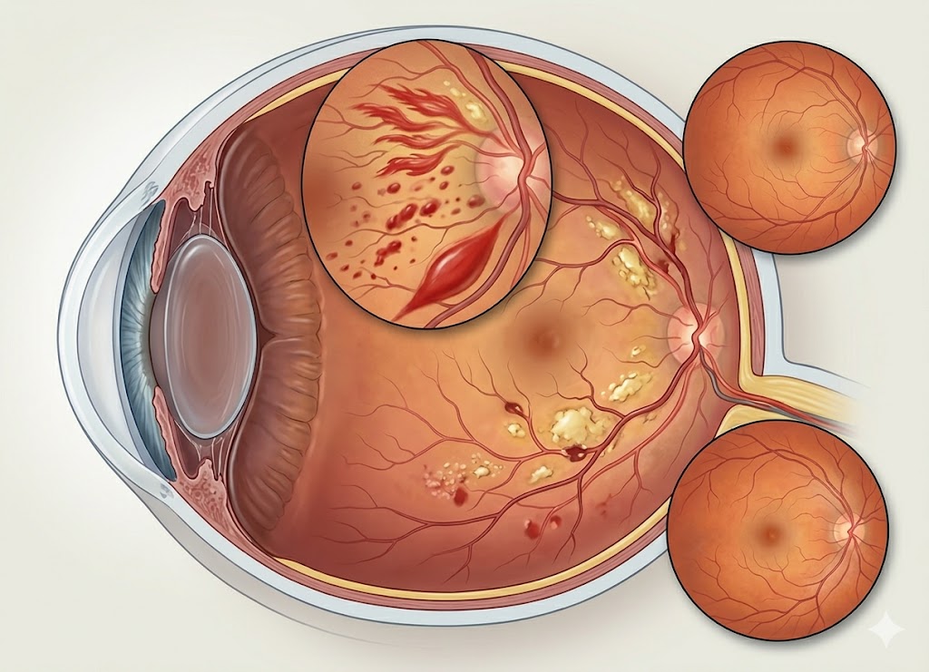
A retinal hemorrhage happens when one or more blood vessels in the retina leak or break, causing blood to collect within the light-sensitive tissue at the back of the eye. Since the retina is essential for vision, bleeding in this area can interfere with eyesight, causing anything from mild visual changes to serious vision loss in severe cases.
A retinal hemorrhage is not a disease on its own. Instead, it is usually a sign of another health problem or an eye injury. Diabetes, high blood pressure, trauma, blood disorders, and other conditions that affect blood vessels can all contribute to retinal bleeding. In some people, a small hemorrhage causes no obvious symptoms and is discovered only during a routine eye examination.
Knowing the possible causes of retinal hemorrhage is important because finding the underlying problem early can help prevent complications and protect vision. In this article, we’ll discuss 10 common causes of retinal hemorrhage and explain when medical evaluation may be necessary.
What Is a Retinal Hemorrhage?
A retinal hemorrhage is bleeding that develops within the retina, the light-sensitive layer of nerve tissue located at the back of the eye. It occurs when the small blood vessels that supply the retina become damaged, leak, or rupture.
You can think of the retina as similar to the film or sensor inside a camera. It receives light and converts it into signals that are sent to the brain to create what you see. When blood collects within or around the retina, it can interfere with the normal passage of light and disrupt the function of retinal cells.
Because the retina depends on a delicate network of blood vessels to receive oxygen and nutrients, damage to these vessels can affect vision.
Is Bleeding Behind the Eye Serious?
When someone notices eye bleeding, one of the first questions is, is bleeding behind the eye serious? The answer depends on where the bleeding occurs, how extensive it is, and what caused it.
Retinal bleeding can sometimes be associated with significant medical conditions, so it should be evaluated by an eye care professional. A small hemorrhage caused by minor trauma may resolve without lasting problems, while bleeding related to conditions such as diabetic retinopathy or uncontrolled high blood pressure may indicate ongoing damage to blood vessels.
The underlying cause is therefore an important part of determining the seriousness of the hemorrhage and the appropriate treatment.
How Retinal Bleeding Impacts Vision
A retinal hemorrhage can affect eyesight by interfering with the retina’s ability to receive and process light. The symptoms depend largely on the amount of blood present and the specific retinal area involved.
Central Vision Blind Spots
When bleeding affects the macula, the central part of the retina responsible for sharp vision, it can cause noticeable changes in eyesight. A person may see blurry text, distorted lines, or a dark spot in the center of their vision.
Sudden Floaters and Cloudiness
If a damaged retinal blood vessel releases blood into the vitreous, the clear gel that fills the inside of the eye, it can cause a sudden increase in floaters. Some people may notice dark spots, cobweb-like shapes, haze, or cloudiness across their vision.
Causes and Structural Identifiers
Finding out what causes bleeding behind the eyes requires a thorough examination. Eye specialists consider both conditions that directly affect the eye and systemic health problems that damage blood vessels.
| Category | Primary Disease Drivers | Visual Impact & Manifestation |
|---|---|---|
| Systemic Conditions | Diabetic Retinopathy, Hypertensive Retinopathy | Diabetes can weaken small retinal vessels and cause bleeding, while high blood pressure can damage and rupture retinal blood vessels. |
| Localized Ocular Issues | Wet Macular Degeneration (AMD), Retinal Vein Occlusion (RVO) | Wet AMD can cause abnormal blood vessels to leak beneath the macula, while blocked retinal veins can lead to pressure buildup and bleeding. |
Determining what causes blood behind your eye or what causes bleeding behind the retina is an important first step in protecting your vision. Once the underlying problem is identified, doctors can recommend treatment aimed at controlling the condition and preventing further retinal damage.
The 10 Primary Causes of Retinal Hemorrhage
Retinal hemorrhage can develop for many different reasons, including chronic diseases, changes within the eye, physical trauma, high-altitude exposure, and blood-related conditions.
Because retinal blood vessels are extremely small and delicate, they can be affected by changes in blood pressure, oxygen levels, circulation, and physical forces. Understanding these potential causes can help explain why retinal bleeding occurs.
Metabolic and Systemic Pressures
Diabetic Retinopathy
Diabetes is a major cause of retinal blood vessel damage. When blood sugar remains high for long periods, the walls of small retinal blood vessels can become weak and develop tiny bulges called microaneurysms. These damaged vessels may leak fluid or small amounts of blood into the retina.
As diabetic retinopathy progresses, some retinal blood vessels may become blocked, reducing the oxygen supply to the retina. In response, the eye can grow abnormal new blood vessels. These vessels are fragile and can bleed easily, sometimes releasing blood into the vitreous and causing sudden changes in vision.
Hypertensive Retinopathy
Long-term high blood pressure can damage the blood vessels supplying the retina. The vessels may become narrower, thicker, and less flexible, which can interfere with normal blood flow.
Severe or prolonged hypertension can also contribute to retinal vessel leakage and bleeding. Retinal changes associated with high blood pressure can be an important sign that blood pressure needs better medical control.
Vascular Blockages and Degeneration
Retinal Vein Occlusion (RVO)
A retinal vein occlusion occurs when a vein responsible for carrying blood away from the retina becomes blocked. The blockage can cause blood and fluid to build up within the retinal circulation.
As pressure increases inside the affected vessels, they may leak or rupture, resulting in retinal hemorrhages and swelling. A central retinal vein occlusion can affect a large portion of the retina, while a branch retinal vein occlusion involves a smaller area.
Wet Age-Related Macular Degeneration (AMD)
Wet age-related macular degeneration can cause bleeding beneath or within the retina.
In this form of AMD, abnormal blood vessels grow underneath the macula. These vessels are fragile and may leak fluid or blood. When the macula is affected, vision can become blurry or distorted, and central vision may be significantly impaired.
Mechanical Friction and Physical Trauma
Head or Eye Trauma
A direct injury to the eye can damage retinal blood vessels and cause bleeding. Injuries may result from accidents, sports-related impacts, falls, or other forms of blunt trauma.
Severe head injuries can also affect the retina through sudden acceleration and deceleration forces. These forces may place stress on the structures inside the eye and potentially contribute to retinal bleeding.
Posterior Vitreous Detachment (PVD)
Posterior vitreous detachment is a common age-related change in which the vitreous gel inside the eye becomes more liquid and gradually separates from the retina.
In many cases, this happens without serious complications. However, if the vitreous remains attached to a retinal blood vessel while pulling away from the retina, the traction can sometimes tear the vessel and cause bleeding.
A person may notice a sudden increase in floaters or cobweb-like shapes as a result.
Blood Chemistry, Elevation, and Medications
High-Altitude Retinopathy (HAR)
High-altitude retinopathy can occur when someone travels to very high elevations where oxygen levels are significantly lower.
The reduced oxygen environment can cause changes in the retinal circulation and blood vessels. In some cases, small retinal hemorrhages may develop. These changes often improve after returning to a lower altitude.
Systemic Hematologic Conditions (Leukemia and Anemia)
Certain blood disorders can increase the risk of retinal bleeding.
Leukemia can affect blood cells and circulation, while severe anemia reduces the amount of oxygen carried by the blood. These changes can place stress on retinal tissues and blood vessels.
Retinal hemorrhages associated with blood disorders may sometimes occur alongside lesions called Roth spots, which are retinal hemorrhages with pale centers.
Anticoagulant and Antiplatelet Medications
Blood-thinning medications generally do not directly cause retinal blood vessels to rupture. However, they can make bleeding last longer or become more noticeable when a blood vessel has already been damaged.
For example, if a person taking an anticoagulant develops a small retinal vessel injury related to another condition, the medication may make it harder for the bleeding to stop quickly.
Understanding these potential risk factors can help doctors determine what causes blood behind your eye. Because retinal bleeding can sometimes be connected to broader health problems, unexplained hemorrhages should be evaluated to identify the underlying cause.
Common Symptoms of a Retinal Hemorrhage
A retinal hemorrhage can cause different changes in vision depending on its size and location. Because the retina itself does not contain pain-sensitive receptors in the same way as many other eye structures, retinal bleeding may occur without eye pain.
Symptoms may appear suddenly and can affect one or both eyes depending on the underlying condition.
Can a Retinal Hemorrhage Occur Without Symptoms?
Yes. A retinal hemorrhage can sometimes develop without causing noticeable symptoms.
Small hemorrhages located toward the outer edges of the retina may not affect central vision. As a result, a person may not realize that bleeding has occurred.
These cases are sometimes discovered during a dilated eye examination. Finding an otherwise unnoticed hemorrhage can be important because it may provide an early clue to conditions such as diabetes, high blood pressure, or other vascular problems.
Direct Visual Signs of Retinal Bleeding
When blood enters the retinal layers or vitreous, it can interfere with the normal passage of light and produce several visual symptoms.
Sudden Onset of Floaters
Blood that enters the vitreous can create new floaters. These may appear as dark dots, spots, lines, strands, or cobweb-like shapes that move as the eye moves.
A larger amount of blood can create a dense haze that significantly reduces vision.
Blurry Central Vision and Distortion
Bleeding that affects the macula can interfere with central vision. Objects may appear blurry, and straight lines can look bent or wavy.
Reading, focusing on small details, or recognizing objects may become more difficult when the macula is involved.
Reddish Tint or Haze
When blood spreads through the vitreous, it can create a reddish, pinkish, or smoky appearance across the visual field. The amount of haze depends on how much blood is present.
Developed Blind Spots (Scotomas)
A concentrated area of blood can block light from reaching retinal cells and create a blind spot called a scotoma.
The location and size of the blind spot depend on where the hemorrhage occurs and how much retinal tissue is affected.
Recognizing sudden visual changes is important. If you experience a sudden increase in floaters, flashes, a dark shadow, distorted vision, or sudden vision loss, seek prompt eye care to determine the cause.
Diagnostic Tests Used to Identify a Retinal Hemorrhage
Diagnosing a retinal hemorrhage usually requires an eye examination that allows the doctor to see the back of the eye.
A comprehensive dilated eye exam is often the first step. Special eye drops enlarge the pupil so the doctor can examine the retina, optic nerve, and retinal blood vessels more clearly.
Additional imaging tests may be used to determine the location and extent of the bleeding.
Optical Coherence Tomography (OCT)
Optical coherence tomography, or OCT, is a non-invasive imaging test that uses light waves to create detailed cross-sectional images of the retina.
It allows doctors to examine the individual retinal layers and identify bleeding, swelling, or fluid accumulation.
Fluorescein Angiography
Fluorescein angiography uses a special dye injected into a vein to examine blood flow through the retinal vessels.
As the dye passes through the eye, photographs can reveal abnormal blood vessels, areas of leakage, and blocked circulation.
Anatomical Classification: The Types of Retinal Hemorrhage
Retinal hemorrhages can be classified according to where the blood collects within or around the retinal tissue. The location can influence the appearance of the hemorrhage and its potential effect on vision.
Preretinal Hemorrhage
A preretinal hemorrhage occurs between the retina and the vitreous. Blood may collect in a characteristic shape because of gravity.
A larger hemorrhage in this area can interfere with the passage of light and cause a significant visual disturbance.
Intraretinal Hemorrhage
Intraretinal bleeding occurs within the layers of the retina itself.
The appearance can vary depending on which retinal layer is affected. Small dot-and-blot hemorrhages are commonly associated with diabetic retinopathy, while flame-shaped hemorrhages can occur with conditions such as hypertension and retinal vein occlusion.
Subretinal Hemorrhage
A subretinal hemorrhage develops beneath the sensory retina.
Blood in this location can interfere with the relationship between the retina and the underlying tissues that provide oxygen and nutrients. Subretinal bleeding can occur with conditions such as wet age-related macular degeneration or significant eye trauma.
When the macula is involved, central vision can be seriously affected.
Retinal Hemorrhage vs. Subconjunctival Hemorrhage
Although both conditions involve bleeding in the eye area, retinal hemorrhage and subconjunctival hemorrhage are very different.
A subconjunctival hemorrhage occurs on the surface of the eye when small blood vessels beneath the clear conjunctival membrane break. It usually appears as a bright red patch on the white part of the eye. In most cases, it does not affect vision and gradually clears as the body reabsorbs the blood.
A retinal hemorrhage occurs much deeper inside the eye, within or around the retina. It cannot usually be seen by simply looking in a mirror and requires an eye examination for proper diagnosis.
Because retinal bleeding can affect vision and may be linked to underlying disease, it requires a different level of medical evaluation.
Potential Long-Term Complications of Untreated Bleeding
The possible complications of a retinal hemorrhage depend on its cause, location, and severity. If the underlying condition continues to damage the retina, long-term vision problems may develop.
Tractional Retinal Detachment
Some retinal diseases can cause abnormal scar tissue to develop on the surface of the retina. As this tissue contracts, it can pull on the retina and potentially cause a tractional retinal detachment.
This complication can result in significant vision loss and requires prompt medical attention.
Neovascular Glaucoma
Severe retinal ischemia, which means inadequate blood and oxygen supply, can stimulate the growth of abnormal blood vessels in the eye.
These vessels may interfere with the eye’s normal drainage system and cause a dangerous increase in eye pressure. Neovascular glaucoma can damage the optic nerve and threaten vision if not treated.
Macular Scarring
When bleeding or an underlying disease seriously damages the macula, scar tissue may develop during the healing process.
Macular scarring can permanently affect central vision and make activities such as reading, recognizing faces, or seeing fine details more difficult.
FAQs
What is retinal hemorrhage?
Retinal hemorrhage is bleeding that occurs within the retina when retinal blood vessels become damaged, leak, or rupture.
What causes retinal hemorrhage?
Common causes include diabetes, high blood pressure, retinal vein occlusion, eye or head trauma, blood disorders, high-altitude exposure, and certain medications.
What are the symptoms of retinal hemorrhage?
Possible symptoms include blurry vision, sudden floaters, blind spots, visual distortion, haze, or sudden changes in vision. Some small hemorrhages may not cause symptoms.
Is retinal hemorrhage an emergency?
Sudden vision loss, a sudden increase in floaters, flashes of light, or a dark curtain or shadow over your vision should be evaluated urgently by an eye care professional.
Can diabetes cause retinal hemorrhage?
Yes. Diabetes can damage the small blood vessels in the retina and may lead to bleeding as diabetic retinopathy progresses.
How is retinal hemorrhage diagnosed?
Diagnosis may involve a dilated eye examination and imaging tests such as OCT or fluorescein angiography, depending on the suspected cause.
Can retinal hemorrhage heal on its own?
Some small hemorrhages may gradually clear, but whether treatment is needed depends on the cause, location, and severity of the bleeding.
What treatments are available for retinal hemorrhage?
Treatment depends on the underlying cause. It may involve managing conditions such as diabetes or high blood pressure, eye injections, laser treatment, surgery, or observation in selected cases.
Can retinal hemorrhage lead to blindness?
Severe retinal bleeding or the disease causing it can result in permanent vision impairment, particularly when the macula or other critical retinal structures are affected.
When should I see a doctor?
Seek prompt medical attention if you develop sudden vision changes, new or numerous floaters, flashes of light, a dark shadow, or unexplained vision loss.
Conclusion
A retinal hemorrhage should not be ignored because it can sometimes signal an underlying eye or systemic health problem. Although some small hemorrhages may resolve without lasting effects, others can threaten vision or indicate conditions that require treatment.
Diabetes, high blood pressure, retinal vein occlusion, trauma, blood disorders, and other factors can contribute to retinal bleeding. Identifying the underlying cause is essential for choosing the appropriate treatment and reducing the risk of further damage.
Regular eye examinations can also help detect retinal changes before they cause noticeable symptoms. If you experience sudden floaters, flashes, blurred or distorted vision, a dark shadow, or sudden vision loss, seek prompt evaluation from an eye care professional.
Early diagnosis and appropriate treatment can help protect retinal health and preserve vision over the long term.