7 Signs of Liver Flukes and How They Can Affect the Bile Ducts
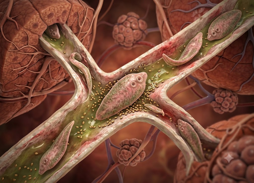
Liver flukes are parasitic flatworms that may infect the liver, gallbladder, and bile ducts. People can become infected by eating raw or undercooked freshwater fish, aquatic plants such as watercress, or other contaminated foods that contain parasite larvae. In some cases, the infection causes little or no noticeable symptoms at first, making it difficult to recognize. As time passes, however, liver flukes can irritate the bile ducts, disturb normal bile flow, and contribute to inflammation or scarring.
Recognizing the potential signs of a liver fluke infection is important because many of its symptoms can resemble common digestive problems. Depending on the parasite and the stage of infection, a person may experience abdominal pain, fever, nausea, vomiting, diarrhea, tiredness, jaundice, or unexplained changes in weight.
Certain species, particularly Clonorchis and Opisthorchis, can live inside the bile ducts and have been associated with long-term inflammation and an increased risk of bile duct cancer. This article looks at seven possible signs of liver flukes and explains how these parasites can affect the bile ducts.
What Is a Liver Fluke?
A liver fluke is a parasitic flatworm that can affect the liver, gallbladder, and bile ducts. These parasites commonly enter the body when someone consumes contaminated raw or undercooked freshwater fish, aquatic plants, or food exposed to infected water. After entering the digestive system, the parasite can move toward the liver or bile ducts and may remain there for an extended period if it is not treated.
At the beginning of an infection, a person may not notice any symptoms. When symptoms do develop, they can include pain in the upper abdomen, fever, nausea, diarrhea, fatigue, jaundice, dark-colored urine, or unexplained weight loss. Some types of liver flukes can also irritate the bile ducts and contribute to inflammation, blockage, infection, or scarring.
Long-lasting infection may become more serious, particularly when the parasites remain in the bile ducts. Chronic infection can cause ongoing bile duct damage and, in certain areas and with certain species, increase the risk of bile duct cancer. Doctors may use stool tests, blood tests, imaging, and other medical evaluations to diagnose the infection. Treatment depends on the type of liver fluke and generally requires an antiparasitic medicine prescribed by a healthcare professional.
7 Key Symptoms of a Liver Fluke Infection
The seven main symptoms associated with a liver fluke infection include fever, pain in the upper right side of the abdomen, nausea, eosinophilia (an increased white blood cell count), jaundice (yellowing of the skin and eyes), long-lasting indigestion, and unexplained weight loss. These symptoms can occur during two different phases of infection: an early acute stage while the parasite is migrating and a later chronic stage when the parasites affect the bile ducts.
Understanding the difference between these stages can help explain why symptoms may change over time. The signs a person experiences can depend on where the parasite is located and how active it is within the liver and biliary system.
The Early Acute Phase: Larval Migration
The infection begins when a person consumes the infective larval stage of the parasite, known as metacercariae. These larvae are commonly acquired from contaminated freshwater plants or raw and undercooked fish. After entering the intestines, the young parasites can pass through the intestinal wall, move across the abdominal cavity, and reach the outer surface of the liver. This migration can trigger the symptoms associated with the early acute phase.
Fever and an overall feeling of illness can develop as the immune system responds to the migrating larvae. This inflammatory reaction may also cause pain in the upper right area of the abdomen, near the liver. The discomfort can range from dull to sharp and may result from inflammation and irritation of the liver’s outer covering. Nausea and occasional vomiting can also occur while the parasite is moving through the tissues.
During this early phase, blood tests may show significant eosinophilia, meaning there is an increased number of eosinophils in the blood. These white blood cells play an important role in the immune response to multicellular parasites. When a high eosinophil count occurs together with abdominal pain and other symptoms, it may provide doctors with an important clue that a parasitic infection is present.
The Late Chronic Phase: Biliary Obstruction
After the initial phase, juvenile parasites can move into the biliary system, where they mature into adult worms, produce eggs, and may remain for years. At this point, symptoms are often related to continued inflammation and physical obstruction of the bile ducts.
Jaundice, which causes the skin and whites of the eyes to appear yellow, can occur during this stage. It may develop when liver flukes interfere with the normal movement of bile. As bile flow becomes disrupted, bilirubin can build up in the bloodstream instead of leaving the body normally through the digestive system. Problems with bile flow can also affect digestion and may contribute to ongoing indigestion, abdominal bloating, gas, and fatty stools.
Over time, problems with nutrient absorption may contribute to poor nutrition and unexplained weight loss. The long-lasting inflammatory response can also leave a person feeling weak and tired. Ongoing irritation caused by the parasite may further contribute to general fatigue.
The Asymptomatic Nature and Advanced Complications
Many people may wonder what liver flukes in humans can do when there are no obvious symptoms. In reality, some infections can remain difficult to detect for a long time. A liver fluke infection may sometimes stay silent or produce only mild symptoms for years.
If only a small number of larvae enter the body, the initial migration may not cause enough inflammation or tissue damage to produce obvious fever or severe abdominal pain. Minor bile duct obstruction may also be tolerated by the body, allowing the infection to appear like occasional or ordinary indigestion. This can make the infection difficult to identify without appropriate testing.
Long-Term Pathological Risks:
Over many years, the continued presence of adult liver flukes can contribute to chronic bile duct inflammation, tissue scarring, and secondary bacterial infections. When the infection remains untreated, the ongoing damage may lead to serious hepatobiliary complications. These can include gallstone formation, advanced liver damage, and an increased risk of a serious bile duct cancer called cholangiocarcinoma.
How Can You Protect Your Bile Ducts From Liver Flukes?
Protecting the bile ducts from liver flukes starts with safe food and water practices that reduce the chance of swallowing infective parasite larvae. Important preventive steps include thoroughly cooking freshwater fish, crustaceans, and aquatic plants. Because infection is mainly associated with consuming contaminated food or water, preventing exposure is an important way to interrupt the transmission cycle.
The following strategies focus on safe food preparation, water treatment, and everyday hygiene to reduce the possibility of infection.
Primary Prevention and Vector-Specific Transmission Routes
Understanding how liver flukes reach humans is an important part of preventing infection. These parasites can enter the body when a person consumes raw, undercooked, or inadequately processed food or water containing the infective larval stage known as metacercariae.
Prevention therefore depends on interrupting the transmission cycle before contaminated food or water is consumed.
Different liver fluke species use different environmental sources to reach humans. Species such as Clonorchis sinensis and Opisthorchis viverrini are mainly associated with freshwater fish. Their larvae can become enclosed within the tissues of freshwater fish, including fish such as carp or tilapia, as well as certain freshwater crustaceans.
In comparison, Fasciola hepatica is associated with aquatic plants. Its larvae can attach to the stems and leaves of plants growing in freshwater that has been contaminated by the feces of infected animals such as sheep and cattle.
Because different species have different transmission routes, prevention should focus on safe preparation and handling of the foods and water sources that may carry the parasite.
Food Safety: Thermal Destruction and Cross-Contamination Firewalls
Safe food preparation is one of the most important defenses against liver flukes. Even when raw ingredients contain parasite larvae, proper preparation can help destroy the organisms before the food is eaten.
                     [Thermal Destruction of Metacercariae]
                                     │
      ┌───────────────────────────────┴───────────────────────────────┐
      ▼                                                               ▼
[Freshwater Fish & Crustaceans]                         [Aquatic Plants & Greens]
 ├── Minimum internal temp: 145°F (63°C)                 ├── Boiling or steaming: 1+ minute
 └── Alternative: Freeze at -4°F (-20°C) for 7 days      └── Heat destroys surface cysts
Thermal Thresholds for Parasitic Elimination
Thorough cooking is one of the most effective ways to destroy parasite larvae in food. Freshwater fish and crustaceans should reach an internal temperature of at least 145°F (63°C), with the flesh becoming opaque and flaking easily with a fork.
Common preparation methods such as pickling, salting, light smoking, or marinating fish in citrus juice may not provide enough heat or other conditions to destroy the protective cysts surrounding the parasite larvae.
For aquatic vegetables and plants that may be exposed to contaminated water, such as watercress, water mint, water chestnuts, or locally grown greens, simply rinsing them with cold water may not be enough to remove firmly attached parasite cysts. Boiling or steaming these plants for at least one minute can provide additional protection.
If freshwater fish is intended to be eaten raw, strict deep-freezing procedures should be followed. Keeping the fish at -4°F (-20°C) or colder for at least seven days can kill encapsulated parasites, although thorough cooking remains the safest preparation method.
Preventing Environmental Cross-Contamination
Cross-contamination can happen when microscopic parasite larvae move from raw ingredients onto clean kitchen surfaces, utensils, or ready-to-eat foods. Good kitchen hygiene can help prevent this from happening.
- Separate Equipment: Use separate cutting boards, knives, and preparation plates for raw freshwater seafood whenever possible. Keep these items away from equipment used for fresh produce, cooked foods, or bread.
- Sanitization Protocol: If the same equipment must be used, clean cutting boards, knives, countertops, and other surfaces thoroughly with hot, soapy water after contact with raw ingredients.
- Hand Hygiene: Wash your hands carefully with soap and warm water after handling raw freshwater fish, crustaceans, or potentially contaminated aquatic plants. Avoid touching clean foods or kitchen surfaces with unwashed hands.
Water Purification and Microscopic Filtration Standards
Although food is a more common source of exposure, contaminated water can also present a risk, particularly in relation to Fasciola hepatica. Parasite larvae may detach from aquatic vegetation and remain in open water, creating a potential risk when contaminated water is swallowed.
                                  [Water Purification Matrix]
                                              │
          ┌────────────────────────────────────┼────────────────────────────────────┐
          ▼                                    ▼                                    ▼
  [Thermal: Rolling Boil]              [Physical: 1-Micron Filter]         [Chemical: Halogen Tablets]
   ├── Time: 1 to 3 minutes             ├── Absolute pore size standard     ├── Secondary alternative
   └── Heat destroys larvae              └── Traps microscopic cysts         └── Depends on water clarity
The Thermal Gold Standard
Bringing potentially contaminated water to a rolling boil for at least one full minute is an effective method for destroying many waterborne pathogens, including liver flukes. At elevations above 6,500 feet (2,000 meters), where water reaches a lower boiling temperature, the boiling period should be extended to three minutes. This approach can help make water safer for drinking, brushing teeth, and washing raw vegetables.
Absolute Micron Filtration Requirements
When boiling is not practical, mechanical filtration can provide another option. A portable or household water filter intended to remove microscopic cysts should have an absolute pore size of 1 micron or smaller.
When selecting a filter, check the manufacturer’s specifications and look for equipment specifically designed for cyst removal, including products that meet relevant NSF/ANSI standards. Basic mesh strainers and ordinary charcoal filters may not be capable of removing organisms of this size from potentially contaminated water.
Limitations of Chemical Disinfection
Chemical disinfectants such as chlorine or iodine tablets can provide another treatment option, but their effectiveness may be reduced under certain conditions. Cold, cloudy, or highly alkaline water can make chemical disinfection less reliable.
If chemical tablets are necessary, first strain cloudy water through a clean cloth or similar material. Then follow the manufacturer’s instructions carefully and allow the treatment to work for the full recommended period before drinking.
Comprehensive Prevention and Exposure Management Framework
When traveling to or living in places where liver flukes are more common, paying attention to everyday food and water exposure can help reduce the risk of infection.
| Transmission Vector | Specific Danger Source | Required Protection Activity | Clinical Rationale |
|---|---|---|---|
| Freshwater Seafood | Raw, pickled, or lightly salted river fish and crabs. | Cook to an internal temperature of 145°F (63°C) or freeze at -4°F (-20°C) for 7 days. | Proper heat can destroy the protective cysts associated with Clonorchis and Opisthorchis larvae. |
| Aquatic Vegetation | Wild watercress, water chestnuts, or greens irrigated with potentially contaminated water. | Boil or steam the plants for at least one full minute and avoid eating raw wild aquatic salads. | Washing alone may not remove firmly attached Fasciola larvae. |
| Open Water Sources | Untreated water from rivers, streams, ponds, or shallow wells. | Bring water to a rolling boil for 1–3 minutes or use an absolute 1-micron filter. | Treatment helps remove or destroy larvae that may be present in contaminated water. |
| Recreational Activities | Swimming or wading in slow-moving freshwater canals or lakes. | Avoid swallowing the water and wash your hands after leaving the water. | Reduces the chance of accidentally swallowing contaminated water. |
Broader Implications of a Liver Fluke Diagnosis
A liver fluke diagnosis involves more than identifying the initial symptoms. Doctors may need to determine the exact parasite, select an appropriate antiparasitic treatment, and evaluate whether the infection has affected the liver or bile ducts. Long-term infections can potentially lead to serious complications, including bile duct cancer, which is why appropriate diagnosis and treatment are important.
Understanding the testing process, treatment options, and differences between liver fluke infections and other liver conditions can help clarify how these infections are managed.
Laboratory and Radiographic Diagnostic Pathways
Confirming an active liver fluke infection generally requires a focused medical evaluation. Because the parasite can move through different areas as it develops, healthcare professionals may combine laboratory tests with imaging studies to identify the infection and check for damage to the biliary system.
                          [Hepatobiliary Diagnostic Protocol]
                                          │
          ┌────────────────────────────────┼────────────────────────────────┐
          ▼                                ▼                                ▼
 [Microscopic Stool Exam]         [Serological Blood Test]         [Advanced Biliary Imaging]
  └── Detects adult fluke eggs      └── Measures immune response      └── Ultrasound, CT, or MRI
  └── Useful in mature infection   └── May identify earlier exposure └── Assesses bile duct changes
Microscopic Stool Examination
A microscopic stool examination can be used to identify a mature liver fluke infection. During this test, a laboratory examines a concentrated stool sample for the characteristic eggs released by adult parasites.
One limitation is the pre-patent period, which refers to the time between initial infection and the point when the parasite becomes mature enough to produce eggs. If testing takes place during the early migration stage, a stool test may be negative even when an infection is present. For this reason, doctors may request multiple stool samples collected on different days to improve the chance of detection.
Serological Blood Testing
When a person asks what liver flukes in humans can do during the first weeks of infection, it is important to consider that the parasites may still be migrating through liver tissue. During this early stage, blood tests such as enzyme-linked immunosorbent assays (ELISA) may be used.
Rather than looking for parasite eggs, serological testing looks for antibodies produced by the immune system in response to the parasite. Depending on the infection and test used, this can help identify exposure before adult worms begin releasing eggs into the stool.
Advanced Medical Imaging
Imaging can help doctors evaluate possible damage to the liver and bile ducts. Abdominal ultrasound, computed tomography (CT), and magnetic resonance imaging (MRI) can reveal changes such as thickened bile duct walls, liver abnormalities, or widening of parts of the biliary system.
In certain complicated cases, doctors may use endoscopic retrograde cholangiopancreatography (ERCP). This minimally invasive procedure allows the biliary system to be examined directly and can sometimes help identify or remove material obstructing the bile ducts.
Species-Specific Antiparasitic Interventions
Treating liver flukes requires medication appropriate for the particular species involved. Ordinary over-the-counter deworming products may not be suitable. A healthcare provider needs to determine the suspected parasite and select the appropriate prescription treatment.
                      [Targeted Antiparasitic Selection]
                                     │
          ┌──────────────────────────┴──────────────────────────┐
          ▼                                                     ▼
 [Praziquantel Therapy]                              [Triclabendazole Therapy]
  ├── Targets: Clonorchis & Opisthorchis              ├── Targets: Fasciola hepatica
  └── Causes parasite paralysis                        └── Acts against larval & adult worms
Praziquantel Protocol
For infections caused by Clonorchis sinensis, also known as the Chinese liver fluke, or Opisthorchis viverrini, the Southeast Asian liver fluke, praziquantel is a standard treatment option.
The medicine affects the parasite’s cellular membranes and causes changes that lead to muscle spasms and paralysis. The weakened parasites can then detach from the bile ducts and pass out of the body through the digestive tract.
Although praziquantel can be highly effective for the appropriate infection, it should be used under medical supervision. Some people may experience side effects such as abdominal discomfort, dizziness, or nausea.
Triclabendazole Protocol
When the infection is caused by Fasciola hepatica, praziquantel is not the preferred treatment. Triclabendazole is the recommended medication for fascioliasis.
This antiparasitic medicine acts on the internal structures and metabolism of the Fasciola parasite and can target both migrating juvenile parasites and mature worms. In severe cases where dead or dying worms contribute to an obstruction in the biliary system, doctors may use an ERCP procedure to help clear the blockage.
Pathological Differentiation: Liver Flukes vs. Viral Hepatitis
Liver fluke infections and viral hepatitis can both cause inflammation and symptoms such as abdominal discomfort or jaundice, but they are very different medical conditions with different causes and treatment approaches.
[Liver Flukes: Multicellular Worms] ──► Food Transmission ──► Mechanical Bile Duct Damage
[Viral Hepatitis: Microscopic Viruses] ──► Blood/Fecal-Oral Routes ──► Liver Cell Damage
Parasitic Worms vs. Viral Pathogens
The primary difference is the organism responsible for the infection. Liver flukes are multicellular parasitic flatworms, while viral hepatitis is caused by microscopic viruses, including hepatitis A, B, C, D, and E.
Their transmission routes also differ. People can acquire liver flukes through contaminated raw or undercooked freshwater fish or aquatic plants. Hepatitis viruses have different transmission routes depending on the type. Hepatitis A and E are commonly spread through the fecal-oral route, while hepatitis B and C can be transmitted through infected blood or bodily fluids.
Sites of Tissue Damage and Treatment Styles
Liver flukes primarily cause structural and mechanical problems by irritating or obstructing the bile ducts. Viral hepatitis, on the other hand, directly affects liver cells, known as hepatocytes.
Because the causes are different, the treatments are also different. Liver fluke infections require specific antiparasitic medications, such as praziquantel or triclabendazole depending on the parasite. Viral hepatitis may be managed with antiviral medication, vaccination for preventable types, or supportive treatment depending on the specific virus.
| Clinical Factor | Liver Fluke Infection | Viral Hepatitis (A, B, C, D, E) |
|---|---|---|
| Causative Agent | Multicellular trematode flatworm (helminth). | Microscopic viral pathogens. |
| Transmission Source | Consuming contaminated raw freshwater seafood or aquatic plants. | Varies by type: contaminated food/water for A/E or blood exposure for B/C. |
| Primary Area of Damage | Mechanical irritation and blockage of the bile ducts. | Direct effects on hepatocytes (liver cells). |
| Medical Treatment | Species-specific antiparasitic medication. | Antiviral treatment, vaccination for preventable types, or supportive care depending on the virus. |
The Long-Term Carcinogenic Risk: Cholangiocarcinoma
Understanding what liver flukes in humans can cause over a long period helps explain why early diagnosis and treatment matter. Chronic infections with Clonorchis sinensis and Opisthorchis viverrini have been associated with an increased risk of cholangiocarcinoma, a serious cancer that develops in the bile ducts.
[Adult Fluke Presence] ──► Mechanical & Chemical Irritation ──► Chronic Tissue Damage ──► CholangiocarcinomaThe International Agency for Research on Cancer (IARC) classifies these particular fish-borne liver flukes as Group 1 carcinogens. This classification reflects evidence that long-term infection with these parasites can contribute to the development of bile duct cancer.
The process can develop gradually over many years. Adult worms attach to the lining of the bile ducts and cause repeated irritation as they move and feed. Their metabolic products can also contribute to irritation of nearby tissue.
Persistent damage can keep the immune system in a state of chronic inflammation. As the cells lining the bile ducts repeatedly repair themselves, continued cell turnover in an inflamed environment can increase the chance of DNA changes. Over a long period, these changes may contribute to malignant transformation and the development of cholangiocarcinoma.
Conclusion
Liver fluke infections may remain unnoticed in the beginning, but they can become more serious when the parasites affect the bile ducts. Symptoms such as upper-right abdominal pain, fever, nausea, diarrhea, jaundice, itching, dark urine, pale stools, or unexplained weight loss deserve medical attention, particularly when they occur after travel or the consumption of raw or undercooked freshwater foods.
A long-term liver fluke infection can contribute to bile duct inflammation, obstruction, infection, and scarring. Certain chronic infections have also been linked to an increased risk of cholangiocarcinoma. When an infection is suspected, appropriate medical testing and species-specific antiparasitic treatment can help reduce the risk of complications.
Frequently Asked Questions
1. What are liver flukes?
Liver flukes are parasitic flatworms that can infect the liver, gallbladder, and bile ducts. They belong to a group of parasites called trematodes, commonly known as flukes. Several species can infect humans, including Fasciola, Clonorchis, and Opisthorchis. Depending on the species and how long the infection lasts, these parasites may cause digestive symptoms, inflammation, or problems with the bile ducts.
2. How do people get liver flukes?
People generally become infected by eating food containing liver fluke larvae. Some species are associated with raw or undercooked freshwater fish, while others can be acquired through aquatic plants such as watercress. The risk is greater in regions where these parasites are common and where food preparation allows the larvae to survive. Thorough cooking and careful food handling can help lower the risk.
3. What symptoms can liver flukes cause?
Liver flukes may cause abdominal pain, fever, nausea, vomiting, diarrhea, fatigue, and reduced appetite. When bile flow is affected, some people may experience jaundice, dark urine, pale stools, or itching. Chronic infections may cause mild or intermittent symptoms for an extended period. In some cases, people have no obvious symptoms and only discover the infection through medical testing.
4. How do liver flukes affect the bile ducts?
Some liver fluke species live inside the bile ducts, where they can cause irritation and inflammation. Over time, this may contribute to thickening, blockage, infection, or scarring of the bile ducts. Chronic infection with certain species has also been associated with an increased risk of bile duct cancer. Persistent symptoms or a known exposure should therefore be discussed with a healthcare professional.
5. How are liver flukes diagnosed and treated?
Doctors may use stool examinations, blood tests, imaging studies, or other tests to look for parasite eggs and signs of liver or bile duct involvement. The most suitable test depends on the suspected species and the stage of infection. Treatment generally involves an antiparasitic medicine, such as triclabendazole for fascioliasis or praziquantel for certain other liver fluke infections. A healthcare professional should determine the appropriate treatment based on the specific parasite and the individual’s condition.