3 Dangerous Stages of Pulmonary Embolism to Watch
Most people have heard of pulmonary embolism. It sounds grave, something involving blood clots in the lungs that doctors caution about. What many don’t realize is that it isn’t a single instant. It often develops in stages, each riskier than the one before if it goes unnoticed.
That is where the real danger lies. A touch of breathlessness, some chest discomfort, or odd tiredness is easy to brush aside as nothing urgent, yet the condition may already be in its early phase and quietly advancing. As it progresses, strain on the heart grows, oxygen levels fall, and the body begins to struggle in ways that aren’t always obvious at first.
What makes pulmonary embolism so frightening is how fast it can escalate. One stage can give way to the next within hours, sometimes minutes, turning a manageable situation into a life-threatening emergency. The body sends signals, but they are often misread or ignored until the condition is critical.
Knowing the three dangerous stages is more than medical trivia. It is a form of protection. If you understand how the condition progresses, you are far more likely to notice that something is wrong and act in time. The sections below explain how each stage develops, which symptoms to watch for, and why timing matters so much for survival.
What Is a Pulmonary Embolism?
A pulmonary embolism (PE) is a sudden blockage in a pulmonary artery. It unfolds from the initial formation of a clot in a distant vein, through that clot’s journey to the lungs, to the eventual obstruction of blood flow there. The process starts when a piece of a blood clot (thrombus) breaks off, most often from a deep leg vein, a condition called deep vein thrombosis (DVT). The detached fragment, now called an embolus, travels through the veins, passes through the right side of the heart, and lodges in the branching arteries of the lungs, disrupting their vital work.
The progression is a cascade that turns a local vascular problem into a body-wide, life-threatening crisis. The whole disease process, covering both the original DVT and the PE that follows, is known as venous thromboembolism (VTE). How severe a PE is depends on the size of the embolus and the patient’s existing heart and lung health. A large clot can block major arteries and cause sudden cardiovascular collapse, while smaller clots may block peripheral vessels and cause subtler but still significant damage.
The Link Between DVT and Pulmonary Embolism
The connection is direct and causal: more than 90% of pulmonary emboli begin as a complication of an existing DVT, when a fragment of a deep-vein clot breaks loose and travels to the lungs. The two conditions are so closely tied that they are regarded as two expressions of the same underlying process, VTE. The embolus follows a clear anatomical route. Once a piece of clot detaches from the wall of a leg or pelvic vein, it enters the venous circulation.
Blood flow then carries it up through the inferior vena cava, the body’s largest vein, into the right atrium of the heart. From there it is pumped into the right ventricle and out into the pulmonary artery, which divides into ever-smaller vessels within the lungs. The embolus keeps moving until it reaches a vessel too narrow to pass through, where it lodges and creates a blockage.
- Where the clot begins: Nearly all emboli come from the deep veins of the legs, such as the popliteal, femoral, or iliac veins. Less often, clots form in deep arm or pelvic veins, or even inside the right side of the heart.
- How embolization happens: The fragment may break away on its own or be set off by physical activity, a shift in blood pressure, or muscle contractions. The part still attached to the vein wall is the thrombus, and the traveling piece is the embolus.
- Clinical meaning: Because of this tight link, anyone diagnosed with a DVT is considered at high risk for PE and is started on anticoagulants promptly to prevent it. Conversely, a patient who presents with a PE often has their legs imaged to look for a DVT.
How Dangerous Is a Pulmonary Embolism?
A PE is always treated as a potentially life-threatening condition needing urgent evaluation and care, although the immediate risk of death varies widely with clot size and the patient’s general health. Not every PE is instantly fatal, but the potential for rapid decline and serious complications makes each one a medical emergency. Sorting PEs into risk categories helps clinicians judge the outlook and choose the right level of treatment.
The danger comes from the combined hit to the respiratory and cardiovascular systems. Severity spans a broad range and is usually classified by hemodynamic stability, meaning whether blood pressure and heart function are compromised.
- Massive PE: The most severe form, defined by sustained low blood pressure (hypotension), shock, or cardiac arrest. It is typically caused by a large clot, such as a saddle embolus sitting at the fork of the main pulmonary artery and blocking flow to both lungs. The right ventricle then fails acutely because it cannot pump against such a blockage, leading to circulatory collapse and a high risk of sudden death.
- Submassive PE: Blood pressure stays stable, but there is evidence of strain or dysfunction in the right ventricle, detectable through imaging such as an echocardiogram or through blood tests showing raised cardiac biomarkers. It doesn’t cause shock right away, but it shows the heart is struggling and the patient could worsen into a massive PE.
- Low-risk PE: Blood pressure is normal and there are no signs of right heart strain. The clot is usually smaller and sits in more peripheral lung arteries. The immediate risk of death is lower, but these PEs can still damage lung tissue (infarction), impair oxygenation, and need prompt anticoagulation to stop the clot from extending or returning. Even a small PE can be fatal if it triggers a deadly heart rhythm disturbance or the patient’s lungs are already severely impaired.
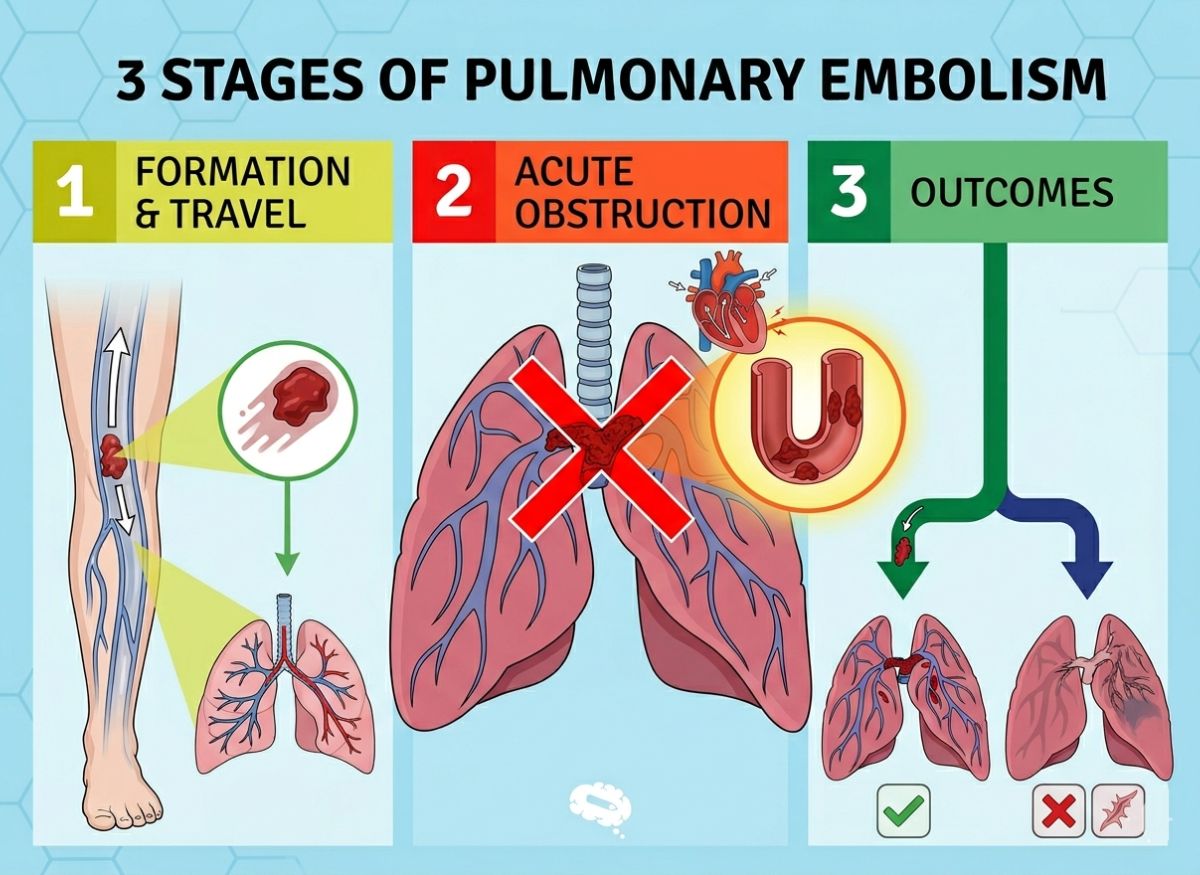
The 3 Stages of Pulmonary Embolism
Stage 1: Embolus Formation and Travel
Stage 1 covers the formation of a clot (thrombus) in a deep vein, governed by three factors known as Virchow’s triad, followed by the clot (or a piece of it) breaking free and beginning to travel through the bloodstream. This is the foundation phase, where the potential for a lung blockage is created.
The thrombus usually forms in the deep veins of the legs or pelvis, where blood moves more slowly and clots more readily. The likelihood rises sharply when one or more parts of Virchow’s triad are present, since together they create a prothrombotic state that makes clotting highly probable.
- Venous stasis: Slowed or stagnant blood flow in the veins. When blood isn’t moving efficiently, clotting factors can build up and start the coagulation cascade. Common causes include prolonged immobility (long-haul flights, bed rest after surgery), paralysis, or heart failure, which reduces overall circulation.
- Hypercoagulability: An increased tendency of the blood to clot. It can be inherited (for example, the Factor V Leiden mutation) or acquired. Acquired causes are more common and include cancer, pregnancy, estrogen-containing medications such as oral contraceptives or hormone replacement therapy, and certain inflammatory diseases. These shift the balance between pro-clotting and anti-clotting proteins.
- Endothelial injury: Damage to the inner lining of a vessel. A healthy lining is smooth and resists clotting, but when injured it exposes underlying tissue that can trigger a thrombus. Causes include direct trauma to a vein, major surgery (especially hip or knee orthopedic surgery), intravenous catheters, and inflammation or infection of the vein wall.
Once the thrombus exists, the last step of Stage 1 is embolization. A clot that is only loosely attached to the vessel wall can break free and become a mobile embolus, swept by venous flow toward the heart and then the lungs.
Stage 2: Acute Pulmonary Obstruction
Stage 2 is the critical moment when the traveling embolus lodges in the pulmonary arteries, physically blocking blood flow, impairing gas exchange, and placing severe, sudden strain on the right side of the heart. This is when a PE becomes clinically obvious and life-threatening. The consequences are immediate and profound, hitting the respiratory and cardiovascular systems at once, and their severity is proportional to the size of the blockage and the patient’s underlying cardiopulmonary reserve.
When the embolus blocks a pulmonary artery, a section of lung continues to receive air (ventilation) but no longer receives blood (perfusion). This is called a ventilation-perfusion (V/Q) mismatch.
- Respiratory effects: The V/Q mismatch is a main cause of hypoxemia (low blood oxygen), because blood cannot pick up oxygen from the blocked segment. The body may try to compensate by redirecting blood to healthier lung areas and breathing faster, producing the classic symptom of sudden, unexplained breathlessness. The blockage can also trigger release of inflammatory substances that constrict the small airways (bronchoconstriction), making breathing harder still.
- Hemodynamic effects: The obstruction sharply raises the resistance the right ventricle must pump against. Pulmonary vascular resistance surges, quickly increasing pressure in the right ventricle and pulmonary artery (acute pulmonary hypertension). The right ventricle is a relatively thin-walled muscle built for low-pressure work and cannot cope with a sudden overload, which can lead to dilation, strain, and eventually failure. When it fails, it can’t move blood effectively to the lungs, so less blood returns to the left side of the heart. Cardiac output then drops sharply, causing systemic low blood pressure, shock, and death.
Stage 3: Resolution or Chronic Complication
In Stage 3 the body follows one of two paths. Either the clot is broken down and absorbed, through the body’s natural fibrinolytic system plus medical treatment (resolution), or it fails to dissolve completely and turns into fibrous tissue, causing lasting obstruction and chronic complications such as Chronic Thromboembolic Pulmonary Hypertension (CTEPH).
This final stage determines long-term outcome after the acute event. Most patients, with proper treatment, see significant or complete clot resolution over weeks to months. The two possible outcomes are a fork in the road for recovery.
- Resolution: The hoped-for result is complete breakdown of the clot, mainly through the body’s own fibrinolytic system, in which an enzyme called plasmin dissolves the fibrin mesh that gives the clot its structure. Anticoagulants such as heparin and warfarin do not actively dissolve an existing clot. They prevent new clots and stop the current one from growing, creating a stable environment in which the body’s natural clot-dissolving mechanisms can work over time. In massive PE, powerful thrombolytic (“clot-buster”) drugs may be given to dissolve the clot quickly and restore flow.
- Chronic complication (CTEPH): In a small but significant share of survivors (around 3 to 5%), the clot doesn’t fully resolve. It becomes organized, scar-like fibrous tissue built into the walls of the pulmonary arteries, forming a permanent, fixed obstruction that leads to CTEPH. The persistent blockages keep pressure in the pulmonary arteries chronically high, causing progressive right heart failure. Symptoms include ongoing breathlessness on exertion, fatigue, and eventually signs of heart failure such as leg swelling. Unlike acute PE, CTEPH is a chronic disease that often needs specialized surgery (pulmonary thromboendarterectomy) or targeted medical therapy.
Acute PE vs. Chronic Thromboembolic Pulmonary Hypertension (CTEPH)
The main differences between acute PE and CTEPH are timeline, underlying pathology, and clinical picture. Acute PE is the immediate event in which a clot, usually from a leg DVT, travels to the lungs and suddenly blocks a pulmonary artery. It brings abrupt symptoms such as sharp chest pain and severe breathlessness and, with large emboli, a fall in blood pressure and possible cardiovascular collapse. Diagnosis and treatment focus on this urgent, life-threatening blockage. CTEPH, by contrast, is a rare but serious long-term complication that develops in a small percentage (2 to 4%) of PE survivors.
In CTEPH, the original clots don’t dissolve fully despite anticoagulation. Over months to years they organize into chronic, scar-like fibrous tissue that becomes part of the pulmonary artery walls. This permanent obstruction steadily raises resistance to blood flow through the lungs, pushing up pressure in the pulmonary arteries (pulmonary hypertension). The high pressure puts heavy strain on the right side of the heart and eventually causes right-sided heart failure.
Both conditions involve clots in the pulmonary arteries, yet they differ fundamentally in timeline, pathology, and consequences.
- Symptoms: Acute PE is a sudden crisis, whereas CTEPH shows up as gradually worsening, often nonspecific breathlessness on exertion, fatigue, and leg swelling over a long period.
- Diagnosis: Acute PE is typically confirmed with a CTPA. CTEPH diagnosis often uses a V/Q scan to find chronic perfusion defects, followed by right heart catheterization to directly measure the raised pulmonary artery pressures.
- Treatment: Anticoagulants are the main treatment for acute PE. CTEPH often requires a highly specialized operation, pulmonary thromboendarterectomy (PTE), to remove the chronic scar tissue from the arteries, or medications designed to treat pulmonary hypertension.
Symptoms and Severity Across the Stages
The symptoms and severity of a PE track its three-stage progression: possible warning signs in the leg during Stage 1 (DVT), intense cardiorespiratory symptoms during Stage 2 (acute obstruction), and possibly lasting exertional limits in Stage 3 (complications). This evolution is a clinical map of the clot’s journey and its effect on the body. Recognizing the signs at each stage is crucial for timely diagnosis and treatment, which strongly influence prognosis.
The picture shifts dramatically as the event unfolds. Early symptoms can be subtle and easy to dismiss, acute symptoms are often sudden and terrifying, and long-term effects can be a lasting reminder, signaling either successful recovery or the start of a chronic secondary condition. Each stage calls for a different level of suspicion and response from patients and clinicians alike.
Warning Signs of Stage 1 (DVT)
The main warning signs of Stage 1, corresponding to the underlying DVT, are symptoms usually confined to one leg: swelling (edema), persistent pain or tenderness (often like a cramp or charley horse), warmth over the affected area, and sometimes red or bluish skin discoloration.
They occur because the clot in the deep vein obstructs flow, causing blood to pool in the limb and triggering inflammation in the vein wall. They are the earliest and most important clues that a person is at risk of developing a PE. Notice that these symptoms almost always affect just one leg.
- Swelling (edema): One of the most common signs. The affected leg, calf, or ankle may look visibly larger than the other. Blood backs up behind the clot, raising pressure in the vein and forcing fluid into surrounding tissue.
- Pain or tenderness: Often a deep ache or persistent cramping that may be most noticeable when standing or walking. The area over the affected vein, especially in the calf, may be tender to the touch.
- Warmth and redness: Inflammation from the clot can make the skin feel warmer than nearby skin or the other leg. It may look red, purplish, or bluish because of congested, oxygen-poor blood.
- Symptom-free DVT: Up to 50% of people with DVT have no symptoms at all. This silence makes DVT especially dangerous, since a PE may be the first sign anything is wrong. That is why knowing risk factors such as recent surgery, immobility, or cancer matters as much as recognizing symptoms.
Symptoms of an Acute Stage 2 Event
The hallmark symptoms of an acute Stage 2 PE are sudden, severe shortness of breath (dyspnea), sharp, stabbing chest pain that worsens with a deep breath (pleuritic pain), an abnormally fast heart rate (tachycardia), and possibly coughing, which may produce blood-streaked sputum (hemoptysis).
These symptoms show that the embolus has lodged in the lungs, creating an immediate crisis for the heart and respiratory system. Their severity often reflects the clot’s size and location. A Stage 2 event is typically dramatic and frightening, prompting people to seek emergency care.
- Sudden shortness of breath: The most common symptom. It can occur at rest or with exertion and is often described as being unable to get enough air. It results directly from the V/Q mismatch and the body’s frantic effort to make up for the lack of oxygenated blood.
- Pleuritic chest pain: Usually sharp and localized, and worse with coughing, sneezing, or deep breaths. It arises from inflammation of the pleura, the lining of the lungs, which can become irritated when a clot causes lung tissue to die (pulmonary infarction).
- Rapid heart rate and palpitations: The heart beats faster to offset reduced oxygen delivery and to push blood past the blockage. People may feel their heart racing or pounding.
- Other severe symptoms: With a large or massive PE, additional critical signs can appear, including profound lightheadedness, dizziness, or fainting (syncope), which point to dangerously low cardiac output and blood pressure. Heavy sweating and intense anxiety or a sense of impending doom may also occur. These indicate a high-risk, life-threatening situation.
Long-Term Effects Signaling Stage 3 Complications
Long-term effects that point to Stage 3 complications, especially CTEPH, include persistent or worsening breathlessness with physical activity, chronic fatigue with much reduced exercise capacity, and occasionally recurring chest pain or swelling in the legs and ankles.
These lingering symptoms suggest the clot has not fully dissolved and has caused permanent damage to the pulmonary arteries, leaving the heart under chronic strain. Many PE survivors return to normal health, but symptoms that persist weeks or months after the acute event deserve further investigation. They differ from the initial acute symptoms and mark a shift from an acute illness to a chronic condition.
- Persistent breathlessness on exertion: The hallmark of CTEPH. Some breathlessness is expected early in recovery, but a failure to improve, or worsening with activities once manageable, is a major red flag. The scarred, blocked arteries can’t handle the extra blood flow exercise demands, so lung pressures soar.
- Chronic fatigue and reduced exercise tolerance: People may feel constantly tired and get exhausted by everyday tasks. The heart is working harder to pump blood through obstructed vessels, and the body is chronically short of oxygenated blood.
- Signs of right heart failure: As CTEPH advances, sustained high pulmonary artery pressure wears down the right ventricle until it weakens and fails. This can cause swelling in the ankles, legs, and abdomen (peripheral edema and ascites) and a racing or fluttering heartbeat.
- Post-PE syndrome: Beyond CTEPH, some patients develop a cluster of problems called Post-PE Syndrome, including functional limitations and psychological effects such as anxiety, depression, and PTSD linked to their near-fatal experience, even without evidence of CTEPH. Careful follow-up is needed to tell normal slow recovery from the onset of serious long-term complications.
How Pulmonary Embolism Is Diagnosed
A PE diagnosis is established through several steps, starting with a clinical assessment of symptoms and risk factors, often using scoring tools such as the Wells score or the Pulmonary Embolism Rule-out Criteria (PERC). If PE is suspected, a D-dimer blood test is commonly used as an initial screen. It measures a substance released when a clot breaks down. A negative result in a low-risk patient makes PE highly unlikely and lets clinicians skip further testing.
A positive D-dimer, however, is not specific to PE and calls for definitive imaging. The gold standard is CT Pulmonary Angiography (CTPA), a specialized CT scan using intravenous contrast dye to produce detailed images of the pulmonary arteries that clearly show any blockages. For patients who can’t have a CTPA (for example, because of kidney disease or a contrast dye allergy), a Ventilation/Perfusion (V/Q) scan is a valuable alternative. This nuclear medicine scan compares airflow (ventilation) with blood flow (perfusion) in the lungs, identifying a PE as an area that is ventilated but not perfused.
Since most PEs begin as clots in the legs, a Doppler ultrasound of the legs is often done to find the source clot, which can support the diagnosis. The tests chosen depend on the patient’s clinical probability of PE and individual risk factors, ensuring an accurate and timely diagnosis.
- An echocardiogram (heart ultrasound) may be used, not to see the clot itself, but to assess its effect by looking for right ventricular strain, which points to a more severe, hemodynamically significant embolism.
- A standard chest X-ray is usually among the first tests, to rule out other causes of chest pain and breathlessness such as pneumonia or a collapsed lung, although it rarely shows direct evidence of a PE.
- Blood tests for cardiac troponins and Brain Natriuretic Peptide (BNP) can help gauge the risk of a diagnosed PE by showing the degree of heart muscle strain.
How Pulmonary Embolism Is Treated
Treatment for a lung clot depends on its size, location, and the patient’s overall stability. The main goals are to stop the clot growing, prevent new clots, and, in severe cases, dissolve the existing blockage. The cornerstone for nearly all PE patients is anticoagulation, commonly called blood thinning. Medications such as heparin, warfarin, or newer Direct Oral Anticoagulants (DOACs) don’t dissolve the existing clot, but they are highly effective at preventing it from enlarging and stopping new ones from forming, giving the body’s natural processes time to gradually break down the embolism.
For a life-threatening massive PE causing shock or low blood pressure, a more aggressive approach is needed. This involves thrombolytics, or clot-busting drugs such as alteplase, given intravenously to actively dissolve the clot and restore flow quickly. These drugs carry a significant risk of major bleeding. When thrombolytics are too risky or have failed, advanced procedures may be used. In catheter-directed thrombolysis, a catheter is guided through the vessels directly to the clot to deliver a lower dose of thrombolytic, reducing the risk of bleeding throughout the body.
In the most critical situations, surgical embolectomy, an open-heart operation to physically remove the clot, may be performed. The choice of treatment depends heavily on the severity of the embolism and the patient’s overall health and bleeding risk.
- Anticoagulant therapy typically lasts at least three to six months but may become lifelong for people with recurrent clots, a persistent risk factor such as cancer, or a genetic clotting disorder.
- An Inferior Vena Cava (IVC) filter is a small cage-like device placed in the body’s largest vein to catch clots traveling from the legs before they reach the lungs. It is reserved for high-risk patients who cannot take anticoagulants.
- Supportive care is also vital, including supplemental oxygen to raise blood oxygen levels and, in severe cases, mechanical ventilation or medications to support blood pressure and heart function.
How to Prevent a Pulmonary Embolism
Many PEs can be prevented by addressing the risk factors that lead to clot formation, particularly DVT. Prevention falls broadly into mechanical methods, pharmacological methods, and lifestyle changes. For people at temporary high risk, such as those who are hospitalized, recovering from major surgery (especially orthopedic), or on long-haul flights, staying mobile is paramount.
Simple actions like getting up and walking often, or doing in-seat exercises such as ankle pumps and calf raises, stimulate leg blood flow and keep blood from stagnating. When mobility is limited, mechanical methods come in. These include graduated compression stockings, which apply gentle pressure to the legs to aid circulation, and intermittent pneumatic compression (IPC) devices, inflatable sleeves that squeeze the legs at intervals to mimic walking.
For higher-risk individuals, doctors prescribe pharmacological prophylaxis with low-dose anticoagulants such as heparin or enoxaparin. This is standard after many types of surgery and for hospitalized patients with limited mobility. Staying well hydrated also matters, since dehydration thickens the blood and raises clotting risk.
- For long trips (flights or car journeys over four hours), get up and move every one to two hours, wear loose clothing, and drink plenty of water while avoiding alcohol.
- People with a personal or family history of clots, or known genetic clotting disorders (thrombophilia), should work closely with their healthcare provider on a long-term prevention plan, which may include lifelong anticoagulants.
- Lifestyle factors such as maintaining a healthy weight, quitting smoking, and managing conditions like heart failure or cancer also play an important part in lowering overall PE risk.
FAQs
1. What are the three types of pulmonary embolism?
PE is often described in three levels based on severity:
- Low-risk (non-massive) PE: Smaller clots with mild or moderate symptoms and stable blood pressure.
- Submassive (intermediate-risk) PE: Clots that begin to strain the heart, causing lower oxygen levels and signs of right heart stress, even with stable blood pressure.
- Massive (high-risk) PE: A life-threatening form in which a large clot causes a sudden drop in blood pressure, severe oxygen deprivation, and risk of collapse.
2. How long does a PE usually last?
The clot can begin breaking down within days to weeks once treatment starts, especially with blood thinners. Full recovery may take several weeks to months, depending on clot size and overall health. Some people have lingering symptoms such as breathlessness during recovery.
3. What are the first signs of a blood clot in the lung?
Early signs often come on suddenly and may include shortness of breath, sharp chest pain (especially when breathing in), a rapid heartbeat, and unexplained coughing. Some people also feel lightheaded or unusually anxious. These symptoms can be subtle at first but should never be ignored.
4. What is the red flag for PE?
A major red flag is sudden, unexplained shortness of breath, especially at rest or if it worsens quickly. Combined with chest pain, fainting, or coughing up blood, it signals a possible medical emergency requiring immediate attention.
5. What is the “rule of 60” in pulmonary embolism?
It is described as a warning sign in severe PE, referring to a heart rate above 110 to 120 beats per minute together with low oxygen levels (below 90 to 92%), which may indicate significant strain on the heart and lungs. It is not a formal diagnostic rule, but it helps flag when a situation may be becoming critical.
Conclusion
Pulmonary embolism is not a single event. It is a condition that can evolve quickly, often passing through stages many people fail to recognize in time. That is why understanding these three dangerous stages matters so much. Each one marks a shift in how the body is coping, from manageable symptoms to life-threatening distress, and the gap between acting early and acting late can be critical. Paying attention to warning signs, especially sudden changes in breathing or chest discomfort, gives you a chance to act before things escalate.
Awareness is your strongest protection. When you understand how a pulmonary embolism progresses, you are better prepared to recognize it, respond fast, and potentially save a life, whether your own or someone else’s.