Bile Duct Blockage: Causes, Symptoms, and Treatment
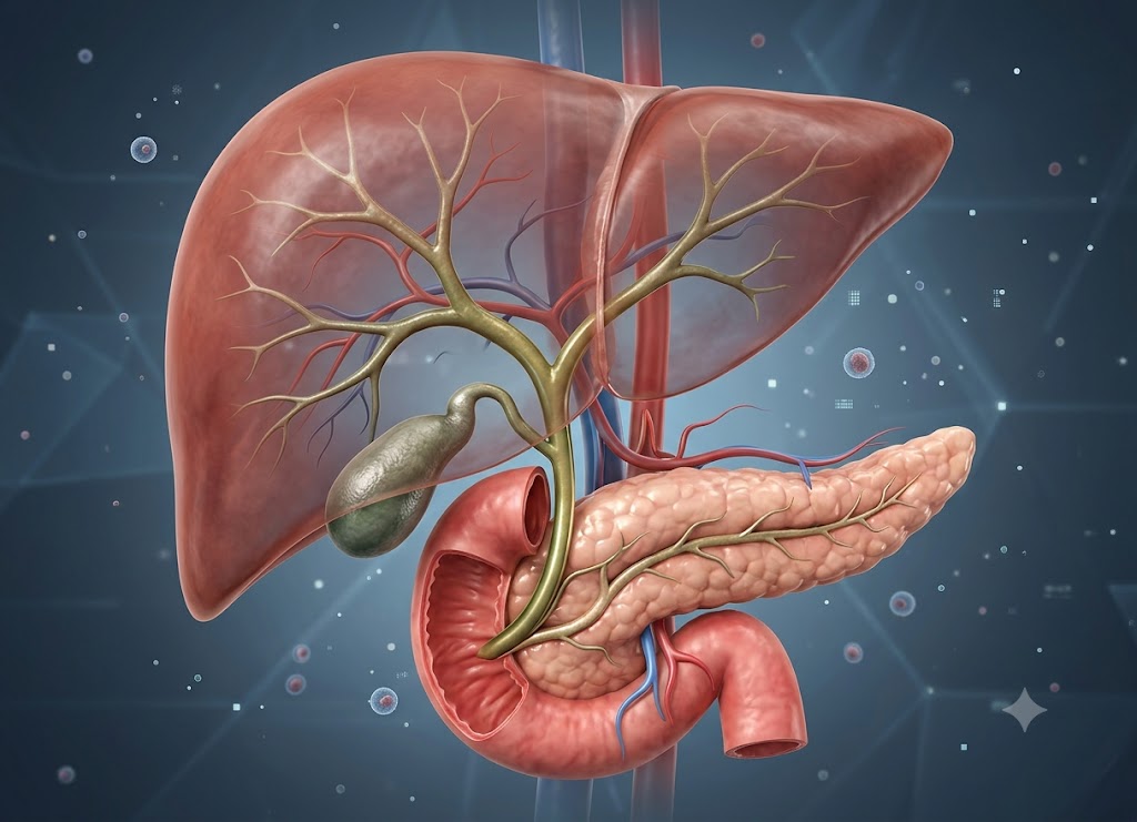
A bile duct blockage occurs when bile can’t flow the way it should from the liver and gallbladder into the small intestine. Bile is a fluid that helps the body break down fat, and it normally moves through a network of small tubes called bile ducts. When one of these tubes gets narrowed or blocked, bile backs up into the liver or gallbladder, leading to pain, yellowing of the skin, itching, infection, or digestive trouble. Gallstones are the most common trigger, but tumors, swelling, scar tissue, injury, parasites, and pancreas problems can also be behind it.
Knowing the warning signs matters, because they shouldn’t be brushed off. Pain in the upper right belly, yellow skin or eyes, dark urine, pale stools, fever, chills, nausea, vomiting, and severe itching can all signal a bile flow problem. How it’s treated depends on the cause options range from medication to endoscopic procedures to surgery. Below, we break down the causes, symptoms, and treatment paths for a blocked bile duct.
Anatomical Architecture: What is a Bile Duct?
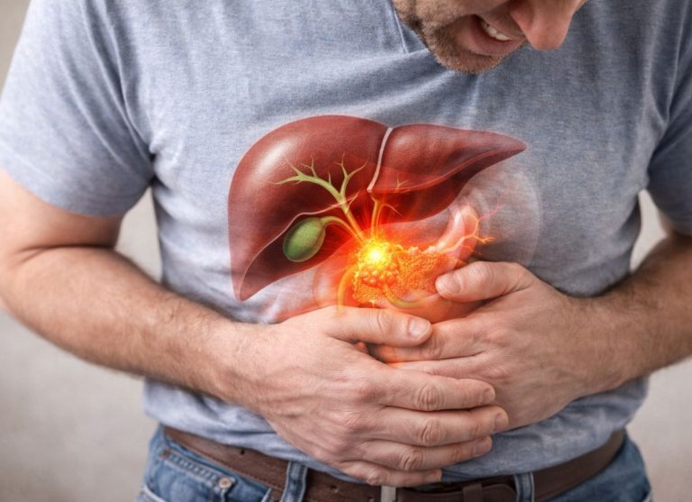
So, what exactly is a bile duct? Think of it as a tube-shaped passage that’s part of a larger network called the biliary tree essentially the body’s internal plumbing for bile.
Your bile ducts fall into two groups: the ones tucked inside the liver (intrahepatic) and the ones outside it (extrahepatic). The intrahepatic ducts gather bile straight from liver cells and feed into two bigger channels the right and left hepatic ducts.
These two ducts leave the liver and join to form the common hepatic duct. A branch duct, the cystic duct, splits off from there and connects to the gallbladder, letting bile flow in for storage between meals.
The common hepatic duct then merges with the cystic duct to create the main pipeline the common bile duct. This duct runs down behind the stomach, cuts through part of the pancreas, and finally empties into the duodenum, the first stretch of the small intestine.
Physiological Role of the Biliary System
The biliary system’s job is to make, concentrate, store, and deliver bile a yellowish-green fluid that’s key for digesting fat and clearing waste. The liver churns out roughly 500 to 1,000 milliliters of bile daily, nonstop.
Bile is a mix of water, cholesterol, fats, electrolytes, and bilirubin (a waste pigment). It also carries bile salts, which act like a natural detergent breaking big fat blobs from food into tiny droplets so pancreatic enzymes can digest them more easily.
Between meals, a valve at the top of the small intestine stays shut. Since bile keeps flowing from the liver anyway, it gets redirected back through the cystic duct into the gallbladder for storage.
Inside the gallbladder, the bile gets concentrated up to ten times stronger as water is drawn out through the organ’s walls. Once a fatty meal reaches the duodenum, the gut releases a hormone called cholecystokinin (CCK).
This hormone tells the gallbladder to squeeze and the intestinal valve to open. Together, this pushes the concentrated bile down the common bile duct and straight into the digesting food.
Pathophysiology and Symptoms of a Blocked Bile Duct
A biliary obstruction happens when something inside the duct system stops bile from moving gallstones that have shifted position, scar tissue narrowing the path, tumors in the pancreas, or even parasites are common culprits.
Once the common bile duct is blocked, bile piles up behind the obstruction, and pressure builds. That pressure pushes bile components out of the liver’s channels and into the bloodstream a condition called cholestasis.
What happens when bile backs up:
- Bile spilling into the blood → yellow eyes and skin, dark urine (kidneys filtering out excess pigment), and itchy skin (bile salts irritating nerves)
- No bile reaching the intestines → fat isn’t broken down properly (greasy stools), the body loses fat-soluble vitamins (A, D, E, K), and stools lose their brown color and turn pale
Here’s what that backup looks like in the body:
Yellow skin and eyes (jaundice). Bilirubin, a yellow pigment left over from old red blood cells, is normally cleared out through bile. When it can’t escape, it builds up in the blood and stains the skin and eyes yellow.
Dark, tea-colored urine. With bilirubin rising in the blood, the kidneys try to flush it out, turning urine a dark brown or cola color.
Pale, clay-colored stools. Normally, gut bacteria turn bilirubin into a brown pigment called stercobilin, which colors stool. Without bile reaching the intestines, that pigment is missing, and stools turn pale or grey.
Fatty, floating stools (steatorrhea). Without bile salts to break down fat, the intestines can’t absorb it well. This leads to greasy, smelly, floating stools and often noticeable weight loss.
Intense itching (pruritus). Bile salts settle into the skin and irritate nerve endings, causing itching that’s hard to relieve with regular creams and often gets worse at night.
If it’s left untreated, stagnant bile becomes a breeding ground for bacteria, which can trigger a dangerous infection called ascending cholangitis, or slowly damage the liver until it scars permanently (cirrhosis) and eventually fails.
The primary causes of Biliary Obstruction
There’s a wide range of reasons a bile duct can get blocked. Doctors sort these causes by where the blockage sits inside the duct (intrinsic) or pressing on it from outside (extrinsic) and by whether it’s non-cancerous (benign) or cancerous (malignant). Because the cause shapes the treatment plan and outlook, figuring out exactly what’s behind the blockage is an essential first step.
Some causes are common and easy to fix, while others are more serious. Sorting causes this way helps doctors narrow down what’s going on and plan the right treatment and the benign-vs-malignant split especially affects how aggressive that treatment needs to be.
Structural vs. Compressive: Intrinsic and Extrinsic Blockages
One way to understand a bile duct blockage is by asking where the obstacle actually sits inside the duct wall or pressing on it from outside. This split changes how a doctor approaches clearing it.
Blockages from inside (intrinsic)
These form directly inside the duct’s inner channel or wall. The most frequent cause here is a gallstone that has migrated into the duct.
Other examples include cholangiocarcinoma a cancer that grows from the cells lining the duct itself as well as rarer issues like blood clots after a liver biopsy, benign growths, or roundworm parasites that end up in the ducts.
Blockages from outside (extrinsic)
These happen when something outside the biliary tree a growth or swelling presses hard enough on the duct to squeeze it shut. The most typical case is a tumor in the head of the pancreas, which sits right where the common bile duct passes through.
Other outside pressures include a pancreatic pseudocyst (a fluid pocket from severe pancreatitis) or swollen lymph nodes near the liver from spreading cancer. There’s also a rare condition called Mirizzi syndrome, where a large gallstone gets stuck in the cystic duct and flattens the nearby hepatic duct.
Pathological Nature: Benign and Malignant Causes
The second way to sort causes is by whether they’re cancerous or not a distinction that matters a lot for outlook and treatment intensity.
Non-cancerous (benign) causes
These are serious but don’t involve cancer cells:
Gallstones stuck in the duct (choledocholithiasis). Hardened deposits that form in the gallbladder can slip out and get lodged in the common bile duct, causing sudden pain and a risk of infection.
Scarring from past surgery. Scar tissue can narrow the duct after procedures like gallbladder removal.
Autoimmune or inflammatory scarring. Primary Sclerosing Cholangitis (PSC) is a long-term autoimmune condition that creates bands of scar tissue throughout the bile ducts. Chronic pancreatitis can do something similar by wrapping the duct in thick tissue.
Birth defects. Choledochal cysts are duct-wall bulges present from birth that let bile pool and stones form.
Cancerous (malignant) causes
These need more complex care and often cause jaundice that builds up slowly and painlessly a red flag doctors watch for:
Pancreatic cancer. Tumors in the head of the pancreas are a leading cause of blockages from outside pressure, simply because of how close the pancreas sits to the duct.
Cholangiocarcinoma. This cancer grows inside the duct itself, gradually thickening the wall until it closes off completely.
Ampullary and gallbladder cancers. Tumors can form at the point where the bile duct meets the small intestine, or advanced gallbladder cancer can spread into nearby ducts.
Cancer spreading from elsewhere. Cancers starting in the colon, breast, or stomach can spread to lymph nodes near the liver and squeeze the ducts shut.
Comparative Matrix of Primary Causes
| Cause Type | Condition | How It Blocks the Duct | Typical Symptoms |
|---|---|---|---|
| Inside / Non-cancerous | Gallstone in the duct | A stone gets wedged in the common duct | Sudden right-side pain, sudden jaundice, fever that comes and goes |
| Outside / Cancerous | Pancreatic cancer | Tumor presses on the duct from outside | Slow, painless jaundice with major weight loss |
| Inside / Non-cancerous | Surgical scarring | Scar tissue narrows the duct after surgery | Jaundice and abnormal liver tests weeks to months after surgery |
| Inside / Cancerous | Cholangiocarcinoma | Cancer grows inside the duct lining | Ongoing jaundice, dark urine, pale stools, itching |
| Inside / Non-cancerous | Primary Sclerosing Cholangitis | Autoimmune scarring bands form in the ducts | Fatigue, intense itching, “beaded” pattern on imaging |
Key signs of Biliary Obstruction
The main signs of a bile duct blockage are jaundice (yellow skin and eyes), dark urine, pale stools, and itching all caused by bilirubin and bile salts building up in the body instead of draining out normally.
When bile can’t flow from the liver to the intestine, its components end up in the bloodstream instead of leaving the body. Seeing this pattern of symptoms together is a strong signal that something’s wrong with the biliary system and needs prompt medical attention. How severe and how these signs combine can also hint at where the blockage is and what’s causing it.
The Classic Symptom Quartet
Because the bile duct plays such a central role in clearing waste, a blockage sets off a chain reaction throughout the body. Once bile flow stalls, it leaks into the bloodstream and settles into tissues producing four classic warning signs.
Yellow skin and eyes (jaundice). This is the most noticeable sign. It happens because bilirubin a pigment from broken-down red blood cells can’t be flushed out through bile, so it builds up in the blood and tints the skin and eye whites yellow.
Dark urine. Often the first thing people notice, sometimes before their skin even turns yellow. Since bilirubin dissolves in water, the kidneys try to filter it out, turning urine dark brown or cola-colored.
Pale or clay-colored stools. As urine darkens, stool loses its normal brown color. That color usually comes from stercobilin, made by gut bacteria processing bile pigments but without bile reaching the gut, that process stops, and stool turns pale or grey. Fat also goes undigested, making stools greasy and foul-smelling, and they may float.
Widespread itching. Many people develop intense itching that can be hard to live with. It’s thought to come from bile salts settling into the skin and irritating nerve endings often worse at night, and it doesn’t respond well to regular anti-itch creams.
Abdominal Pain Dynamics and Biliary Colic
Why does one blockage cause severe pain while another causes none at all? It comes down to how quickly the blockage develops.
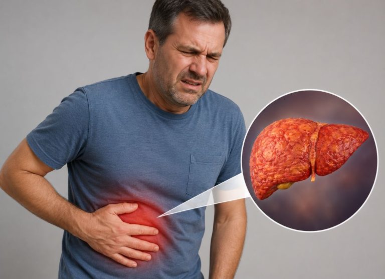
When a gallstone suddenly lodges in the main duct, it triggers sharp, intense pain called biliary colic felt in the upper right belly, just under the ribs, and often spreading to the back, between the shoulder blades, or up to the right shoulder.
This pain tends to come in strong waves lasting hours, often triggered by a fatty meal since fat prompts the gallbladder to contract hard against the stuck stone, spiking pressure and pain. It’s one of the most recognizable signs of a gallstone-related blockage.
The Ominous Nature of Painless Jaundice
No pain doesn’t mean no danger. When a blockage builds slowly from a tightening scar or a growing tumor, for example the picture looks completely different. Since the tumor grows gradually, the ducts have time to stretch and adjust, so the sudden pain trigger never fires.
Watch for this: Gradual jaundice, dark urine, and pale stools showing up with no pain at all is called “painless jaundice.” Combined with unexplained weight loss and appetite loss, it’s a classic red flag for cancer often pancreatic or gallbladder cancer.
Because of this, understanding how a blockage develops over time quickly and painfully, or slowly and silently helps doctors catch dangerous, progressive disease before it causes lasting liver damage.
How is Biliary Obstruction treated?
Treating a bile duct blockage starts with relieving the pressure and restoring bile flow usually through an endoscopic or radiologic procedure followed by treatment aimed at the actual cause, which might mean surgery or other therapy.
The Multi-Tiered Therapeutic Goals
When someone shows the classic signs of a blocked duct, treatment follows a three-part plan. Because a full blockage can overload the liver with bile, there’s a real risk of tissue damage and serious infection.
The three priorities:
- Relieve the pressure fast reduce backup, clear jaundice, prevent sepsis
- Fix the root cause remove stones, treat tumors, widen strictures
- Manage complications antibiotics if needed, vitamin support, monitor blood clotting
Restoring flow quickly
The first priority is decompressing the bile ducts to get flow moving again. This clears toxic bilirubin from the blood, easing jaundice, itching, and pain.
Just as important, clearing the stagnant bile prevents ascending cholangitis a fast-moving infection that can turn into sepsis. Doctors typically do this with minimally invasive procedures, using plastic or metal mesh stents to hold the duct open.
Treating what caused it
Once the patient is stable and bile is flowing again, doctors turn to the underlying problem, so the blockage doesn’t come back:
Gallstones are removed directly, and the gallbladder is often taken out afterward to prevent new stones.
Cancerous tumors may require major surgery like the Whipple procedure for pancreatic cancer sometimes paired with chemotherapy.
Benign strictures are treated with repeated stretching (dilation) or a surgical bypass to reroute bile flow permanently.
Managing the aftermath
The third goal is addressing the effects of prolonged bile backup. Without bile reaching the gut, the body can’t absorb vitamins A, D, E, and K.
Left unchecked, low vitamin K can affect blood clotting, and low vitamin D can weaken bones over time. Doctors typically manage this with vitamin supplements and regular liver monitoring to catch scarring early.
Advanced Endoscopic and Percutaneous Interventions
Doctors have non-surgical ways to clear blockages from the inside, without needing a large incision.
Endoscopic Retrograde Cholangiopancreatography (ERCP)
This is the go-to non-surgical procedure. A doctor threads a flexible scope down the throat, through the stomach, and into the small intestine, right where the bile duct opens.
From there, they inject contrast dye into the biliary tree and use live X-ray to map out the blockage. A small cut is made in the exit valve to widen the opening.
Through this wider opening, doctors can pull out stones with tiny baskets, take biopsies, or place a stent to bypass a tumor. ERCP combines diagnosis and treatment in one visit, making it a lower-risk option especially for patients too weak for major surgery.
Percutaneous Transhepatic Cholangiography (PTC)
If a patient’s anatomy has been altered by past surgery, or a tumor completely blocks the path an endoscope would take, ERCP might not work. In these cases, radiologists use PTC instead.
Guided by ultrasound, a thin needle is passed through the skin, through the liver, and into an enlarged bile duct inside the liver. From there, doctors can place an external drainage bag or push a stent through the blockage from above.
Surgical Strategies: Curative Resection and Rerouting
While stents provide quick relief, surgery offers a lasting fix for more complex structural problems and cancers.
Removing the gallbladder
For recurring gallstone problems, laparoscopic gallbladder removal (cholecystectomy) eliminates the source entirely, stopping new stones from forming. If a stone is too large to pull out endoscopically, surgeons may open the duct directly to remove it.
Major cancer surgery
For cancers in the pancreas or gallbladder, surgery may be the only real chance at a cure. For pancreatic cancer, that often means a Whipple procedure removing the head of the pancreas, part of the intestine, the gallbladder, and the bile duct, then reconnecting the digestive tract.
Surgical bypass
When a tumor can’t be safely removed, or scarring is too extensive to stretch open, surgeons can reroute bile flow entirely:
Choledochojejunostomy connects the bile duct, above the blockage, directly to the middle part of the small intestine.
Hepaticojejunostomy is used for blockages higher up, near the liver bypassing the main duct and connecting straight to the intestine.
Both create a new path for bile to flow, bypassing the blockage and resolving symptoms for good.
What are the diagnostic processes and long-term considerations for Biliary Obstruction?
Diagnosing a bile duct blockage combines blood tests, imaging, and sometimes procedures. Long-term care focuses on treating the root cause, preventing it from happening again, and managing any lasting effects like liver damage.
Recovery also depends on lifestyle changes alongside medical treatment understanding both the diagnostic path and the day-to-day adjustments matters for staying healthy afterward.
Cascading Diagnostic Framework
Doctors typically follow a step-by-step process starting simple and moving to more detailed imaging and, if needed, treatment.
Blood tests first
A liver function panel is usually the first step. A blockage typically shows up as:
High bilirubin. A sharp rise shows the liver is processing waste but can’t get it out of the body, so it spills into the blood.
High ALP and GGT levels. These enzymes live in the cells lining the bile ducts. When the ducts are under pressure or damaged, the enzymes leak into the bloodstream.
Imaging next
Once blood work suggests a blockage, doctors use imaging to pinpoint it:
Ultrasound is usually first it’s great at spotting gallstones and measuring how wide the ducts are. A dilated duct is a clear sign something’s blocking flow downstream.
CT scan gives detailed cross-sectional images and is especially good at catching outside causes, like a pancreatic tumor or swollen lymph nodes.
MRCP (a specialized MRI) creates a detailed map of the entire biliary tree without needing dye or radiation making it the top choice for mapping complex strictures or stones before treatment.
Treatment during diagnosis
If imaging shows a blockage that needs physical removal, doctors turn to ERCP which lets them see the duct on X-ray and immediately treat it, whether that means pulling out stones, taking a biopsy, or placing a stent.
Pathological Complications of Delayed Treatment
Putting off treatment can turn a manageable blockage into a medical emergency. Trapped, stagnant bile creates pressure and a breeding ground for problems.
Serious infection (sepsis). Stagnant bile lets bacteria multiply, causing ascending cholangitis marked by fever, jaundice, and belly pain (known as Charcot’s Triad). Left untreated, bacteria can spread into the bloodstream, causing sepsis, organ failure, or death.
Liver scarring (secondary biliary cirrhosis). Months of untreated backup irritates the liver, causing it to replace healthy tissue with scar tissue. Over time, this damages liver function permanently and may eventually require a transplant.
Pancreatitis. If a stone or tumor blocks the shared exit point where the bile duct and pancreatic duct meet, digestive enzymes back up into the pancreas and become active too early causing painful, damaging inflammation.
Differential Pathology: Biliary Obstruction vs. Cholecystitis
Biliary obstruction and cholecystitis share some symptoms, like upper right belly pain, but they’re different conditions needing different treatment.
The key difference is location. A biliary obstruction can happen anywhere along the bile duct system from tiny ducts inside the liver to the main duct and can be caused by stones, scarring, or tumors.
Cholecystitis, on the other hand, is inflammation of the gallbladder itself, usually from a stone stuck in the cystic duct (the side branch leading to the gallbladder). This traps bile inside and stretches the gallbladder, triggering inflammation.
How they differ:
Symptoms. Cholecystitis causes a steady ache and fever. A duct obstruction causes wave-like pain along with jaundice, dark urine, and pale stools.
Treatment. Cholecystitis is usually fixed by removing the gallbladder. A duct blockage is treated by clearing the duct itself often with ERCP while the gallbladder may be left alone during the initial emergency.
Post-Obstructive Recovery and Lifestyle Adaptation
After treatment clears a blockage, certain diet and lifestyle changes help the liver heal and lower the risk of another blockage.
[Low-Fat Nutrition Plan] ──► [Strict Alcohol Abstinence] ──► [Gradual Weight Management]Eating light on fat
A low-fat diet is central to recovery. Since bile’s main job is breaking down fat, overloading a healing biliary system with greasy or fried food can trigger painful spasms.
Lean proteins like chicken, baked fish, and lentils are good choices, along with whole grains, fruit, and vegetables. Healthy fats like those in avocado or walnuts are fine in small amounts.
Protecting the liver
Cutting out alcohol. Since the liver filters alcohol, a liver that’s already under stress from a blockage needs a break from added toxins to heal properly.
Staying hydrated. Drinking enough water keeps bile flowing smoothly and helps prevent sludge or crystals from forming again.
Managing weight carefully
Keeping a stable, healthy weight helps prevent future gallstones, since excess weight is a major driver of cholesterol-based stones. Regular, moderate exercise paired with balanced eating is the safest approach.
Importantly, weight loss should be gradual. Crash dieting can actually backfire it makes the liver release extra cholesterol into bile and slows gallbladder contractions, which raises the risk of new stones forming.
Conclusion
A blocked bile duct can turn serious quickly, since trapped bile can lead to infection, liver stress, gallbladder inflammation, or worsening jaundice. Gallstones are the most common cause, but tumors, scarring, inflammation, injury, and other duct conditions can also be responsible. Warning signs like severe belly pain, fever, chills, yellow skin or eyes, dark urine, pale stools, vomiting, or confusion call for prompt medical care. If a blockage is suspected, doctors typically rely on blood tests, ultrasound, CT, MRI, or ERCP to find the cause and get bile flowing again.
Frequently Asked Questions
1. What is a bile duct blockage?
It means bile can’t move normally through the bile ducts. Since bile helps digest fat, when it can’t drain properly, it backs up into the liver, gallbladder, or ducts leading to pain, jaundice, itching, infection, or digestive issues.
2. What causes a bile duct blockage?
Gallstones are the most common cause. Others include duct narrowing, inflammation, tumors, pancreatic disease, surgical injury, parasites, or scarring. Some autoimmune or chronic duct conditions can also interfere with bile flow. Identifying the exact cause matters, since it determines treatment.
3. What are the symptoms of a bile duct blockage?
Common symptoms include upper right belly pain, yellow skin or eyes, dark urine, pale stools, nausea, vomiting, fever, chills, and itching. Pain can spread to the back or right shoulder. If infection sets in, symptoms can worsen quickly and need urgent care.
4. How is a bile duct blockage diagnosed?
Doctors typically start with blood tests to check bilirubin and liver enzymes, then move to imaging like ultrasound, CT, MRI, or MRCP. ERCP can be used to both diagnose and treat blockages, especially when stones need removal.
5. How is a bile duct blockage treated?
Treatment depends on the cause, location, and severity. Gallstones are often removed endoscopically, sometimes followed by gallbladder removal. Narrowed ducts may need stents or dilation, while tumors or serious inflammation require specialist care. Infections may need antibiotics along with urgent drainage.