8 Warning Signs of Vascular EDS Complications
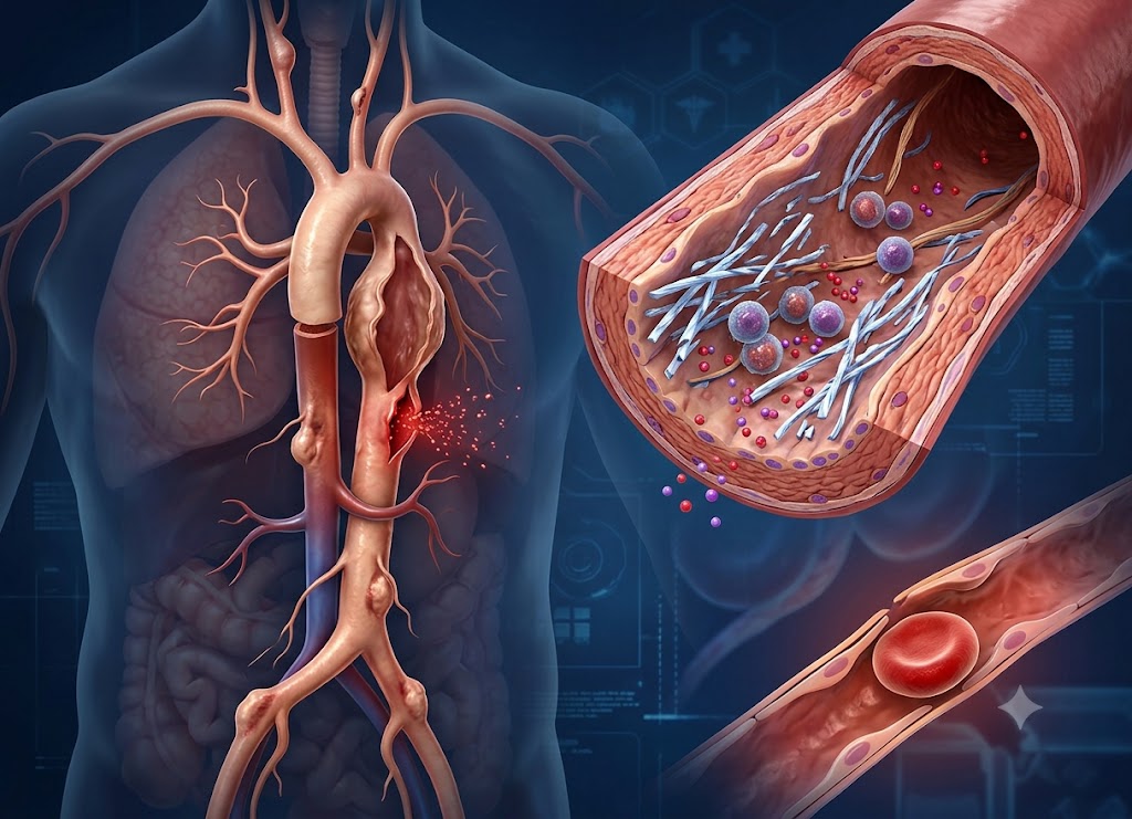
Vascular EDS is a rare and severe subtype of Ehlers-Danlos syndrome that weakens connective tissue in blood vessels and internal organs. It’s typically caused by mutations in the COL3A1 gene, which is responsible for producing type III collagen a protein that reinforces arteries, intestines, the uterus, skin, and other tissues. Because these structures become fragile, people with vascular EDS face elevated risks of arterial tears, organ rupture, bowel perforation, lung collapse, and serious internal bleeding. Complications can strike suddenly, even in individuals who seemed perfectly healthy moments before.
Learning to spot the warning signs of a vascular EDS complication can mean the difference between a fast emergency response and a delayed one. Sudden chest pain, intense abdominal pain, unexplained fainting, breathing difficulty, stroke-like symptoms, a severe headache, unusual swelling, or sharp pain in the back, neck, or limbs are all red flags that demand immediate attention. Other indicators include easy bruising, thin see-through skin, prominent veins, and a family history of sudden unexplained death or arterial rupture. Below, we walk through eight critical warning signs of vascular EDS complications and explain why acting fast can save a life.
What is Vascular EDS and Why Are Its Complications Life-Threatening?
Vascular Ehlers-Danlos Syndrome (vEDS) is a rare, autosomal dominant connective tissue disorder that mainly disrupts type III collagen, leaving blood vessels and hollow organs dangerously fragile which is exactly why its complications turn life-threatening so fast. To understand why this condition is so serious, it helps to look at both its genetic origin and the essential job type III collagen performs throughout the body.
The Genetic Roots of Vascular EDS
Understanding vascular EDS starts with its genetic foundation. It’s a rare, dominantly inherited condition that falls under the wider umbrella of Ehlers-Danlos syndromes.
Where other EDS subtypes mostly cause loose joints or overly stretchy skin, vascular EDS zeroes in on the circulatory system and internal organs. The culprit is a mutation in the COL3A1 gene, which provides the blueprint for type III collagen a strong, fibrous protein that forms the structural backbone of tissues that need to stretch and flex.
Normally, type III collagen lets blood vessels withstand the constant pressure of pumping blood and allows the intestines to expand during digestion. But in someone with vascular EDS, the mutation triggers what’s called a “dominant negative” effect.
In practical terms, the body manufactures malformed collagen strands that tangle with and weaken the healthy collagen around them. The result: thinning artery walls, brittle organ tissue, and translucent skin a combination that can lead to sudden structural failure.
Why Tissue Fragility Creates Medical Emergencies
Because defective type III collagen leaves the body’s internal “scaffolding” so weak, complications tend to arrive as abrupt, severe emergencies. Major arteries and hollow organs can tear or burst spontaneously under everyday pressure no accident or injury required.
                  [COL3A1 Gene Mutation]
                             │
                             ▼
               [Malformed Type III Collagen]
                             │
     ┌───────────────────────┼───────────────────────┐
     ▼                       ▼                       ▼
[Arterial Dissection]   [Hollow Organ Rupture]   [Uterine Fragility]
 ├── Inner wall tear     ├── Spontaneous bowel tear ├── Rupture during labor
 ├── Wall layers split   ├── Fecal peritonitis      ├── Severe pelvic bleeding
 └── Fatal bleeding      └── Widespread sepsis      └── High maternal risk
The main structural dangers include:
Sudden Arterial Tearing and Rupture: Weak artery walls can’t reliably handle normal blood pressure, so the inner lining may tear without warning. This is an arterial dissection blood pushes between the vessel’s layers, and the tear can progress to a full rupture with massive internal bleeding. These tears can occur anywhere but are especially dangerous in the aorta and the major vessels feeding the brain, abdomen, and limbs.
Bowel Wall Rupture: The colon relies heavily on collagen support to withstand the mechanical strain of digestion. Without it, the colon can tear on its own, releasing intestinal contents into the abdominal cavity. This triggers peritonitis, an infection that can escalate quickly into life-threatening sepsis.
Uterine Rupture in Pregnancy: In pregnant patients, the uterine wall lacks the structural resilience to withstand labor. The strain of childbirth can cause the uterus to tear, leading to severe pelvic hemorrhage that endangers both mother and baby.
Identifying the Clinical Signs and Symptoms of Vascular EDS
Spotting the physical features and early symptoms of vascular EDS is essential for catching the condition before a major internal crisis develops. The visible signs differ noticeably from other EDS subtypes and are often detectable during a routine exam.
Many patients share a recognizable facial pattern: large, wide-set eyes, a narrow nose, thin lips, sunken cheeks, and small earlobes. The skin also offers a telltale clue it’s usually thin and translucent enough that veins are clearly visible on the chest, abdomen, and limbs.
Bruising from minor knocks tends to be frequent and pronounced, and the skin on the hands and feet may appear prematurely aged or wrinkled. Unlike most other Ehlers-Danlos types, joint flexibility in vascular EDS is usually confined to the small joints of the fingers and toes rather than larger joints like knees or elbows.
When these physical traits appear alongside a family history of early strokes, aneurysms, or organ ruptures, genetic testing of the COL3A1 gene should follow promptly, along with the creation of a protective care plan.
8 Urgent Warning Signs of a Vascular EDS Complication
There are eight core warning signs of a vEDS emergency, and they naturally cluster around three underlying events: arterial rupture or dissection, organ rupture, and a handful of other critical events including collapsed lung. Each of these signals a life-threatening process already underway, and each calls for immediate emergency care.
Vascular Red Flags: Signs of Arterial Rupture or Dissection
Because vascular EDS leaves blood vessels so fragile, recognizing an active vascular crisis quickly can be lifesaving. Four specific signs point toward a possible vessel tear or full arterial rupture.
Sudden, Intense, Unrelenting Pain. The most striking sign of a vascular emergency is pain that hits at full intensity almost instantly. Where it’s felt depends on which vessel has failed. A torn aorta, for instance, often produces a tearing or ripping sensation across the chest, back, or abdomen. A torn limb artery causes sharp, localized pain, and the limb may turn cold, pale, and lose its pulse. This kind of abrupt, unyielding pain signals a structural breakdown within the vascular system.
Signs of Hemorrhagic Shock. When an artery bursts internally, rapid blood loss forces the body into hemorrhagic shock as it tries to protect vital organs. Watch for a fast but weak pulse alongside a sudden drop in blood pressure. The skin turns pale, cold, and clammy, breathing becomes quick and shallow, and reduced oxygen to the brain can bring on anxiety, confusion, or fainting.
Stroke-Like Neurological Symptoms. A dissection in the carotid or vertebral arteries can cut off blood flow to the brain and trigger a stroke. The FAST checklist is useful here: facial drooping on one side, weakness or numbness in an arm, and slurred speech. Other warning signs include sudden loss of balance, double vision, or a headache so severe it’s described as the worst of the person’s life.
A Rapidly Growing Hematoma. Easy bruising is common with vascular EDS, but a bruise that swells, becomes painful, and visibly expands under the skin is a different matter entirely. This points to an underlying vessel that has ruptured and is actively bleeding into surrounding tissue. The area will feel firm and noticeably grow within minutes a sign of significant internal bleeding requiring urgent evaluation.
Visceral Red Flags: Signs of Spontaneous Organ Rupture
Beyond the vascular system, defective collagen leaves internal organs prone to tearing under ordinary, everyday strain. Recognizing these signs matters just as much, since they represent some of the most dangerous gastrointestinal and reproductive complications tied to vascular EDS.
                   [Organ Rupture: Two Failure Patterns]
                                   │
     ┌─────────────────────────────┴─────────────────────────────┐
     ▼                                                           ▼
[Peritoneal Inflammation]                              [Internal Hemorrhage]
 ├── Rigid, board-like abdomen                          ├── Visible abdominal swelling
 ├── Extreme tenderness to touch                        ├── Sharp blood pressure drop
 └── Spilled contents trigger sepsis                     └── Blood pooling in the pelvis
Rigid, Board-Like Abdomen. A spontaneous rupture of a hollow organ most often the sigmoid colon, or the uterus during pregnancy causes sudden, intense pain in the lower abdomen or pelvis. As blood or intestinal contents leak into the abdominal cavity, the lining becomes inflamed (peritonitis), and the surrounding muscles tighten into a hard, board-like wall that’s extremely tender to touch. High fever, chills, and persistent vomiting typically follow as infection spreads.
Swelling Abdomen With Shock Symptoms. Rupture of a highly vascular organ like the spleen or a severe uterine tear during labor causes significant hidden bleeding inside the abdomen. Alongside the signs of hemorrhagic shock, the belly will visibly swell as it fills with blood. Sudden abdominal pain paired with a rapidly expanding midsection and shock symptoms points to a critical internal emergency.
Other Critical Emergencies: Lung and Eye Complications
A few vascular EDS complications appear outside the digestive and circulatory systems entirely, with symptoms that call for specialized emergency treatment.
Sudden One-Sided Chest Pain and Breathlessness: Sharp chest pain on one side that worsens with deep breathing, paired with sudden shortness of breath, suggests a spontaneous pneumothorax (collapsed lung). Fragile lung tissue can develop small air blisters that rupture, letting air escape into the chest cavity. The trapped air compresses the lung, and without prompt decompression, breathing and circulation can quickly deteriorate.
Carotid-Cavernous Sinus Fistula. In rare cases, a fragile carotid artery tears into the venous network behind the eye, forming an abnormal connection called a fistula. High-pressure arterial blood then floods the low-pressure eye veins, causing the eye to bulge, turn dark red, and swell, along with a constant pulsing or whooshing sound in the head that matches the heartbeat. This requires urgent neurovascular treatment to avoid permanent vision loss or brain hemorrhage.
What Emergency Actions Should You Take If You Suspect a vEDS Complication?
If you suspect a vEDS complication, the single most important step is to call emergency medical services right away and clearly state the diagnosis of Vascular Ehlers-Danlos Syndrome. Beyond that, having an emergency plan in place ahead of time ensures responders understand the patient’s specific risks, which can meaningfully improve the outcome.
Immediate Response Steps During a Crisis
When a life-threatening vEDS complication is suspected, speed and precision matter enormously. Since the condition causes extreme structural fragility in blood vessels and organs, a patient’s status can worsen within minutes.
[Suspected vEDS Emergency] ──► Call Emergency Services Right Away ──► State the Diagnosis Clearly
                                                                             │
     ┌───────────────────────────────────────────────────────────────────────┴───────────────────────────────────────┐
     ▼                                                                                                               ▼
[What to Tell Dispatch]                                                                                 [Caring for the Patient On-Site]
 ├── Ask for Advanced Life Support (ALS)                                                                  ├── Lay the patient flat on their back
 ├── Say: "Vascular Ehlers-Danlos Syndrome"                                                               ├── Keep them warm
 └── Warn of possible spontaneous rupture                                                                 └── Limit all movement
Key steps to follow:
Call for Help Immediately. Dial your local emergency number without hesitation never attempt to drive the patient to the hospital yourself. A personal car lacks life-support equipment, and any delay from driving can turn catastrophic if shock sets in. An ambulance means trained paramedics can begin stabilizing the patient oxygen, vital monitoring en route to the hospital.
Name the Condition Clearly. Tell the dispatcher and responders exactly what’s happening: “This is a critical emergency. The patient has a diagnosed genetic condition, Vascular Ehlers-Danlos Syndrome (vEDS), and we suspect an arterial or organ rupture.” This wording prompts dispatch to send an Advanced Life Support unit.
Repeat the Diagnosis to Paramedics on Arrival. When the crew arrives, say it again. This protects the patient from procedures that could otherwise cause harm it alerts responders to handle blood pressure cuffs and IV lines gently, and to route the patient to a hospital with vascular and general surgery teams on call.
Keep the Patient Still and Calm. While waiting for help, have the patient lie flat and stay as calm and warm as possible. Reducing stress and movement helps keep heart rate and blood pressure down, easing pressure on fragile vessel walls.
Building a Proactive Emergency Plan
Because serious vascular EDS symptoms can appear suddenly sometimes leaving a patient unable to communicate a pre-built emergency plan is essential to long-term management.
A solid emergency plan should include:
A Visible Medical Alert. Anyone with this condition should wear a medical alert bracelet or necklace at all times, engraved with “Vascular Ehlers-Danlos Syndrome (vEDS)” and “Risk of Spontaneous Arterial/Organ Rupture.”
A Detailed Medical ID Card. Carry a crisis card in your wallet, or save a digital medical ID on your phone, listing your name, confirmed COL3A1 mutation, primary risks, and contact numbers for your vascular specialists.
A Ready-to-Go Hospital Bag. Keep a bag packed near the door with recent imaging scans, a current medication list, copies of your emergency care plan, and spare medical alert cards to hand to ER staff.
Treatment Precautions Emergency Teams Must Know
Part of the emergency card’s job is warning physicians away from standard procedures that are safe for most patients but dangerous for someone with fragile tissue. Unfamiliar emergency teams need to know:
Avoid Invasive Arteriography. Traditional catheter angiograms threading a physical tube through the arteries carry a high risk of puncturing or tearing vessels at the insertion point.
Instead, non-invasive imaging should be used to locate internal bleeding:
CT Angiography (CTA): Maps blood vessels using an IV line in the arm, with no arterial catheter required.
MR Angiography (MRA): Produces detailed vascular images without catheters or radiation.
[Suspected Arterial Tear] ──► Skip Catheter Angiography ──► Use CTA or MRA Instead
Sharing this plan with family, friends, coworkers, and local emergency services helps make sure the people around you can act fast when it counts.
What are the Diagnostic and Management Strategies for Vascular EDS?
Diagnosing and managing vascular Ehlers-Danlos syndrome (vEDS) combines non-invasive imaging, preventive medical therapy, lifestyle adjustments, and genetic testing to confirm the underlying collagen defect and guide ongoing care. These strategies matter not just for responding to emergencies, but for reducing the odds one happens in the first place.
A multidisciplinary care team geneticists, cardiologists, vascular surgeons, and emergency physicians is key to addressing the specific vulnerabilities that come with vEDS. This shifts the focus from reactive crisis response toward proactive surveillance, which meaningfully improves outcomes and quality of life.
Emergency Room Diagnostics: Safe Vascular Imaging
When a patient with suspected vascular EDS symptoms arrives at the ER, choosing the right diagnostic path quickly is critical. Because tissue fragility makes standard invasive tests risky, hospital teams rely almost entirely on non-invasive imaging to spot dissections, ruptures, or aneurysms.
CT Angiography (CTA) is the go-to tool in an acute crisis. It pairs fast X-ray imaging with IV contrast dye to generate a detailed 3D map of the vascular system within seconds, letting physicians quickly pinpoint active bleeding or a torn vessel wall.
MR Angiography (MRA) offers a similar non-invasive option using magnetic fields rather than radiation. It takes longer, making it less suited to unstable emergencies, but it’s the preferred method for routine, lifelong monitoring.
Important Surgical Rule: Traditional catheter arteriography should never be used on anyone with known or suspected vascular EDS. Threading a catheter through fragile arteries risks slicing through thin vessel walls, potentially causing a new dissection or rupture right at the insertion site.
Long-Term Prevention and Management
Managing vascular EDS long-term means combining daily medication, activity limits, and regular imaging to protect fragile tissue.
       [Long-Term Vascular Protection Plan]
                           │
     ┌─────────────────────┼─────────────────────┐
     ▼                     ▼                     ▼
[Medication]            [Activity Limits]      [Regular Imaging]
 ├── Celiprolol (β1)     ├── No contact sports  ├── Yearly head-to-pelvis MRA
 ├── Tight BP control    ├── Avoid heavy lifting ├── Track silent aneurysms
 └── Lower vessel stress └── No straining/Valsalva└── Schedule repairs early
The main goal of drug therapy is reducing strain on thin arterial walls through tight blood pressure control, often using the beta-blocker Celiprolol, shown to reduce arterial tearing.
When Celiprolol isn’t an option, doctors may turn to Carvedilol or ARBs like Losartan to keep blood pressure low and steady.
Lifestyle changes matter just as much. Patients need to avoid contact sports, high-impact activity, and anything that triggers the Valsalva maneuver heavy weightlifting or intense isometric exercise included since these cause sharp blood pressure spikes that fragile vessels can’t tolerate. Regular surveillance imaging, such as an annual head-to-pelvis MRA, helps catch silent aneurysms early so they can be repaired on a planned, elective basis rather than in an emergency.
Comparative Pathology: vEDS vs. Other EDS Variants
Every form of Ehlers-Danlos syndrome stems from a flaw in connective tissue formation, but the clinical picture varies widely depending on which type of collagen is affected.
| EDS Subtype | Genetic/Collagen Defect | Main Vulnerable Systems | Typical Complications |
|---|---|---|---|
| Vascular EDS (vEDS) | COL3A1 mutation affecting Type III Collagen | Circulatory system, GI tract, pregnant uterus | Arterial dissection, colon perforation, uterine rupture during labor |
| Hypermobile EDS (hEDS) | Genetic cause not yet identified | Joints, joint capsules, peripheral nerves | Chronic joint dislocations, instability, chronic pain, POTS |
| Dermatosparaxis EDS (dEDS) | ADAMTS2 mutation affecting collagen processing | Skin structure and integrity | Severe bruising from minor contact, sagging skin, wide atrophic scars |
This comparison shows why it’s risky to confuse vascular EDS symptoms with those of other subtypes. Hypermobile and dermatosparaxis forms bring real daily challenges pain, joint instability, skin fragility but they don’t carry the same risk of sudden arterial or organ failure that defines vascular EDS.
The Gold Standard: Genetic Testing and COL3A1 Analysis
Sequencing the COL3A1 gene remains the gold standard for confirming vascular EDS. Because physical signs can be subtle or overlap with other connective tissue disorders, a physical exam alone isn’t reliable enough. A confirmed mutation in COL3A1 removes the ambiguity and reshapes how a patient’s care moves forward.
A confirmed genetic diagnosis changes care in several key ways:
Starting Preventive Treatment Right Away: A positive result signals it’s time to begin blood pressure medication, set firm activity limits, and establish a lifelong imaging surveillance schedule.
Safer Emergency Care: Documenting the COL3A1 mutation in a patient’s chart warns ERs and surgeons ahead of time, prompting gentler techniques, avoidance of catheter-based procedures, and extra caution during surgery.
Family Planning and Cascade Testing: Since vEDS is inherited in an autosomal dominant pattern, each child of an affected parent has a 50% chance of inheriting the mutation. A confirmed diagnosis opens the door to cascade screening, letting relatives get tested before experiencing a sudden, silent vascular event so preventive care can start early.
Conclusion
Vascular EDS leaves arteries and organs fragile, which is why sudden symptoms should never be brushed off. Severe chest, abdominal, back, or head pain can point to arterial dissection, rupture, bowel perforation, or internal bleeding. Breathlessness, fainting, stroke-like symptoms, coughing up blood, severe swelling, or signs of shock all warrant urgent medical attention. Anyone diagnosed with vascular EDS should have a clear emergency plan in place, carry medical identification, and stay closely connected with specialists who understand this condition.
Read more: How a Cardiac CT Scan Helps Detect Coronary Artery Disease
Frequently Asked Questions
1. What is vascular EDS?
Vascular EDS is a rare subtype of Ehlers-Danlos syndrome that primarily affects blood vessels and hollow organs. It usually stems from a genetic change impacting type III collagen, which can leave arteries, intestines, the uterus, and other tissues more fragile than normal. Because complications can be severe, people with vascular EDS typically need ongoing specialist care.
2. What complications can vascular EDS cause?
Vascular EDS can lead to arterial tears, ruptures, aneurysms, bowel perforation, organ rupture, spontaneous lung collapse, and serious internal bleeding. Some patients also have thin translucent skin, easy bruising, visible veins, and distinctive facial features. In adults, a sudden vascular or organ event is sometimes the first noticeable sign of the condition which is why early diagnosis and emergency awareness matter so much.
3. What warning signs require emergency care?
Seek emergency care immediately for severe chest, abdominal, back, or neck pain, or a sudden headache. Other urgent signs include fainting, difficulty breathing, coughing blood, sudden weakness, confusion, vision changes, or stroke-like symptoms. Severe swelling, unexplained bruising, a rapid heartbeat, clammy skin, or shock symptoms should also be treated as emergencies. Anyone with vascular EDS should inform emergency staff of their diagnosis right away.
4. How is vascular EDS diagnosed?
Diagnosis generally combines clinical evaluation with genetic testing. Providers look for features like thin translucent skin, easy bruising, prior arterial events, bowel rupture, or a family history of the condition. Genetic testing can confirm disease-causing changes in COL3A1 in most cases. Diagnosis is best handled by specialists experienced with inherited connective tissue disorders.
5. How can people with vascular EDS lower their complication risk?
Working closely with specialists, following recommended vascular monitoring, and avoiding high-risk trauma or unnecessary invasive procedures all help reduce risk. Blood pressure control is often central to care, since lower vessel stress reduces strain on fragile arteries. Patients should review medications, surgical risks, pregnancy planning, and emergency preparedness with their care team, and a medical alert card or bracelet can help emergency responders act safely and quickly.