Cholangiocarcinoma: Causes, Symptoms, and Treatment Options
Cholangiocarcinoma is an uncommon but dangerous cancer that starts in the bile ducts the narrow tubes that move bile from the liver into the small intestine. Bile helps the body break down fat and clear out waste. Because tumors in these ducts usually grow slowly and stay silent early on, most people aren’t diagnosed until the disease has already advanced. Knowing what to watch for can help someone get checked out sooner.
A number of things raise the odds of developing this cancer long-standing bile duct inflammation, certain liver conditions, structural abnormalities in the ducts, chronic infections, and simply getting older. Common warning signs include jaundice, itching, dark-colored urine, pale stools, belly pain, sudden weight loss, and tiredness though these can point to other liver or bile duct problems too.
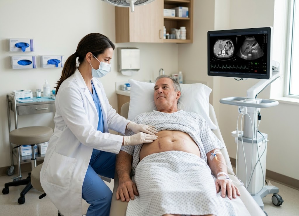
Doctors typically confirm the disease through blood work, scans, endoscopic exams, and a biopsy. Below is a breakdown of what causes cholangiocarcinoma, how it shows up, how it’s diagnosed, and what treatment looks like.
Pathophysiology and Mechanisms of Biliary Obstruction
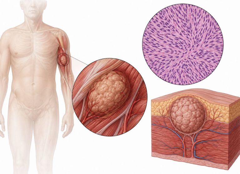
Cholangiocarcinoma also called bile duct cancer develops from cholangiocytes, the cells that form the lining of the biliary tree. This “tree” is really a branching network of tubes that carries bile, the fat-digesting fluid made by the liver, down into the small intestine.
The disease begins when these lining cells start multiplying out of control. That unchecked growth eventually forms a mass that narrows, and then blocks, the inside of the duct.
Tumor forms in the duct → blocks bile flow → bilirubin backs up into the bloodstream → jaundice spreads through the body
Once the duct is blocked, bile can no longer move normally, so its components pile up. Bilirubin the yellow pigment in bile leaks back into the liver and then into the blood, which is what triggers the classic body-wide symptoms. Underneath it all, ongoing inflammation is thought to be the real engine of the disease: repeated irritation and cell damage push the cholangiocytes toward the genetic mutations that eventually turn them cancerous.
Anatomical Classification: The Three Main Types
Doctors sort bile duct cancer into three categories based on exactly where along the biliary tree it forms. This matters a lot, because location determines which surgery is possible, which organs are at risk, and how the outlook looks.
Intrahepatic (iCCA) Perihilar (pCCA) Distal (dCCA)
                     [Cholangiocarcinoma Structural Types]
                                      │
     ┌────────────────────────────────┼────────────────────────────────┐
     ▼                                ▼                                ▼
[Intrahepatic (iCCA)]           [Perihilar (pCCA)]               [Distal (dCCA)]
 ├── Inside the liver tissue     ├── At the main hepatic junction ├── Near the small intestine
 ├── Vague, hidden early signs   ├── Causes early, acute jaundice ├── Pass through pancreatic head
 └── Requires partial hepatectomy└── Highly complex liver surgery└── Requires Whipple procedure
Intrahepatic Cholangiocarcinoma (iCCA)
This form grows in the tiny bile ducts buried deep inside the liver itself, and makes up roughly 1 in 10 cases. Because it’s tucked away inside liver tissue, it rarely causes noticeable symptoms early on it’s often found by accident during a scan done for something else. It can also look a lot like other liver cancers, such as hepatocellular carcinoma. Surgery usually means removing part of the liver (a partial hepatectomy).
Perihilar Cholangiocarcinoma (pCCA)
Sometimes called a Klatskin tumor, this is the most frequent type, making up over half of all cases. It forms at the hilum the point just outside the liver where the left and right hepatic ducts join to form the common hepatic duct. Because everything funnels through this one spot, even a small tumor here can shut down bile drainage from the whole liver.
These tumors sit close to the major blood vessels feeding the liver, which makes surgery technically demanding. Removing one typically means taking out the affected ducts, part of the liver, the gallbladder, and nearby lymph nodes.
Distal Cholangiocarcinoma (dCCA)
This type forms lower down, in the part of the common bile duct nearest the small intestine, and accounts for about 20–30% of cases. It develops where the duct passes through the head of the pancreas. Like perihilar tumors, it tends to cause obstruction symptoms early. Because it sits so close to the pancreas and duodenum, treating it surgically requires a major operation known as a Whipple procedure.
| Type | Location | Presentation | Surgery |
|---|---|---|---|
| Intrahepatic (iCCA) | Inside the liver lobes | Solid mass, upper belly pain, weight loss | Partial liver resection |
| Perihilar (pCCA/Klatskin) | Main duct junction outside the liver | Narrowing of main drainage ducts, early severe jaundice | Extended liver resection + duct reconstruction |
| Distal (dCCA) | Lower common bile duct | Blockage near pancreatic head, early jaundice, digestion issues | Whipple procedure |
Early Symptoms of Cholangiocarcinoma
Most early symptoms trace back to one thing: bile getting trapped by the tumor. That typically shows up as jaundice, dark urine, pale stools, and intense itching. These signs tend to show up sooner and more clearly in perihilar and distal tumors, since they block the main bile pathway directly.
Intrahepatic tumors are different because they grow inside the liver tissue itself, they may not trigger these classic signs until much later, showing up instead as vague belly pain or unexplained weight loss. This quiet early behavior is a big reason the cancer is so often caught late.
Primary Signs of Biliary Obstruction
The first symptoms come straight from the physical blockage. As the duct narrows, bile can’t reach the intestine, so its components build up in the blood and tissues instead.
Jaundice: The hallmark sign yellowing of the skin and the whites of the eyes. It happens because bilirubin can’t get past the blockage and accumulates in the body. Importantly, this jaundice is usually painless, which is a red flag doctors take seriously, since a painful blockage is more often just a gallstone.
                      [Biliary Obstruction Indicators]
                                        │
     ┌──────────────────────────────────┼──────────────────────────────────┐
     ▼                                  ▼                                  ▼
[Scleral Jaundice]               [Excretory Discoloration]        [Intense Pruritus]
 ├── Yellow skin and eye tissue   ├── Dark, tea-colored urine      ├── Bile salts deposit in skin
 ├── Bilirubin buildup in blood   ├── Gray, clay-colored stools    ├── Irritates dermal nerves
 └── Painless onset is a red flag └── Bilirubin routing is altered └── Unrelieved by scratching
Dark urine and pale stools: As blockage worsens, the kidneys try to filter out the excess bilirubin, turning urine dark brown or tea-colored. At the same time, since bilirubin can’t reach the intestine to color the stool normally, stools turn pale, gray, or clay-like.
Intense itching (pruritus): Bile salts that can’t be cleared build up in the skin and irritate nerve endings, causing itching that doesn’t respond to scratching or typical anti-itch creams.
Abdominal Pain and Constitutional Decline
Belly pain: Unlike the sudden, sharp pain of a gallstone attack, this is usually a dull, constant ache in the upper right abdomen, caused by the tumor stretching the liver’s outer capsule or pressing on nerves nearby.
General decline: Weight loss, appetite loss, and fatigue are common, driven by poor fat absorption (since bile isn’t flowing) and the metabolic changes cancer causes throughout the body.
Systemic “B-Symptoms”
Beyond the local blockage, the disease can also trigger a set of whole-body symptoms usually associated with lymphoma but sometimes seen here too driven by inflammatory chemicals (cytokines) the tumor releases.
                       [Systemic Inflammatory Triad]
                                 │
     ┌───────────────────────────┼───────────────────────────┐
     ▼                           ▼                           ▼
[Persistent Fever]          [Drenching Night Sweats]    [Severe Cachexia]
 ├── Low-grade, steady heat  ├── Soaks pajamas and linen ├── Loses >10% weight in 6 months
 ├── Tumor cytokine release  ├── Metabolic deregulation  ├── Muscle and fat wasting
└── No source of infection └── Temperature imbalances └── Systemic metabolic shiftFever: Low-grade and persistent, without any obvious infection. If a blockage becomes severe enough to cause a secondary infection (acute cholangitis), fever can spike suddenly with chills a medical emergency.
Night sweats: Heavy sweating that soaks through clothes and sheets, caused by the tumor disrupting normal temperature regulation.
Significant weight loss (cachexia): Losing more than 10% of body weight within six months, as the cancer forces the body to break down muscle and fat for energy.
Symptom summary:
| Category | What it looks like | Why it happens |
|---|---|---|
| Blocked bile flow | Painless jaundice, dark urine, pale stools, itching | Bilirubin and bile salts backing up into blood and tissue |
| Local tumor effects | Dull, steady upper-right abdominal ache | Tumor stretching the liver capsule or pressing on nerves |
| Whole-body symptoms | Low fevers, night sweats, muscle wasting | Inflammatory chemicals released by the tumor |
Because these signs overlap with more common conditions like gallstones or hepatitis, doctors rely on the full picture. Any combination of painless jaundice, dark urine, or sudden weight loss calls for prompt evaluation.
What Causes Cholangiocarcinoma?
There’s often no single identifiable cause most cases happen sporadically. But the disease is strongly tied to anything that keeps the bile duct lining chronically inflamed and damaged over time.
The Inflammatory Microenvironment: Relentless Cellular Damage
Ongoing inflammation → constant cell irritation → overactive tissue repair → DNA damage → cancer
Long-term inflammation forces the duct-lining cells into a repeating cycle of damage and repair. Over years, this wears down their DNA, and the accumulated errors eventually let some cells escape normal growth controls, turning cancerous.
Primary Risk Factors and Biliary Pathology
                    [Primary Biliary Risk Factors]
                                    │
     ┌──────────────────────────────┼──────────────────────────────┐
     ▼                              ▼                              ▼
[Primary Sclerosing Cholangitis] [Parasitic Liver Flukes]       [Congenital Biliary Cysts]
 ├── Autoimmune duct fibrosis    ├── Ingestion of raw river fish ├── Structural bile stagnation
 ├── Lifetime cancer risk 10-20% ├── Intense mechanical friction  ├── Recurrent toxic infections
 └── Primary Western driver      └── Leading Southeast Asian cause└── Requires preventive removal
Primary Risk Factors and Biliary Pathology :The leading risk factor in Western countries. This autoimmune condition causes progressive scarring and narrowing of the bile ducts, giving affected people a 10–20% lifetime risk of developing cancer hundreds of times higher than average.
Liver flukes: In parts of Southeast Asia, eating raw or undercooked freshwater fish can transmit parasitic flukes (Opisthorchis viverrini or Clonorchis sinensis) that live inside the bile ducts for decades, causing constant mechanical and chemical irritation. This is the leading cause in these regions.
Congenital duct abnormalities: Conditions like choledochal cysts or Caroli’s disease, present from birth, cause parts of the biliary tree to widen abnormally. Bile pools and stagnates there, leading to chronic irritation, infections, and stones which is why doctors often recommend removing these cysts early.
Chronic liver disease and cirrhosis: Long-term damage from hepatitis B or C, heavy alcohol use, or fatty liver disease raises risk, especially for the intrahepatic type, since widespread scarring damages the small internal bile ducts.
Past toxin exposure: A well-known historical example is Thorotrast, a radioactive imaging contrast agent used in the 1930s–50s. Its radiation exposure led to a spike in cases decades later among people who’d received it.
Genetic Syndromes and Inherited Predispositions
Most cases stem from mutations picked up over a lifetime (not inherited), but a small share are linked to genetic syndromes passed down in families:
                 [Inherited Predisposition Syndromes]
                                   │
     ┌─────────────────────────────┼─────────────────────────────┐
     ▼                             ▼                             ▼
[Lynch Syndrome (HNPCC)]      [BAP1 Tumor Syndrome]         [Multiple Papillomatosis]
 ├── Defective DNA repair genes├── Inherited germline mutation ├── Wart-like biliary growths
 ├── High colorectal risk      ├── Risk of skin/eye melanoma ├── High rate of transformation
 └── Elevates cholangiocarcinoma└── Elevates intrahepatic risk └── Rare structural genetic link
- Lynch syndrome (HNPCC): Caused by faulty DNA-repair genes like MLH1 or MSH2 better known for raising colorectal cancer risk, but it raises bile duct cancer risk too.
- BAP1 tumor syndrome: Inherited mutations in the BAP1 gene raise risk for several cancers, including uveal melanoma, mesothelioma, and intrahepatic cholangiocarcinoma.
- Multiple biliary papillomatosis: A rare condition with numerous wart-like growths inside the bile ducts, many of which can turn cancerous over time.
Family History
Most patients have no family history of the disease at all. Still, having a close relative (parent, sibling, or child) who’s been diagnosed slightly raises your own risk likely from a mix of shared environment and subtle inherited traits, which is why doctors ask about family history during evaluation.
Risk factor overview:
| Risk Factor | Where it’s most common | How it drives inflammation |
|---|---|---|
| Primary Sclerosing Cholangitis | Western countries | Autoimmune scarring and duct irritation |
| Liver flukes | Southeast Asia | Parasite movement + toxic byproducts |
| Choledochal cysts | Rare, congenital | Bile pooling → infection and tissue damage |
| Lynch syndrome | Inherited | Breakdown of the body’s DNA-repair system |
Advanced Treatments for Cholangiocarcinoma
Treatment is shaped by the cancer’s stage, location, and genetic profile, and can involve surgery, liver transplant, chemotherapy, radiation, and newer precision options like targeted therapy and immunotherapy.
Surgery
Surgical removal is still the only real shot at a cure but because this cancer is usually caught late and often grows near major blood vessels, only about 20–35% of patients qualify for curative surgery at diagnosis.
Tumor location → choice of surgery → goal of clean (R0) margins
The aim is always an R0 resection removing the tumor with no cancer cells left at the edges. The exact operation depends on location:
- Partial hepatectomy (liver resection): Standard for intrahepatic tumors, and a major part of surgery for perihilar tumors too removing the affected liver segment, plus ducts and lymph nodes for perihilar cases.
- Whipple procedure (pancreaticoduodenectomy): Required for distal tumors, given their closeness to the pancreas involves removing the pancreatic head, duodenum, gallbladder, and lower bile duct, then reconnecting the digestive tract.
- Bile duct resection alone: In rare early cases limited to the middle of the common bile duct, only the damaged segment is removed and reconnected directly to the small intestine.
                        [Surgical Treatment Matrix]
                                     │
     ┌───────────────────────────────┼───────────────────────────────┐
     ▼                               ▼                               ▼
[Partial Hepatectomy]       [Whipple Procedure]             [Liver Transplantation]
 ├── Standard for iCCA & pCCA├── Standard for distal dCCA    ├── Highly selected perihilar cases
 ├── Removes affected liver   ├── Removes pancreatic head/duo ├── Follows strict Mayo protocol
 └── Achieves clean R0 margin └── Reconstructs GI tract flow  └── Aggressive pre-neoadjuvant care
Liver Transplantation
For a carefully selected group with early-stage, unresectable perihilar tumors (under 3 cm, no spread), a liver transplant is sometimes an option, following strict protocols like Mayo Clinic’s. Before transplant, patients go through intensive treatment radiation, internal radiation (brachytherapy), and chemotherapy to shrink cancer cells first. Patients who complete this successfully can see 5-year survival rates of 65–70%.
Targeted Therapy and Immunotherapy
When surgery isn’t an option, treatment shifts to systemic therapies guided by the tumor’s genetic makeup.
Targeted therapy blocks specific mutations driving the cancer’s growth, found through genomic testing more common in intrahepatic tumors:
- FGFR2 fusions (10–15% of intrahepatic cases): treated with drugs like pemigatinib or futibatinib.
- IDH1 mutations (15–20% of intrahepatic cases): treated with ivosidenib.
- Other rarer targets: BRAF V600E mutations (dabrafenib + trametinib) and NTRK fusions (larotrectinib).
Immunotherapy works differently instead of attacking the cancer directly, it helps the immune system see and destroy it, by blocking checkpoint proteins (PD-1/PD-L1) that tumors use to hide from immune cells.
- The TOPAZ-1 trial showed adding the immunotherapy drug durvalumab to standard chemo (gemcitabine + cisplatin) improved survival and cut death risk by 20% compared to chemo alone now a standard first-line approach.
- A small group of patients (1–3%) with highly mutated tumors (MSI-H or dMMR) respond especially well to immunotherapy.
| Treatment | How it works | Best for | Examples |
|---|---|---|---|
| Surgery | Physically removes the tumor | Early, localized disease | |
| Targeted therapy | Blocks specific growth mutations | Advanced intrahepatic tumors with FGFR2/IDH1 changes | Pemigatinib, Ivosidenib |
| Immunotherapy | Helps immune cells attack the tumor | Advanced disease (with chemo) or MSI-H/dMMR tumors | Durvalumab, Pembrolizumab |
The Prognosis and Management Outlook for Cholangiocarcinoma
Outlook varies widely depending on stage at diagnosis, tumor location, and whether it can be fully removed. Good management combines diagnostics, a multidisciplinary treatment team, and supportive/palliative care.
Diagnosis and Staging
Blood panel & markers → imaging scan (MRCP) → biopsy (ERCP) → TNM staging
Blood tests: Elevated bilirubin, ALP, ALT, and AST typically show up with a blocked duct. Doctors also check tumor markers:
- CA 19-9: The main marker used to track this cancer elevated levels are suggestive, though not definitive on their own since benign conditions can cause spikes too.
- CEA: A less specific marker also used to help confirm and monitor.
Imaging: Ultrasound is often used first to spot dilated ducts or a liver mass. CT and MRI give more detailed views for mapping the tumor and checking for vessel involvement. MRCP is a specialized, non-invasive MRI technique that maps the entire biliary tree without contrast dye.
Biopsy: Usually collected during an ERCP, where a flexible scope is passed through the mouth into the small intestine, a small catheter threaded into the bile duct, dye injected to visualize the blockage under X-ray, and tissue samples collected with brushes or small tools.
TNM staging then classifies the cancer by:
- T (Tumor): size and depth of invasion, including into nearby blood vessels
- N (Node): whether nearby lymph nodes are involved
- M (Metastasis): whether it’s spread to distant organs like lungs, bone, or the abdominal lining
Dynamic Prognostic Determinants
                       [Prognostic Impact Variables]
                                     │
     ┌───────────────────────────────┼───────────────────────────────┐
     ▼                               ▼                               ▼
[Surgical Resectability]        [Tumor Differentiation]         [Performance Status]
 ├── Primary indicator of cure   ├── Evaluates cellular grade    ├── Assesses overall baseline health
 ├── Requires clear R0 margins   ├── Low-grade grows slowly      ├── Dictates systemic tolerance
└── Limited by vascular growth └── High-grade spreads rapidly └── Preserves ongoing liver reserve- Resectability: Whether the tumor can be fully removed with clean margins is the single biggest factor in long-term survival.
- Location: Intrahepatic tumors need liver resection; perihilar tumors are surgically complex due to proximity to major vessels; distal tumors require a Whipple procedure.
- Tumor grade: Well-differentiated (low-grade) cells look closer to normal and grow slower; poorly differentiated (high-grade) cells are more aggressive and spread faster.
- Overall health: How well a patient tolerates surgery or systemic treatment, along with how the tumor responds to targeted therapies, shapes the long-term picture.
Cholangiocarcinoma vs. Hepatocellular Carcinoma (HCC)
Both are primary liver cancers, but they’re quite different diseases:
| Factor | Cholangiocarcinoma | Hepatocellular Carcinoma |
|---|---|---|
| Cell of origin | Cholangiocytes (bile duct lining) | Hepatocytes (main liver cells) |
| Main risk factors | PSC, liver flukes, choledochal cysts | Hepatitis B/C, alcohol-related liver disease |
| Key tumor marker | CA 19-9 | AFP |
| Typical symptoms | Jaundice, itching, pale stools | Abdominal fluid buildup, internal bleeding |
Comprehensive Palliative and Supportive Care Protocols
Palliative care isn’t the same as hospice it runs alongside active treatment from the point of diagnosis, focused on comfort and quality of life.
                    [Palliative Care Integration]
                                  │
     ┌────────────────────────────┼────────────────────────────┐
     ▼                            ▼                            ▼
[Mechanical Interventions]  [Nutritional Counseling]     [Psychosocial Guidance]
 ├── Places metal/plastic stents├── Adjusts enzyme levels    ├── Offers emotional counseling
 ├── Restores normal bile flow ├── Manages cancer cachexia ├── Connects family support groups
└── Relieves severe skin itch └── Protects systemic fitness└── Coordinates advance directives- Biliary stenting: A plastic or metal stent placed via ERCP to reopen a blocked duct, restoring bile flow and relieving jaundice and itching.
- Pain management: Targeted medications combined with nerve blocks (like a celiac plexus block) to manage deep abdominal pain.
- Nutrition support: Dietitians help manage weight loss and muscle wasting through diet adjustments and enzyme replacement therapy.
- Emotional and practical support: Counseling, family support resources, and help with advance care planning to make sure a patient’s wishes are documented and respected.
Conclusion
Cholangiocarcinoma is a rare cancer that’s genuinely hard to catch early, since symptoms often only show up once it’s already progressed. Persistent jaundice, itching, belly pain, sudden weight loss, or changes in urine and stool color shouldn’t be brushed off especially for anyone with a known liver or bile duct condition.
Treatment depends heavily on where the cancer is and how advanced it is, ranging from surgery to chemotherapy, radiation, targeted therapy, immunotherapy, or procedures to relieve blockage. Outcomes vary, but ongoing advances in both diagnosis and treatment keep improving care making early evaluation genuinely important whenever concerning symptoms appear.
Frequently Asked Questions
1. What is cholangiocarcinoma?
It’s a cancer of the bile ducts, the tubes carrying bile from liver to intestine. It can form inside the liver (in the smaller ducts) or outside it (in the larger ducts). It’s rare but often aggressive, and early symptoms can be easy to miss.
2. What are the early symptoms?
Early on, there may be no symptoms at all. As it grows, signs can include jaundice, itching, dark urine, pale stools, upper abdominal pain, fatigue, and unexplained weight loss though these can have other causes too, so evaluation is needed.
3. What causes it?
Often there’s no clear single cause, but chronic inflammation and bile duct injury play a major role. Risk factors include PSC, bile duct cysts, liver flukes in some regions, chronic liver disease, cirrhosis, and certain inherited conditions though many patients have no identifiable risk factor at all.
4. How is it diagnosed?
Through a combination of blood tests, imaging (ultrasound, CT, MRI, MRCP), and biopsy via endoscopic procedures like ERCP, with a lab confirming cancer cells under a microscope.
5. What treatments are available?
Depends on location, stage, and resectability options include surgery, chemotherapy, radiation, targeted therapy, immunotherapy, or procedures to relieve blocked ducts. Many patients get a combination tailored to their case.
6. Can it be cured?
Yes, in some cases particularly when caught early and fully removed surgically. Many cases, though, are found after spreading, when a cure isn’t possible. Even then, modern treatments can slow progression and improve quality of life, with regular follow-up being an important part of ongoing care.