CT Angiogram: 10 Important Facts You Should Know
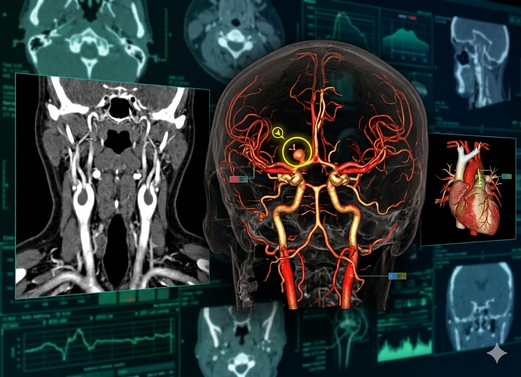
A CT angiogram is a specialized imaging test that combines computed tomography with contrast dye to produce detailed pictures of blood vessels throughout the body. It allows doctors to examine arteries and veins closely and look for problems such as blockages, narrowing, aneurysms, or other abnormalities.
Because CT angiography can produce detailed images quickly, it is often used in emergencies as well as for planned diagnostic evaluations.
Your doctor may recommend a CT angiogram if your symptoms suggest a problem with blood flow. Chest pain, shortness of breath, symptoms of a stroke, or unexplained swelling may all require further evaluation. CT angiograms are also commonly used to examine the coronary arteries, blood vessels in the brain, and circulation in the arms and legs.
Although the test is generally considered safe, it involves radiation exposure and, in many cases, an iodine-based contrast dye. Knowing what to expect can help you prepare and understand why certain precautions are necessary.
Here are 10 important facts to know before having a CT angiogram, including preparation, what happens during the scan, possible risks, and how the results are handled.
What Is a CT Angiogram and Why Might You Need One?
A CT angiogram is a specialized diagnostic imaging test that uses X-ray technology and injectable contrast dye to create detailed images of blood vessels and blood flow.
To understand a CT angiogram, it helps to compare it with a standard CT scan. A regular CT scan can provide detailed pictures of organs, bones, and soft tissues. A CT angiogram is specifically designed to highlight blood vessels, making it easier for doctors to examine their structure and identify abnormalities.
The term “angio” refers to blood vessels.
Doctors may order a CT angiogram when they suspect a problem involving circulation, blood vessel structure, or an organ supplied by a particular vessel. Symptoms such as unexplained chest pain, severe headaches, dizziness, or signs of a stroke may lead to this type of imaging.
What Specific Conditions Does a CT Angiogram Help Diagnose?
CT angiography can help doctors identify many different vascular conditions. These include aneurysms, arterial blockages, blood clots, abnormal blood vessel connections, tumors involving blood vessels, and injuries affecting major vessels.
Because the images provide detailed information about the size, shape, and condition of blood vessels, they can also help doctors plan treatments such as surgery or stent placement.
Some of the main conditions evaluated with CT angiography include:
Aneurysms
An aneurysm is a weakened area in an artery wall that causes the vessel to bulge. A CT angiogram can show the size and shape of an aneurysm and help doctors determine where it is located.
Aneurysms can occur in the aorta, brain arteries, and arteries supplying the legs. Detecting an aneurysm is important because a rupture can cause a life-threatening emergency.
Blockages (Atherosclerosis and Stenosis)
CT angiography can identify narrowed or blocked arteries caused by plaque buildup.
It may be used to evaluate:
- Coronary arteries for signs of heart disease
- Carotid arteries in the neck
- Renal arteries supplying the kidneys
- Peripheral arteries in the legs
The images help doctors determine whether blood flow is being restricted and how severe the narrowing may be.
Blood Clots (Thrombosis and Embolism)
A CT angiogram of the chest is commonly used to detect a pulmonary embolism, which occurs when a blood clot blocks an artery in the lungs.
CT angiography can also be used to evaluate blood vessels for other types of clots and abnormalities that may interfere with normal circulation.
Vessel Malformations and Dissections
CT angiograms can reveal unusual blood vessel formations, including arteriovenous malformations, in which arteries and veins are connected abnormally.
The test can also detect an aortic dissection. This serious condition occurs when a tear develops in the inner layer of the aorta, allowing blood to enter the wall of the artery.
Tumors and Vascular Supply
CT angiography can show the blood vessels supplying a tumor. This information can be especially helpful when surgeons are planning an operation.
Knowing how a tumor is connected to nearby blood vessels can help the medical team anticipate blood flow and plan how to manage bleeding during treatment.
How Does the Iodine-Based Contrast Dye Work?
A CT angiogram with contrast depends on an iodine-based contrast material to make blood vessels easier to see.
The contrast temporarily makes the blood more visible to X-rays. As a result, blood vessels appear brighter on the images, allowing the radiologist to examine them in greater detail.
Think of the body’s blood vessels as a network of roads. A regular CT scan can show the surrounding landscape, including organs and tissues, but it may not clearly outline every road. The contrast dye acts like a highlighter that makes the vascular network stand out.
The contrast is usually injected through an IV placed in your arm. Once it enters the bloodstream, it travels throughout the body.
Iodine absorbs X-rays more effectively than surrounding soft tissues. As the CT scanner captures numerous cross-sectional images, the contrast-filled vessels become clearly visible.
A computer then combines the individual images to create detailed views and three-dimensional reconstructions of the blood vessels. This allows the radiologist to examine the vessels for narrowing, blockages, bulges, abnormal connections, and other changes.
10 Key Preparation Steps for Your CT Angiogram
Preparing for a CT angiogram usually involves following instructions about fasting, medications, medical conditions, and what to bring to your appointment.
These precautions help protect you from possible complications related to contrast dye and help the imaging team obtain clear, useful images.
Your preparation instructions may include what you can eat and drink, whether certain medications need to be adjusted, and whether blood tests are required beforehand.
It is especially important to tell the medical team about allergies, kidney problems, diabetes, and the possibility of pregnancy. This information helps them determine whether the contrast material and CT scan are appropriate for you and whether additional precautions are needed.
Do You Need to Fast Before the Procedure?
You may be asked to avoid solid food for several hours before a CT angiogram, often around four to six hours. The exact instructions can vary between imaging centers, so always follow the directions given to you.
Fasting can reduce the risk of complications if the contrast causes nausea or vomiting. Having an empty stomach can lower the chance of stomach contents entering the lungs if vomiting occurs while you are lying on the scanner table.
Your facility may provide instructions such as:
No Solid Food: You may be told to avoid meals, snacks, candy, and chewing gum for a specified period before the scan.
Clear Liquids Are Often Permitted: Some facilities allow clear liquids, such as water, black coffee or tea, clear broth, or apple juice, until a certain time before the scan. Dairy products and drinks containing pulp may not be allowed.
Follow the exact fasting and drinking instructions provided by your medical facility because requirements can differ.
Health Information You Must Share with Your Doctor
Tell your doctor and the radiology team about important health information before your scan. This includes previous reactions to contrast dye, allergies, kidney disease, diabetes, and any possibility that you may be pregnant.
This information helps the medical team assess the potential risks and decide whether you need additional precautions or a different imaging test.
Important details to report include:
Disclose All Allergies: Tell the staff if you have previously reacted to contrast dye or other medications. A shellfish allergy is not considered the same as an allergy to iodine contrast, but you should still report all significant allergies and previous reactions. If you have had a previous reaction to contrast material, your doctor may recommend specific precautions or pre-medication.
Report Kidney Disease, Diabetes, and Pregnancy: The kidneys remove contrast material from the body, so kidney function may need to be checked before the scan. Diabetes is also important to report because certain diabetes medications may require special instructions around the time of contrast administration. You should also tell your doctor or technologist if you are pregnant or could be pregnant because CT scans involve ionizing radiation.
What Should You Wear and Bring to Your Appointment?
For your CT angiogram appointment, wear comfortable clothing and avoid clothing with metal whenever possible. You should also bring the documents requested by the imaging facility, such as identification, insurance information, and your doctor’s referral or imaging order.
Metal can interfere with imaging and create artifacts that make the pictures harder to interpret.
- Wear Comfortable, Metal-Free Clothing: Simple clothing without metal zippers, buttons, snaps, or underwire can make the process easier. Depending on the area being scanned, you may be asked to change into a hospital gown.
- Leave Jewelry and Valuables at Home: You may need to remove necklaces, earrings, watches, rings, piercings, and other metal objects before the scan.
- Bring Necessary Documents: Have your photo ID, insurance card, and any referral or order forms required by the imaging center.
Are Pre-Scan Tests Required?
In some cases, a blood test is performed before a CT angiogram to check kidney function. This is particularly important when contrast dye will be used.
Because the kidneys help remove the contrast material from the body, doctors may want to confirm that they are functioning adequately before the scan.
A common test measures creatinine, a waste product normally filtered out by the kidneys. The creatinine result can be used to calculate the estimated glomerular filtration rate, or eGFR, which provides information about kidney function.
If kidney function is significantly reduced, the imaging team may need to modify the plan, delay the scan, or consider an alternative test without contrast.
Kidney function testing may be especially important for people with known kidney disease or other conditions that can affect kidney health.
What Can You Expect During and After the Scan?
A CT angiogram is usually a quick and non-invasive imaging procedure. You will lie on a motorized table that moves through the circular opening of the CT scanner while X-ray images are taken.
If contrast is required, it will be injected through an IV during the scan.
The active scanning portion generally takes only a few minutes, although you may spend more time in the imaging department getting prepared and positioned.
The technologist will explain each step and communicate with you through an intercom. You may also be asked to hold your breath for short periods to help prevent movement from affecting the images.
After the scan, you can usually return to your normal activities unless your medical team gives you different instructions.
What Is the Step-by-Step Process of the Scan Itself?
A CT angiogram generally follows a series of straightforward steps. These include placing the IV, positioning you on the scanner table, injecting contrast, capturing the images, and removing the IV.
Although you may spend 15 to 30 minutes in the scanning area, the actual X-ray exposure is much shorter.
Intravenous Line Insertion
A nurse or technologist will place a small IV catheter into a vein, usually in your arm or hand. This allows the contrast material to be delivered directly into your bloodstream.
You may feel a brief pinch when the IV is inserted, similar to having blood drawn.
Positioning on the Scanner Table
You will lie on a padded table that moves into the center of the CT scanner, called the gantry.
The technologist will position your body carefully so that the area being examined is aligned correctly. Pillows, foam supports, or other positioning aids may be used to help you remain comfortable and still.
Contrast Dye Injection
The IV is connected to an injector that delivers the contrast material at a controlled rate.
As the contrast enters your bloodstream, you may experience a sudden warm or flushing sensation. Some people notice a metallic taste or a brief sensation similar to needing to urinate.
These sensations are common and usually disappear within a short time.
Imaging and Breath-Holding
The technologist controls the scanner from a nearby room but can communicate with you through an intercom.
As the table moves through the scanner, the X-ray equipment rotates around your body to capture the required images.
You may hear instructions asking you to hold your breath for several seconds. Staying still and following these instructions helps produce clearer images and reduces motion-related blurring.
Procedural Completion
Once all the necessary images have been obtained, the table moves out of the scanner.
The technologist removes the IV and places a small bandage over the insertion site.
Immediate Post-Scan Instructions
After a CT angiogram with contrast, you may be encouraged to drink fluids to help your body eliminate the contrast material through the kidneys.
Unless your doctor has placed limits on how much fluid you can drink because of a heart or kidney condition, staying well hydrated after the scan may be recommended.
Most people do not need a special recovery period after a CT angiogram.
Resuming Normal Diet and Activities
Unless you have received different instructions, you can generally return to your usual meals and daily activities after the scan.
Sedation is not normally required for a routine CT angiogram, so there are generally no restrictions on driving unless you were given medication that affects alertness.
Monitoring the IV Access Site
Check the area where the IV was inserted. Mild tenderness or minor discoloration can occur.
Contact your healthcare provider if you develop increasing pain, significant swelling, spreading redness, warmth, or other concerning changes around the IV site.
Receiving Your Imaging Results
The images from your CT angiogram are reviewed by a radiologist. The radiologist prepares a report describing the findings and sends it to the doctor who ordered the scan.
The technologist performing the scan generally cannot provide a diagnosis during the appointment. Your doctor will explain the results and what they mean for your individual situation.
Deeper Insights: Risks, Alternatives, and Results
Understanding a CT angiogram involves more than knowing how the scanner works. It is also useful to understand possible side effects, how the test compares with other vascular imaging methods, and how results are reviewed.
Knowing what to expect can help reduce anxiety and make it easier to discuss concerns with your healthcare team.
Potential Risks and Side Effects
CT angiography is generally considered safe, but it does involve some risks related to contrast material and radiation exposure.
The most common reactions to contrast are mild and temporary. You may notice a warm sensation, flushing, or a metallic taste immediately after the injection.
Some people may experience nausea, headache, itching, or hives. Tell the technologist if you notice any unusual symptoms.
A severe allergic-type reaction to contrast is rare but can cause symptoms such as difficulty breathing or a significant drop in blood pressure. Imaging facilities are equipped to respond to serious reactions if they occur.
Contrast can also affect kidney function in people who already have significant kidney problems. This is one reason kidney function may be checked before the scan.
CT angiography also exposes the body to ionizing radiation. The radiation dose is higher than that of a standard X-ray, although modern CT scanners use techniques designed to limit exposure while still producing useful diagnostic images.
Radiology teams generally follow several safety principles:
- ALARA Principle: Radiation exposure is kept As Low As Reasonably Achievable while still obtaining the images needed for diagnosis.
- Modern Technology: Newer CT scanners use dose-reduction technologies to help limit radiation exposure.
- Justification: CT angiography is ordered when the expected diagnostic benefit is considered important for the patient’s care.
How Does a CT Angiogram Compare to an MRA?
Both CT angiography and Magnetic Resonance Angiography, or MRA, can produce detailed images of blood vessels, but they use different technologies.
A CT angiogram uses X-rays and often requires an iodine-based contrast material. The resulting images can be reconstructed into detailed views of the blood vessels.
An MRA uses a magnetic field and radiofrequency waves instead of X-rays, so it does not involve ionizing radiation.
The choice between the two depends on factors such as the part of the body being examined, the medical question being investigated, the patient’s health, and whether contrast can safely be used.
Effective Ways to Manage Anxiety or Claustrophobia
It is normal to feel nervous about medical imaging, especially if you are uncomfortable in enclosed spaces.
A CT scanner has a large circular opening that you pass through. Unlike some MRI scanners, the CT scanning area is relatively short and open, but the equipment and sounds can still feel unfamiliar.
Several strategies may help.
Direct Communication and Pre-Medication
Tell your doctor about significant anxiety before your appointment. Knowing what will happen can make the procedure feel more predictable.
If your anxiety is severe, your doctor may discuss whether medication is appropriate. If you take a sedative, you will need to follow instructions about transportation and should not drive yourself afterward.
Mindful Breathing Exercises
Slow, controlled breathing can help you relax before and during the scan.
One approach is to breathe in slowly through your nose, pause briefly, and then exhale gradually through your mouth. Repeating this pattern may help reduce physical tension and nervousness.
Visual and Auditory Distraction
Some people find it easier to close their eyes and focus on a calming mental image before entering the scanner.
Depending on the facility, headphones or other audio options may also be available. Listening to calming music can provide a distraction from the sounds of the equipment.
When and How Will You Receive Your Scan Results?
After the CT angiogram is completed, the images are sent to a radiologist for review.
The technologist who operates the CT scanner is responsible for obtaining high-quality images but generally does not interpret them for the patient.
A radiologist, who is a physician trained in medical imaging, reviews the images for abnormalities such as blockages, narrowing, aneurysms, dissections, or other vascular changes.
The final report is then sent to the doctor who ordered the scan.
Standard Turnaround
The time needed to complete and report the scan varies. In many routine cases, the radiology report may be available within one to three business days.
Urgent examinations may be reviewed much more quickly. For example, when a CT angiogram is ordered because of sudden symptoms or suspected pulmonary embolism, the results may be communicated to the medical team promptly.
The Follow-Up Consultation
Your ordering doctor will explain the results and discuss what they mean in relation to your symptoms, medical history, and other test results.
Depending on the findings, your doctor may recommend additional testing, lifestyle changes, monitoring, medication, or another form of treatment.
Accessing Your Records
You may be able to access your imaging report and scan images through a patient portal or another medical records system.
The images themselves can be difficult to interpret without medical training, so it is best to discuss them with your doctor or another qualified healthcare professional rather than trying to interpret individual images on your own.
Conclusion
A CT angiogram is an important diagnostic tool that gives doctors detailed information about blood vessels throughout the body. It can help identify conditions such as blockages, aneurysms, blood clots, dissections, and other vascular abnormalities.
Knowing how to prepare can make the experience easier. You may need to fast, provide information about your medications and medical history, have your kidney function checked, and follow specific instructions from the imaging center.
The scan itself is generally quick and straightforward. Contrast dye may cause a temporary warm sensation or metallic taste, while serious reactions are uncommon. Because CT angiography involves both contrast material and radiation, your healthcare team will consider your individual risks before proceeding.
If your doctor has recommended a CT angiogram, the test is intended to provide detailed information that can help guide diagnosis and treatment. Following the preparation instructions and discussing any health concerns with your medical team can help ensure the procedure is performed as safely and effectively as possible.