7 Signs of Paget’s Disease of the Breast People Often Mistake for a Skin Problem
Paget’s disease of the breast is an uncommon form of breast cancer that shows up on the nipple and, in most cases, spreads to the areola around it. The tricky part is that its early warning signs look almost identical to everyday skin complaints eczema, dermatitis, irritation, or a rash that just won’t quit. Because of this, people often notice itching, redness, flaking, crusting, or discharge and write it off as a surface-level skin issue, especially if the symptoms seem to fade in and out at first.
Recognizing this condition matters because it’s frequently a signal of breast cancer developing underneath the surface, in the same breast. Unlike ordinary skin irritation, the changes almost always show up in just one nipple and tend to get worse rather than better with time. Catching these changes early can mean getting evaluated and tested sooner rather than later. Below are seven signs of Paget’s disease of the breast that are commonly confused for ordinary skin conditions.
Defining Paget’s Disease of the Breast
Paget’s disease of the breast is a rare, specific type of cancer that develops in the skin of the nipple and the areola surrounding it. It’s an important condition to recognize precisely because it almost never appears on its own it’s usually a visible clue pointing to a cancer growing somewhere deeper in the breast tissue.
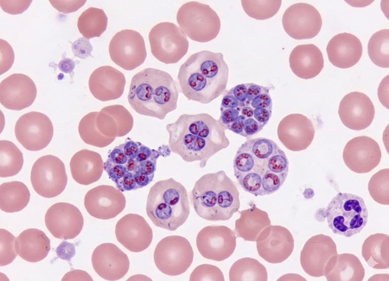
At the cellular level, doctors identify it by finding unusually large, abnormal cells called Paget cells sitting inside the epidermis, the skin’s outer layer. Viewed under a microscope, these cells stand out for their pale interior and oddly shaped, prominent nuclei. Sometimes they appear alone, and sometimes in small clumps scattered through the skin.
Paget cells are a type of adenocarcinoma, meaning they come from glandular tissue, which raised the question of how they end up on the skin’s surface at all. The leading explanation, known as the migration theory, holds that the cancer starts inside the milk ducts and the abnormal cells slowly travel upward through those ducts until they reach the nipple’s surface, causing the visible irritation people notice.
Somewhere between 90% and 97% of people diagnosed with Paget’s disease of the nipple turn out to have an underlying cancer as well. That hidden cancer is usually either ductal carcinoma in situ (DCIS), which stays confined inside the milk ducts, or invasive ductal carcinoma, where the cancer cells have pushed through the duct walls into surrounding tissue.
Roughly half of these patients also have a lump that can be felt during a physical exam a sign that usually points to an invasive cancer. When no lump is present, the hidden cancer is more likely to be the non-invasive DCIS type. Because the link between the skin changes and a deeper cancer is so strong, finding Paget cells on the skin is reason enough for doctors to thoroughly investigate the rest of the breast.
What Causes the Condition and Who Is at Risk?
The factors that lead to Paget’s disease of the breast largely overlap with what drives ordinary breast cancer, since this condition is essentially an extension of cancer that started in the milk ducts.
The Impact of Age and Lifespan Exposures
Age is the single biggest risk factor that can’t be changed. Most people diagnosed are over 50, with the typical age at diagnosis landing around 62. It’s very unusual to see this disease in anyone under 30. This pattern reflects the way cellular damage accumulates over decades, combined with a lifetime of shifting hormone levels that can gradually push vulnerable cells toward becoming cancerous.
Genetic Predispositions and Family History
Personal or family cancer history raises the odds significantly. Someone who has already had cancer in one breast faces a higher chance of a new, separate cancer forming in the other breast or elsewhere in the same one.
Having a close relative a mother, sister, or daughter who’s had breast cancer can roughly double a person’s baseline risk, and that risk climbs further if the relative was diagnosed young.
Much of this family risk traces back to inherited mutations. Changes in the BRCA1 and BRCA2 genes weaken the body’s ability to repair damaged DNA, sharply raising lifetime risk for breast, ovarian, and pancreatic cancers. Mutations in other genes, including PALB2, CHEK2, and ATM, can have a similar destabilizing effect.
On top of genetics, standard breast cancer risk factors also apply here: dense breast tissue on a mammogram, starting periods before age 12, reaching menopause after 55, or having a first full-term pregnancy after 30 can all add to a person’s overall risk.
The 7 Key Symptoms of Paget’s Disease of the Breast
Because this condition so closely resembles ordinary skin problems, its warning signs are easy to overlook. It almost always affects just one breast, with changes that build up gradually around the nipple and areola.
The seven signs to watch for:
- Persistent itching, tingling, or a burning feeling
- Redness and visible inflammation
- Flaky, scaly, or thickened skin
- Crusting, oozing, or weeping from open areas
- A nipple that’s recently flattened or turned inward
- Discharge from the nipple that happens on its own (clear, bloody, or straw-colored)
- A lump that can be felt deeper in the breast
Skin-Related Symptoms on the Nipple and Areola
In its earliest stage, Paget’s disease looks a lot like eczema, psoriasis, or contact dermatitis which is exactly why it’s so often missed at first.
Localized Itching, Tingling, or Burning
A lingering itch or tingling deep in the nipple skin is often one of the first things people notice. Unlike ordinary dryness or a mild allergic reaction, it doesn’t fade with time. Many describe it as a persistent “pins-and-needles” sensation or a dull burning. One key warning sign: this discomfort won’t budge no matter how much hydrocortisone cream or moisturizer is applied.
Redness, Inflammation, and Flaky Skin
The skin in the affected area turns visibly red and inflamed, usually in a clearly outlined patch. As abnormal cells build up in the skin, they interfere with its normal renewal process, causing flaking and rough peeling. Over time, the area can start to feel thick and leathery, losing the smooth texture of healthy skin.
Crusting, Oozing, and Open Ulceration
As things progress, the skin’s surface can break down completely, leaving raw patches or open sores. These may weep a clear or yellowish fluid, sometimes mixed with a small amount of blood, which dries into a crust. This isn’t discharge from the milk ducts it’s fluid coming directly from the damaged skin. A sore on the nipple that won’t heal is a strong signal that something more than a rash is going on.
Structural and Discharge-Related Symptoms
As the underlying cancer grows, its effects go beyond the surface and start to change the physical shape and function of the breast tissue.
Nipple Flattening or Inversion
A nipple that used to point outward can gradually flatten or pull inward. This happens because cancer cells trigger scar-like fibrous tissue to form inside the milk ducts, and that tissue acts like a drawstring, tugging the nipple back. Some people are simply born with inverted nipples, but any new or one-sided change in shape deserves prompt medical attention.
Spontaneous Ductal Nipple Discharge
This is different from the oozing on the skin’s surface it’s fluid coming out of the milk ducts themselves, often without any squeezing involved. It can look clear, straw-colored, or bloody. Bloody discharge from just one duct is a well-known warning sign of an underlying ductal cancer.
A Palpable Breast Lump or Mass
Around half of people with this condition also have a firm lump that can be felt during an exam. This usually marks the site of an invasive cancer, and it may sit right beneath the nipple or be tucked deeper in another part of the breast. When a lump shows up alongside skin changes, it often signals a more advanced stage, which makes mammograms and ultrasounds essential for pinpointing exactly where the cancer is.
Deep-Dive Diagnostic Protocols: Tracing the Malignant Blueprint
Confirming this diagnosis takes more than a quick look doctors need a layered approach combining hands-on exams, imaging, and lab work, since the visible skin problem is often just the tip of a cancer sitting deeper in the breast.
It typically starts with a thorough clinical exam. A breast specialist checks both breasts for symmetry, texture, and any irregularities, and also feels for swelling in the lymph nodes under the arm and near the collarbone, since invasive cancers can spread through the lymphatic system. Any firmness or reduced mobility in these nodes raises concern that the cancer may have started to spread.
[Visible Skin Changes] --> [Deeper Tissue Imaging] --> [Cell-Level Confirmation]
  - Redness on nipple         - Diagnostic mammogram      - Punch or wedge biopsy
  - Scaling or crusting       - Breast ultrasound          - Pathology review
  - Nipple pulling inward     - Contrast breast MRI        - Immunohistochemistry
To find the tumor hiding beneath the surface, doctors typically order:
Diagnostic Mammogram More detailed than a routine screening mammogram, this takes close, magnified images of the nipple area and the tissue behind it. Even though the rash itself may not be visible on X-ray, the scan can reveal tiny calcium deposits or distortions in tissue structure that point to DCIS or a deeper mass.
Targeted Breast Ultrasound Paired with a mammogram, ultrasound uses sound waves to tell the difference between a harmless fluid-filled cyst and a solid, potentially cancerous mass. It’s especially useful for spotting small tumors near the nipple that a mammogram alone might miss.
Contrast-Enhanced Breast MRI When results from mammography and ultrasound aren’t conclusive, an MRI with contrast dye can help. Because tumors build new blood vessels to feed their growth, the dye highlights these areas clearly, giving doctors a detailed picture of the disease’s extent and ruling out additional hidden tumors elsewhere in the breast.
Imaging alone isn’t enough, though a biopsy is required to confirm the diagnosis under a microscope. This is usually a quick outpatient procedure done under local anesthesia. Depending on what the skin looks like, the surgeon might use a punch biopsy (removing a small cylinder of full-thickness skin), a shave biopsy (taking just the top affected layer), or a wedge biopsy (a small, V-shaped piece of nipple tissue).
Diagnosis is confirmed once a pathologist spots the characteristic Paget cells scattered through the skin’s outer layer. Additional staining checking for markers like cytokeratin 7 (CK7) and the HER2 protein helps rule out other rare skin conditions or melanoma.
Paget’s Disease vs. Eczema: The Vital Clinical Distinctions
Confusing this cancer with ordinary areolar eczema or contact dermatitis is one of the most common and riskiest mistakes in breast health. Both conditions cause red, itchy, scaly skin on the nipple, which is exactly why people and even doctors sometimes brush off the early signs as a routine rash.
That mix-up can let a hidden cancer grow unchecked for months. Fortunately, there are clear differences once you know what to look for.
The biggest giveaway is how the skin responds to treatment. Eczema is driven by inflammation or irritation, so it typically clears up quickly with steroid creams, barrier ointments, and moisturizers.
Paget’s disease, on the other hand, is driven by cancer cells multiplying steadily steroids won’t touch it. A cream might calm the surface temporarily, but the underlying disease keeps progressing regardless.
| Clinical Feature | Areolar Eczema (Dermatitis) | Paget’s Disease of the Breast |
|---|---|---|
| Which breast is affected | Usually both | Almost always just one |
| Reaction to steroid cream | Clears up quickly | No improvement; keeps worsening |
| Nipple structure | Stays intact | Can flatten or invert permanently |
| Fluid from the area | Clear fluid from scratching | Spontaneous discharge, often bloody |
| Type of discomfort | Sharp, surface-level itch | Deep burning and tingling |
Another clear difference is which breast is involved. Eczema tends to appear on both sides at once, since it’s a broader skin condition. Paget’s disease, by contrast, comes from cancer developing in one breast’s duct system, so it almost always stays confined to that single side.
As the cancer spreads through the skin, it can cause lasting damage eczema simply doesn’t open sores that don’t heal, and structural changes like a flattened or inward-turning nipple, something eczema never causes no matter how irritated the skin gets.
The type of discomfort also differs. Eczema tends to cause a sharp, surface itch that makes you want to scratch. Paget’s disease more often produces a deeper burning or tingling sensation. Any rash confined to one nipple that doesn’t clear up with standard skin treatment within a few weeks should be biopsied to rule out cancer.
The Biological Link to Hidden Breast Malignancies
The connection between the visible skin changes and a hidden tumor underneath is strong enough that it shapes the entire treatment plan. In more than 90% of cases, that red, crusty patch on the nipple isn’t skin cancer at all it’s a sign of breast cancer growing beneath the surface.
Per the migration theory, the cancer doesn’t start on the nipple’s skin. It begins deep inside the milk ducts, and over time, cells break off from that original tumor and travel upward through the ducts to reach the nipple’s surface, where they cause the irritation and rash people eventually notice.
[Cancer in Milk Ducts] --> [Cells Travel Upward] --> [Cells Reach the Skin]
  - DCIS or invasive cancer   - Move through ducts        - Cause rash, crusting
What kind of hidden cancer is found and how advanced it is largely determines a person’s outlook.
Non-Invasive Underlying Cancer (DCIS)
When testing shows no lump and the cancer is confined entirely to the milk ducts, the outlook is generally very good. This is ductal carcinoma in situ (DCIS) the cells haven’t gained the ability to spread beyond the ducts, so treatment mainly focuses on local removal and preventing recurrence.
Invasive Underlying Cancer (Invasive Ductal Carcinoma)
If a lump is present, there’s a strong chance over 90% that the hidden cancer has become invasive, meaning it has broken through the duct walls into surrounding tissue. Once that happens, the cancer can potentially reach blood vessels and lymph nodes, so treatment decisions depend heavily on the tumor’s size, whether it’s reached nearby lymph nodes, and its hormone or genetic markers.
Comprehensive Treatment Framework: From Surgery to Systemic Care
Treatment is tailored to each patient by a team that typically includes breast surgeons, oncologists, and radiation specialists, with the goal of clearing all cancer cells while keeping the risk of recurrence low.
Surgical Intervention Options
Breast-Conserving Surgery (Lumpectomy) When the cancer is localized without a large deep tumor, surgeons can remove the nipple-areola complex along with a wedge of the underlying ducts, making sure the surrounding tissue is free of cancer cells. This approach is almost always followed by radiation therapy to destroy any remaining microscopic cells.
Total Mastectomy If the cancer is invasive, spread across multiple areas, or large relative to the breast, removing the entire breast including skin, tissue, and the nipple-areola complex is usually recommended. Reconstruction can happen at the same time or later, depending on the patient’s preference.
Lymph Node Evaluation
When invasive cancer is found, or during a mastectomy, surgeons often check the lymph nodes under the arm using a sentinel lymph node biopsy. A tracer dye is injected near the tumor and follows the natural path lymphatic fluid takes, leading to the first node it reaches. Only that node is removed and tested if it’s clear, no further nodes need to come out, which lowers the risk of lasting arm swelling (lymphedema).
[Tracer Injected Near Tumor] --> [Follows Lymph Drainage] --> [Test First Node Only]
  - Done during surgery          - Natural fluid pathway         - Clear: no further nodes removed
                                                                  - Positive: more nodes checked
Adjuvant Systemic Therapies
When invasive cancer is confirmed, additional treatments may target cancer cells anywhere in the body:
Chemotherapy Recommended when the tumor is large, fast-growing, or has reached the lymph nodes, chemotherapy works through the bloodstream to stop rapidly dividing cells.
Hormone Therapy If the tumor tests positive for estrogen or progesterone receptors, daily medication (like tamoxifen or an aromatase inhibitor) can block the hormones fueling its growth, usually taken for 5 to 10 years.
Targeted Biological Therapy Tumors that overexpress the HER2 protein tend to grow faster, so drugs like trastuzumab and pertuzumab are used to specifically target and shut down that growth pathway.
Conclusion
Paget’s disease of the breast is easy to overlook because it so often mimics eczema, dry skin, or mild irritation. Persistent flaking, itching, redness, crusting, burning, discharge, or a change in nipple shape shouldn’t be dismissed, especially if it’s one-sided or isn’t improving with basic skin care. Most nipple rashes aren’t cancer, but only a medical evaluation can confirm that for sure. If you notice changes around the nipple or areola that stick around, getting checked out early is always the safer choice.
Frequently Asked Questions
1. What is Paget’s disease of the breast?
It’s a rare form of breast cancer that mainly affects the nipple and areola, causing skin changes that can resemble eczema or a persistent rash. It’s often connected to an underlying breast cancer, so ongoing nipple changes are worth having checked out.
2. Why does it get mistaken for a skin condition?
Because the redness, itching, flaking, and crusting it causes look a lot like eczema or contact dermatitis. The difference is that Paget’s-related symptoms tend to stick around, worsen over time, and usually affect just one side.
3. What symptoms should raise concern?
Flaky or crusted skin, oozing, burning, persistent itching, nipple discharge, a flattened nipple, a palpable lump, or thickened skin are all worth mentioning to a doctor especially bloody or unexplained discharge or changes limited to one side.
4. How is it diagnosed?
Diagnosis usually starts with a breast exam, followed by imaging such as a mammogram, ultrasound, or MRI, and finally a biopsy to confirm the presence of cancer cells under a microscope.
5. Is it treatable?
Yes. Treatment depends on whether an underlying cancer is present and how advanced it is, and can include surgery, radiation, chemotherapy, hormone therapy, or targeted therapy. Catching it early generally leads to better outcomes.