8 Warning Signs of a Chiari Malformation Most People Ignore
Have you ever had unexplained headaches, dizziness, or a feeling of pressure in your neck and put it down to stress or tiredness? These symptoms are common, but they can sometimes point to something more serious: Chiari malformation.
This rare condition occurs when brain tissue extends into the spinal canal. That disrupts the normal flow of cerebrospinal fluid and puts pressure on the brain and spinal cord. Many people don’t realize their symptoms could be linked to it, so they dismiss them as minor problems that will pass.
Chiari malformation is hard to diagnose because its symptoms overlap with common conditions like migraines, neck pain, and even anxiety. It can go unnoticed for years, leaving people with chronic pain, discomfort, and neurological problems without knowing why.
Severity varies. Some people have only mild symptoms, while others develop serious complications such as paralysis, breathing problems, or loss of coordination.
The usual symptoms include persistent headaches at the back of the head, dizziness, vision problems, and difficulty swallowing. They may come and go or worsen over time, which makes it easy to blame stress or something unrelated. Recognizing them early can make a big difference to diagnosis and treatment.
In this article, we look at eight warning signs of Chiari malformation that are often ignored or misdiagnosed, so you can seek the right care before the condition causes more serious problems.
8 Warning Signs of a Chiari Malformation
Headache and Neck Pain
The headache of a Chiari malformation is quite distinctive. It is usually a pressure-like pain at the back of the head (the occipital region) that is triggered or made worse by anything that raises pressure inside the skull, such as coughing, sneezing, laughing, or straining. Doctors often call it a Chiari headache, and it is one of their main diagnostic clues.
The pain can be sharp, stabbing, or throbbing, and it often spreads from the base of the skull into the neck and shoulders. Unlike tension headaches or migraines, it is tied directly to straining (Valsalva maneuvers). These actions briefly raise the pressure of the cerebrospinal fluid (CSF), pressing the herniated cerebellar tonsils against the brainstem and spinal cord in the crowded space of the foramen magnum.
These headaches can be debilitating, though they are often short, lasting from a few seconds to several minutes after the trigger. They are almost always felt at the junction of the head and neck, and people often describe them as crushing or bursting pressure.
Neck pain, stiffness, and reduced range of motion are common alongside the head pain. So is a “coat hanger” pattern of pain across the tops of the shoulders and upper back, caused by irritation of nerves in that area.
Some people also develop a constant background headache or a feeling of steady pressure in the head. When the link to straining isn’t noticed, this can lead to a misdiagnosis of chronic migraine or tension headache.
Dizziness and Vertigo
Dizziness is a general feeling of being off balance, lightheaded, or woozy. Vertigo is more specific: the sensation that you or the room around you is spinning. Both happen when the signals between the cerebellum and the vestibular system (the balance center of the inner ear) are disrupted. These two systems normally work together to keep you oriented in space.
Ataxia (Unsteady Gait)
Ataxia means a lack of voluntary muscle coordination. In people with Chiari, it often shows up as a clumsy, wide-based, staggering walk. They may struggle to walk in a straight line, trip often, and find sudden turns difficult. The cerebellum can no longer coordinate the complex muscle movements that steady walking requires.
Dysmetria and Fine Motor Impairment
Dysmetria is the inability to judge distance or range of movement, so a person overshoots or undershoots their target. Reaching for a glass, buttoning a shirt, or writing can become difficult and clumsy. Poor hand-eye coordination and intention tremors (shaking that gets worse when reaching for something) are also common and make fine motor tasks harder still.
Visual Disturbances
The brainstem is compressed by the herniated cerebellum, and it acts as a key relay station for nerve signals traveling to and from the eyes and ears. When the nerves that control the eye muscles are affected, the eyes can become misaligned and cause double vision (diplopia).
Nystagmus, in which the eyes make repetitive, uncontrolled movements (side to side or up and down), is another classic sign of cerebellar or brainstem dysfunction. Many people also report blurred vision, spots or floaters, and strong sensitivity to bright light, which can make night driving or well-lit rooms uncomfortable. Pain behind the eyes is another common complaint.
Auditory and Vestibular Symptoms
Tinnitus, the perception of ringing, buzzing, or hissing with no external source, is a frequent symptom. It can result from pressure on the auditory nerve (the eighth cranial nerve). Because this nerve also plays a part in balance, hearing symptoms often come with vertigo or dizziness.
Some people notice reduced hearing, muffled sounds, or a feeling of fullness in the ears. These symptoms are sometimes misdiagnosed as Meniere’s disease or another inner ear disorder.
Paresthesia and Dysesthesia
Paresthesia refers to abnormal sensations such as pins and needles, tingling, or numbness, usually in the hands, arms, feet, or legs. Dysesthesia is more unpleasant and is often described as burning, aching, or electric-shock sensations.
These occur because the nerve tracts that carry touch, temperature, and pain signals are squeezed at the craniocervical junction. Some people also lose the ability to feel temperature properly, which makes them more prone to burns.
Progressive Muscle Weakness
Muscle weakness (paresis) tends to affect the arms more than the legs. You may notice your grip getting weaker, making it hard to open jars or hold objects, and fine skills like writing or fastening buttons may decline. Over time, the weakness can lead to muscle wasting (atrophy). It results from damage to the motor nerve pathways running through the compressed part of the spinal cord.
What is a Chiari Malformation?
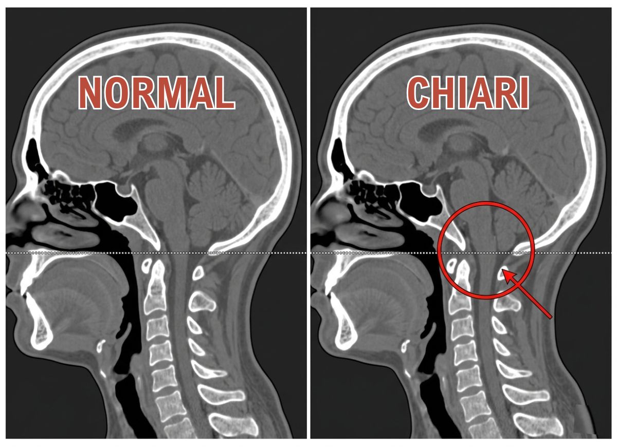
A Chiari malformation is a structural defect of the cerebellum, the part of the brain that controls balance. Brain tissue extends into the spinal canal through the foramen magnum, the natural opening at the base of the skull. It happens because the part of the skull that holds the cerebellum (the posterior fossa) is smaller or more misshapen than normal, which crowds the brain and pushes it downward.
The condition mainly affects two structures at the base of the brain. The cerebellum, in the posterior cranial fossa, coordinates voluntary movement, posture, balance, and speech so that muscle activity is smooth and balanced. The brainstem connects the cerebrum and cerebellum to the spinal cord and controls essential automatic functions such as breathing, heart rate, consciousness, and sleep cycles.
In a Chiari malformation, parts of the cerebellum called the cerebellar tonsils are pushed down through the foramen magnum, the large opening through which the spinal cord passes. This compresses both the brainstem and the upper spinal cord, which explains why the symptoms are so varied.
When the tonsils are squeezed, the functions they help regulate suffer. That leads to the classic symptoms: ataxia (poor coordination and unsteady gait), vertigo, dysmetria (misjudging distance), and nystagmus (involuntary eye movements). Pressure on the brainstem can also disrupt vital functions, causing swallowing problems (dysphagia), sleep apnea, a hoarse voice, and, in severe cases, abnormal heart rhythms.
The herniated tonsils also act like a cork in a bottle, blocking the normal circulation of CSF between the brain and spinal canal. This can raise pressure inside the skull and cause the typical Valsalva-induced headaches. It can also force CSF into the spinal cord itself, forming a fluid-filled cavity called a syrinx (syringomyelia), which can lead to progressive pain, weakness, and loss of sensation.
Types of Chiari Malformations
Chiari malformations are classified into several types according to the anatomy of the malformation and how much tissue passes through the foramen magnum.
Type I is the most common and often the mildest. It is usually diagnosed in adolescence or adulthood. The cerebellar tonsils descend at least 4 millimeters below the foramen magnum. It may cause no symptoms, but when it does, they often include headaches (especially after coughing or straining), neck pain, and balance problems.
Type II, also called the Arnold-Chiari malformation, is more severe and is almost always linked to a form of spina bifida called myelomeningocele. Both the cerebellar tonsils and parts of the brainstem extend into the foramen magnum. It is usually diagnosed at birth or in infancy and is frequently accompanied by hydrocephalus.
Type III is very rare and severe. Part of the cerebellum and/or brainstem herniates through an abnormal opening at the back of the skull (an encephalocele). It causes serious, often life-threatening neurological problems and needs immediate surgery.
Type IV is also rare, and its classification is debated. It involves an incomplete or underdeveloped cerebellum (cerebellar hypoplasia). Parts of the cerebellum are missing, but there is no significant herniation of brain tissue through the foramen magnum.
Is a Chiari Malformation Present From Birth?
Usually, yes. A Chiari malformation is typically congenital, meaning it is present at birth because of abnormal development of the brain and skull before birth. The two most common types (Type I and Type II) are considered primary, or congenital. They are thought to result from a posterior fossa, the space at the back of the skull that houses the cerebellum, that is too small or misshapen. That space cannot hold the cerebellum properly, so the cerebellar tonsils are pushed down through the foramen magnum.
Congenital (primary) Chiari malformation is the most common form. Its underlying cause is related to genetic factors or unknown problems during fetal development that affect how bone and brain form. It is often associated with other congenital conditions, such as spina bifida (in Type II), hydrocephalus, and connective tissue disorders like Ehlers-Danlos syndrome.
Acquired (secondary) Chiari malformation develops after birth and is not related to a skull defect present from the start. It can occur when too much cerebrospinal fluid is drained from the lumbar or thoracic spine, often because of injury, infection, or a shunt used to treat hydrocephalus. The loss of CSF volume makes the brain sag downward, pulling the cerebellar tonsils into the foramen magnum and creating a malformation that works much like the congenital type. In rare cases, tumors or other lesions that raise pressure inside the skull can also lead to an acquired Chiari malformation.
Even though the structural defect is there from birth, symptoms of Type I often don’t appear until late childhood, adolescence, or adulthood, which can be confusing for patients. It is also possible, though much less common, for a Chiari malformation to be acquired later in life.
What are the Causes of a Chiari Malformation?
Abnormal Fetal Development and Acquired
The main difference between the two forms is where they come from. Congenital malformations are present at birth because of developmental abnormalities. Acquired malformations develop later in life because of an outside event or condition.
Congenital, or primary, Chiari is a developmental disorder. It starts with a problem during fetal growth that leaves the posterior fossa abnormally small or misshapen. The lack of space crowds the cerebellum and pushes the tonsils down through the foramen magnum. It is often linked to genetic factors, though the specific genes involved are not fully understood. It is a structural problem a person is born with, even if symptoms appear much later.
Acquired, or secondary, Chiari does not come from a pre-existing skull abnormality. Instead, another condition changes the pressure balance inside the skull and spinal column. The most common cause is excessive drainage of cerebrospinal fluid (CSF) from the lumbar spine, which can follow a spinal tap, spinal trauma, or a CSF leak. Less fluid means less buoyancy for the brain, so it sags downward and the tonsils herniate. Brain tumors, cysts, or hematomas that create pressure and push the cerebellum down can also be causes. In these cases the skull structure starts out normal, and a later event creates the malformation.
Genetic Predisposition and Family History
Chiari malformation doesn’t always follow a simple inheritance pattern, but there is growing evidence that it can run in families. Having a close relative with Chiari malformation or a related condition like syringomyelia may raise your risk. Researchers are studying specific genetic mutations, but for most cases a clear genetic link has not been established.
Associated Connective Tissue Disorders
Conditions like Ehlers-Danlos syndrome (EDS) and other hypermobility disorders are strongly associated with Chiari malformation. They affect collagen in the body, leading to overly flexible joints and loose connective tissue. This weakness may affect the stability of the craniocervical junction and could contribute to, or worsen, the cerebellar herniation and its symptoms.
Physical Trauma and Strain
Head or neck trauma, such as whiplash from a car accident, a fall, or contact sports, can trigger symptoms in someone who previously had none. These events can shift the brain suddenly or increase inflammation at the craniocervical junction, turning a silent anatomical variation into a painful, symptomatic condition. Activities that involve straining or repeated Valsalva maneuvers, such as heavy weightlifting or chronic coughing, can also make symptoms worse by raising pressure inside the skull.
Chiari Malformation Treatment
Treatment ranges from conservative, non-surgical management for mild symptoms to surgery for severe or progressive cases. The choice is highly individual and depends on how severe the symptoms are, how far the cerebellum has herniated, whether a syrinx (syringomyelia) is present, and how much the condition affects the patient’s quality of life.
People with no symptoms, or only minor ones like mild headaches, are often first managed with “watchful waiting” and regular monitoring by a neurologist or neurosurgeon. If symptoms are present but manageable, treatments aimed at symptom relief are used. Surgery is generally kept for cases where conservative treatment fails or there are signs of significant neurological decline.
Non-surgical Treatments
Non-surgical treatment focuses on managing symptoms and improving quality of life. It mainly involves pain medication, physical therapy, and lifestyle changes. These approaches are usually the first choice for people with mild to moderate symptoms and no progressive neurological problems.
The goal is not to correct the structural defect but to ease the pain and limitations it causes. Combining several strategies often works best, and many patients are able to avoid or delay surgery while keeping a good level of daily function.
Over-the-counter pain relievers such as ibuprofen or naproxen can help with mild headaches and neck pain. For more severe or persistent pain, doctors may prescribe stronger medication, including muscle relaxants for neck stiffness and spasms, or neuropathic pain drugs like gabapentin or pregabalin for nerve-related pain, tingling, and numbness.
Physical therapy can improve balance, strengthen the neck muscles so they support the head better, and teach posture-correcting exercises. Therapists can also guide gentle stretching to relieve neck stiffness and improve range of motion. Occupational therapy helps patients adapt to their limitations by teaching new ways to do daily tasks, which saves energy and reduces strain.
Avoiding triggers is another key part of conservative care. Patients should steer clear of activities that involve straining, such as heavy lifting, high-impact sports, or playing certain wind instruments. They are also advised to avoid positions that hyperextend the neck, and to manage conditions that cause chronic coughing or constipation so that pressure inside the skull doesn’t rise.
Surgery Treatment
Surgery becomes necessary when there is progressive neurological deterioration, severe pain that doesn’t respond to conservative care, or a syrinx (syringomyelia). The decision is a significant one, made by a neurosurgeon together with the patient. It is not recommended for people with no symptoms or only mild, stable ones.
The main goal is not to cure the malformation but to decompress the crowded area at the base of the skull. That relieves pressure on the brainstem and spinal cord and restores normal CSF flow. Surgery is recommended when the condition is clearly causing significant and potentially irreversible harm.
Progressive neurological deficits are among the strongest reasons for surgery. These include worsening muscle weakness, significant balance problems (ataxia), loss of sensation, difficulty swallowing (dysphagia), or changes in bladder or bowel control. They suggest that ongoing compression is damaging neural tissue.
Surgery may also be the best option when headaches, neck pain, or other symptoms are so severe that they seriously interfere with work, school, or daily activities and haven’t improved with thorough non-surgical treatment.
A syrinx, a fluid-filled cyst within the spinal cord, is a serious complication of Chiari malformation that can cause progressive pain, weakness, and sensory loss. If an MRI confirms one, surgery is almost always recommended to stop it from growing and prevent permanent spinal cord damage.
The main operation is called posterior fossa decompression. A small piece of bone is removed from the back of the skull (and sometimes the top vertebra) to make more room for the brain and restore CSF flow.
Chiari Malformation Diagnosis
Diagnosis is a multi-step process. It starts with a thorough clinical evaluation and is confirmed with medical imaging.
A physician, usually a neurologist or neurosurgeon, begins with a detailed medical history and a full neurological exam. The exam tests balance, coordination, reflexes, muscle strength, and sensation to find deficits that fit Chiari malformation. Because symptoms can be vague or overlap with other conditions, imaging is the deciding step.
The gold standard is Magnetic Resonance Imaging (MRI) of the brain and spine. It produces detailed cross-sectional images that clearly show where the cerebellar tonsils sit in relation to the foramen magnum, and it can measure how far they have herniated, a key criterion especially for Type I. A specialized version called Cine MRI can also show how CSF flows around the brain and spinal cord, helping to identify blockages caused by the malformation.
A Computed Tomography (CT) scan may be used in some cases, particularly in emergencies or to assess the bones of the skull and spine, but it shows less soft-tissue detail than MRI. Together, the clinical findings and MRI results allow a conclusive diagnosis and guide treatment decisions.
Chiari Malformation and Other Medical Conditions
Telling Chiari malformation apart from other neurological conditions is a critical part of diagnosis, because symptoms such as headache, dizziness, fatigue, and chronic pain overlap so much. The differential diagnosis relies heavily on neurological imaging, which reveals the structural abnormality that other disorders lack.
Multiple sclerosis (MS), for example, can cause numbness, weakness, and balance problems similar to Chiari. But MS is an autoimmune disease in which the body attacks the myelin sheath of nerves. An MRI in someone with MS shows lesions or plaques in the brain and spinal cord, not the cerebellar tonsil herniation typical of Chiari.
Fibromyalgia is another condition with overlapping symptoms. It involves widespread musculoskeletal pain, fatigue, and cognitive difficulties (“fibro fog”). The pain and fatigue can resemble Chiari, but fibromyalgia is considered a central pain sensitization disorder and has no identifiable structural cause on MRI.
Chronic migraines can also cause severe, debilitating headaches, but Chiari headaches usually have a specific trigger. They are frequently brought on or worsened by coughing, sneezing, or straining (a Valsalva-induced headache).
In the end, a patient’s symptoms guide the initial investigation, but the definitive diagnosis comes from seeing the anatomical abnormality on an MRI, a finding that is absent in these other conditions.
Associated Conditions like Syringomyelia and Hydrocephalus
Two of the most significant conditions associated with Chiari malformation are syringomyelia and hydrocephalus. Both stem from the same underlying problem: disrupted circulation of cerebrospinal fluid (CSF).
Syringomyelia is the development of a fluid-filled cyst, called a syrinx, inside the central canal of the spinal cord. In someone with Chiari malformation, the herniated cerebellar tonsils block the normal, pulsing flow of CSF from the brain down into the spinal canal. The resulting pressure can force CSF into the spinal cord tissue itself and form a syrinx. The cyst can expand over time, compressing and damaging nerve fibers in the cord. The symptoms depend on its location and size but often include chronic pain, progressive weakness in the arms and legs, stiffness, and loss of sensation, especially for pain and temperature.
Hydrocephalus is an excessive buildup of CSF in the brain’s ventricles. The same blockage at the foramen magnum that can cause syringomyelia can also stop CSF from being absorbed properly, so fluid backs up in the brain. The increased pressure inside the skull can cause an enlarged head in infants, as well as headaches, cognitive problems, vision problems, and poor coordination.
Hydrocephalus can occur with Type I, but it is far more common in Type II and is one of its defining features. Both syringomyelia and hydrocephalus are serious complications that often need surgery to restore normal CSF flow and relieve pressure on the brain and spinal cord.
FAQs
1. Is a Chiari malformation serious?
Chiari malformation ranges from mild to severe, and in some cases it can be life-threatening. Left untreated, severe forms can cause significant neurological problems such as paralysis, difficulty breathing, or loss of coordination.
The condition can disrupt the normal flow of cerebrospinal fluid (CSF) and put pressure on the brain and spinal cord. Early diagnosis and treatment are crucial to prevent severe complications. In some cases it can be managed effectively with surgery and ongoing care, which improves the outlook.
2. Can you live a normal life with Chiari malformation?
Yes, many people with Chiari malformation live normal lives, especially when it is diagnosed early and managed well. Symptom severity varies, and people with a mild form may not have significant limitations.
More severe cases can cause chronic headaches, dizziness, balance problems, or coordination difficulties. With proper treatment, including surgery and therapy, many people manage their symptoms and stay active. It is important to follow your treatment plan and work closely with your healthcare providers.
3. How do you fix Chiari malformation?
Treatment depends on how severe the condition and its symptoms are. For many people, surgery is the most effective option. The most common operation is decompression surgery, which removes part of the skull or upper spine to relieve pressure on the brain and spinal cord. This helps restore CSF flow and reduce symptoms.
Non-surgical treatments such as pain management, physical therapy, and lifestyle changes may also be recommended, especially when surgery isn’t needed. Regular follow-up with a specialist is essential to track progress and adjust the plan.
4. Is Chiari a permanent disability?
Not always. The impact varies a lot from person to person. Some people have long-term neurological effects, while others recover fully or have only mild, manageable symptoms.
In severe cases, especially if untreated, Chiari malformation can lead to permanent disability such as paralysis or ongoing neurological damage. With early diagnosis and appropriate treatment, though, many people can limit the long-term effects and lead productive, independent lives.
5. Can Chiari malformation get worse with age?
Yes, it can, especially if left untreated. Over time, pressure on the brain and spinal cord can increase and make symptoms stronger. Symptoms such as headaches, neck pain, balance problems, or trouble swallowing may gradually get worse.
Not everyone’s symptoms progress, however. In some cases they stabilize, particularly when the condition is managed with surgery or other treatment. Regular monitoring by a healthcare provider is essential to keep the condition under control and prevent complications.
6. What should people with Chiari not do?
People with Chiari malformation should avoid activities that put excessive strain on the neck and spine, especially heavy lifting, twisting motions, and high-impact activities. Sports like football or gymnastics, or anything involving sudden movements or impacts, could worsen symptoms or cause further complications.
It’s also wise to avoid positions that increase pressure on the spine, such as prolonged bending or sitting in one position for too long. Every case is different, so work with your healthcare provider to work out which activities are safe for you.
7. Will a brain MRI show Chiari malformation?
Yes. A brain MRI is the most effective imaging tool for diagnosing it. MRI scans give detailed images of the brain and spinal cord and can show how far brain tissue has moved into the spinal canal. They are also useful for spotting related problems such as fluid buildup or structural changes.
An MRI can help assess severity and guide treatment decisions too. If Chiari malformation is suspected, a doctor will likely order one to confirm the diagnosis.
8. Do people with Chiari malformation have bigger brains?
No. In fact, Chiari malformation involves displacement of brain tissue, often because there is less room for the brain inside the skull. That displacement puts pressure on the brain and spinal cord and causes the symptoms of the condition.
People with Chiari may have some unusual structural features in the brain or spinal canal, but it isn’t accurate to say their brains are bigger. The condition affects how the brain and spinal cord interact, not the size of the brain.
9. Can you fly with Chiari malformation?
Flying is generally safe for most people with Chiari malformation, but it may worsen symptoms for some, especially those with severe cases. Changes in air pressure during a flight can increase headaches, dizziness, or pressure in the head and neck.
If you plan to fly, take precautions such as staying hydrated, avoiding lifting heavy bags, and resting as much as possible during the flight. If you have significant symptoms or a history of serious complications, it’s best to check with your healthcare provider before traveling by air.
Conclusion
Chiari malformation is complex and often misunderstood, but with early diagnosis and proper management, many people with the condition lead fulfilling lives.
Experiences range from mild symptoms managed with conservative treatment to severe cases that need surgery. Understanding the warning signs, risks, and treatment options is essential to managing the condition well.
If you or someone you know has symptoms of Chiari malformation, seeking medical advice early can make a real difference in limiting complications and improving quality of life. With the right care, people with Chiari malformation can manage their condition and thrive for years to come.