9 Facts About Optic Neuritis and Multiple Sclerosis Link
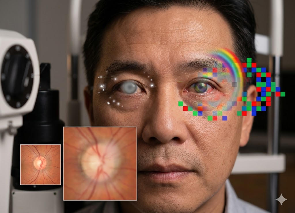
Optic neuritis is an inflammatory disorder involving the optic nerve, which carries visual information from the eyes to the brain. When inflammation affects this important nerve, a person may experience sudden changes in vision, discomfort when moving the eyes, or temporary and sometimes partial vision loss in one eye. Although many people experience improvement over time, optic neuritis can sometimes point toward an underlying neurological condition.
One of the strongest medical associations is between optic neuritis and multiple sclerosis (MS), a chronic autoimmune disorder affecting the central nervous system. For some people, optic neuritis becomes the first noticeable indication of MS, which is why recognizing its symptoms and receiving an appropriate evaluation can be important.
In this article, we’ll discuss 9 important facts about optic neuritis and its relationship with multiple sclerosis, including its causes, symptoms, diagnosis, recovery, and possible long-term implications.
What Is the Fundamental Link Between Optic Neuritis and Multiple Sclerosis?
The connection between optic neuritis and multiple sclerosis is largely related to demyelination, a process in which inflammation damages the myelin covering surrounding nerve fibers. This same autoimmune process is central to MS, which means inflammation of the optic nerve can sometimes represent an early sign or an episode associated with the disease.
During an autoimmune reaction, the immune system mistakenly attacks myelin, the protective coating surrounding nerve fibers within the central nervous system. This system includes the brain, spinal cord, and optic pathways. When the inflammatory response targets the optic nerve, inflammation and damage occur, resulting in optic neuritis.
For this reason, an episode of optic neuritis may not always be an isolated eye problem. In some individuals, it can represent a localized manifestation of a wider demyelinating process.
What Is Optic Neuritis?
The optic nerve is a major part of the visual pathway. It contains more than a million nerve fibers that carry information from the retina toward the brain’s visual centers. These fibers are protected by myelin, which helps electrical signals travel rapidly and efficiently.
When inflammation damages this protective layer, communication between the eye and brain can become disrupted. This interruption can lead to common optic neuritis symptoms such as reduced vision, faded colors, and blind spots.
Inflammation can also affect the underlying nerve fibers themselves. While early damage may primarily involve myelin, more severe or prolonged inflammation can result in axonal injury and potentially lasting visual problems.
A typical presentation often includes a sudden decrease in vision in one eye, discomfort when moving the eye, and gradual visual recovery over time.
Atypical cases can look different. Both eyes may be affected, pain may be absent, or vision loss may be much more severe and permanent. Such presentations can be associated with conditions including Neuromyelitis Optica Spectrum Disorder (NMOSD) or Myelin Oligodendrocyte Glycoprotein Antibody Disease (MOGAD).
Is Optic Neuritis Always a Sign of Multiple Sclerosis?
No. Experiencing optic neuritis does not automatically mean that a person will develop multiple sclerosis. However, MS is an important underlying cause, particularly among younger adults.
Because several other disorders can produce similar symptoms, doctors generally need to conduct a detailed evaluation before establishing a connection with MS. Identifying the actual cause is important because different conditions may require different forms of treatment and long-term monitoring.
Neuromyelitis Optica Spectrum Disorder can affect the optic nerves and spinal cord and may produce severe, bilateral vision problems. MOGAD can also cause significant inflammation of the optic nerve. Blood tests for specific antibodies can help doctors distinguish these conditions.
Other possible causes include infections such as mumps, shingles, Lyme disease, and syphilis. Conditions such as lupus and sarcoidosis may also affect the nervous system. In less common cases, vitamin B12 deficiency, toxic substances, or medication-related reactions may produce similar nerve damage.
Even with these possibilities, a typical episode of unilateral optic neuritis in a younger adult can have a particularly strong association with MS.
How Is Optic Neuritis Often the First Clinical Sign of MS?
For some people with multiple sclerosis, optic neuritis is the first neurological symptom they notice. When a single neurological episode occurs because of inflammation or demyelination in the central nervous system, it may be described as a Clinically Isolated Syndrome.
The optic nerve is considered part of the central nervous system, so inflammation affecting it can represent a demyelinating event.
Whether an isolated episode eventually meets the diagnostic criteria for MS depends on evidence showing that demyelination has occurred in different areas and at different points in time. Brain MRI is particularly useful because it can identify lesions in areas that may not have produced noticeable symptoms, including the brain’s white matter or spinal cord.
If characteristic lesions are present during the initial evaluation, the likelihood of a later MS diagnosis can be considerably higher. In certain situations, MRI findings combined with clinical evidence may allow MS to be diagnosed after the first neurological episode.
Diagnostic Findings and Symptoms Connect Optic Neuritis to an MS Diagnosis
Several findings can help physicians determine whether optic neuritis may be associated with multiple sclerosis. A typical pattern includes vision loss in one eye, discomfort with eye movement, and reduced color perception, along with characteristic lesions on brain MRI.
Patient-reported symptoms provide important clinical information, while MRI findings offer objective evidence of possible demyelination. The discovery of lesions elsewhere in the central nervous system can be especially useful when determining whether the episode is part of MS rather than an isolated event.
Classic Symptoms of Optic Neuritis
Symptoms generally develop over several hours or a few days. The most recognizable features include:
Unilateral Vision Loss
Vision loss is one of the most noticeable symptoms and commonly affects one eye in MS-associated cases. The severity can range from mild blurring or a small blind spot to significant loss of vision.
People may describe the affected eye as though they are looking through fog, mist, or a dirty window. Vision can continue getting worse for approximately one to two weeks before reaching a stable point, after which gradual improvement may begin.
Pain with Eye Movement
Pain during eye movement is a common feature of optic neuritis. It is often experienced as an aching or uncomfortable sensation behind the eye and may become more noticeable when looking upward, sideways, or in another direction.
The discomfort is related to inflammation around the optic nerve and can occur before or alongside the visual changes. In many cases, the pain gradually improves within a week or two.
Loss of Color Vision (Dyschromatopsia)
Changes in color perception are another important sign. Bright colors, especially red, may appear faded or less vivid when viewed with the affected eye.
This reduced intensity is sometimes called red desaturation and can provide a useful clinical clue. A simple comparison can be made by looking at a red object separately with each eye.
Color perception may remain somewhat altered even after visual sharpness improves. Some people can also experience flashing or flickering sensations, particularly when moving the affected eye.
What Do Brain MRI Scans Reveal About the Link?
Brain MRI is an important part of evaluating the relationship between optic neuritis and multiple sclerosis. While orbital MRI can show inflammation involving the optic nerve, a brain MRI can reveal lesions elsewhere in the central nervous system.
Neurologists commonly examine MRI images for T2-hyperintense lesions, which appear as bright areas on particular imaging sequences. These lesions can reflect areas of inflammation, demyelination, or scarring.
Important locations include:
- The periventricular region around the brain’s ventricles
- The juxtacortical region close to the cerebral cortex
- The infratentorial region involving the brainstem or cerebellum
- The spinal cord
The number and location of lesions can provide useful information about future MS risk. Long-term data from the Optic Neuritis Treatment Trial found that people with one or more lesions on their initial brain MRI had an estimated 72% risk of developing clinically definite MS within 15 years, compared with approximately 25% among those whose initial MRI showed no lesions.
What Is Uthoff’s Phenomenon in Patients?
Uhthoff’s phenomenon refers to a temporary worsening of previous neurological symptoms when body temperature increases. In someone who has experienced optic neuritis, this may appear as temporary blurring or dimming of vision in the previously affected eye.
It does not necessarily represent a new relapse or permanent worsening of the disease. Instead, increased temperature can temporarily interfere with signal transmission through previously damaged or demyelinated nerve fibers.
Possible triggers include:
- Physical exercise
- Fever
- Hot baths or showers
- Saunas
- Hot or humid weather
- Emotional stress
Symptoms generally improve once body temperature returns to normal. Staying cool, drinking cold fluids, avoiding excessive heat, and using other cooling measures may help reduce these temporary changes.
Long-Term Risk of Developing MS After Optic Neuritis
The overall long-term risk of developing multiple sclerosis after a first episode of optic neuritis has been estimated at approximately 50% over 15 years. However, the individual risk can vary substantially depending on findings from the initial brain MRI.
Much of the long-term information comes from the Optic Neuritis Treatment Trial (ONTT), which followed patients over an extended period and provided important information about the relationship between optic neuritis and MS.
Understanding the ONTT Risk Stratification
One of the most useful predictors of future MS is whether characteristic white matter lesions are visible on the initial brain MRI.
Patients With One or More Lesions
Individuals with one or more characteristic T2-hyperintense lesions on their initial MRI had an estimated 72% chance of developing MS within 15 years according to the long-term ONTT findings.
Patients With a Normal Brain MRI
Those whose initial brain MRI did not show lesions had a considerably lower estimated risk, with approximately 25% developing MS over the same 15-year period.
A normal MRI therefore does not eliminate the possibility of MS, but it can provide important information about long-term risk.
Clinical Implications of the Risk Data
These differences demonstrate why brain MRI is an important part of evaluating a first episode of optic neuritis. Rather than relying only on a general risk estimate, physicians can use imaging findings to develop a more individualized assessment.
The results may also help doctors and patients discuss monitoring and possible disease-modifying treatment when appropriate.
Does Vision Fully Recover After an Optic Neuritis Episode?
Most people experience substantial improvement in vision following optic neuritis, although some subtle visual changes may remain.
Recovery often begins within two to three weeks and can continue for many months. More than 90% of patients may eventually achieve visual acuity of 20/40 or better in the affected eye.
However, having good visual acuity does not necessarily mean that every aspect of vision has returned completely.
The Nuance of “Full Recovery”
A person may be able to read a 20/20 line on an eye chart while still noticing small differences in visual quality.
Possible long-term changes include:
- Reduced Contrast Sensitivity: Difficulty distinguishing similar shades, particularly in dim lighting, fog, or glare.
- Impaired Color Vision: Colors may continue to look less vivid or somewhat washed out in the affected eye.
- Visual Field Defects: Small blind spots may remain in the central or peripheral visual field.
- Uhthoff’s Phenomenon: Vision can temporarily become blurry or dim when body temperature rises.
- Pulfrich Phenomenon: In uncommon cases, moving objects may appear to follow a curved or elliptical path because of differences in signal transmission between the two eyes.
Detecting Residual Changes
Some visual changes are difficult to detect with a standard eye chart. Specialized tests can provide additional information.
These may include contrast sensitivity testing, automated visual field examinations, and Optical Coherence Tomography (OCT), which can measure the thickness of the retinal nerve fiber layer.
These tests can help identify subtle effects that remain after the main episode has improved.
Other Conditions Related to or Mimicking MS-Associated Optic Neuritis
Several inflammatory and demyelinating disorders can produce symptoms similar to MS-related optic neuritis. Two important examples are Neuromyelitis Optica Spectrum Disorder (NMOSD) and Myelin Oligodendrocyte Glycoprotein Antibody Disease (MOGAD).
Because these conditions can look similar during their initial stages, distinguishing between them is important for diagnosis, treatment, and long-term monitoring.
The Difference Between Typical and Atypical Optic Neuritis
Recognizing whether the presentation is typical or atypical can help doctors determine which underlying causes should be investigated.
Typical Optic Neuritis
Typical optic neuritis, which is commonly associated with MS, generally affects one eye and causes discomfort that becomes worse with eye movement. Vision usually declines gradually over several days rather than disappearing instantly.
The severity can vary, but patients generally retain some level of light perception. Recovery commonly begins within a few weeks.
Brain MRI may also show asymptomatic lesions associated with demyelination.
Atypical Optic Neuritis
Certain features suggest that a broader investigation may be necessary. These include:
- Bilateral Involvement: Both eyes become affected at the same time or within a short interval.
- No Pain: The usual discomfort associated with eye movement is absent.
- Severe Vision Loss: Vision becomes profoundly impaired or disappears completely.
- Significant Optic Disc Swelling: Examination reveals marked swelling at the back of the eye.
- Limited Treatment Response: Vision does not improve as expected after steroid treatment or symptoms quickly return after treatment is reduced.
How Does Optic Neuritis in Neuromyelitis Optica Differ From MS?
Both multiple sclerosis and Neuromyelitis Optica are autoimmune disorders affecting the central nervous system, but their optic nerve attacks can have different characteristics.
NMO-related optic neuritis can cause rapid and severe vision loss and may affect both eyes. Attacks can recur and may result in more permanent damage with less complete visual recovery.
Several diagnostic tests can help distinguish NMO from MS.
- Aquaporin-4 Antibodies (AQP4-IgG): A blood test used to identify antibodies associated with NMO.
- Optic Nerve Lesion Length: MRI may reveal unusually long lesions involving a substantial portion of the optic nerve.
- Brain MRI: Brain imaging may be normal or show findings that do not fit typical MS criteria.
Correctly distinguishing NMO from MS is particularly important because treatments used for MS are not necessarily appropriate for NMO and, in some circumstances, may be ineffective or harmful.
The Role of MOG Antibodies in Diagnosing Optic Neuritis
Myelin Oligodendrocyte Glycoprotein Antibody Disease, or MOGAD, is another condition that can resemble both MS and NMO.
MOG is a protein associated with myelin in the central nervous system. When the immune system produces MOG-IgG antibodies, inflammation and demyelination can occur.
Optic neuritis may be an early manifestation of MOGAD, making MOG-IgG testing particularly useful when symptoms are atypical or when standard MS and NMO investigations do not provide a clear explanation.
Common features of MOG-associated optic neuritis include:
- Presentation: Both eyes may be affected, and significant optic disc swelling can occur.
- Steroid Response: Vision can improve substantially following corticosteroid treatment, although future attacks may occur.
- Diagnostic Testing: A positive MOG-IgG result together with appropriate clinical findings can support a diagnosis of MOGAD.
Can Children Develop Optic Neuritis?
Yes, children can develop optic neuritis, although it is less common than in adults. Pediatric cases can have different causes and characteristics, and their relationship with future multiple sclerosis can differ from adult cases.
In some children, optic neuritis occurs as an isolated event after an infection or, in some cases, a vaccination. This may be described as post-infectious optic neuritis.
Children are also more likely than adults to experience inflammation affecting both eyes. Significant optic disc swelling can also occur.
Because optic neuritis in a child can have several possible causes, a thorough neurological and ophthalmological evaluation is important. The long-term likelihood of developing MS is generally lower for children who experience bilateral, post-infectious episodes.
Some children with severe or recurrent optic neuritis may eventually be diagnosed with MOGAD, making appropriate antibody testing an important part of the diagnostic process.
Conclusion
Optic neuritis is an inflammatory condition that can affect vision and may sometimes provide an early indication of an underlying neurological disorder. Although many people experience substantial recovery, its association with multiple sclerosis makes appropriate evaluation and follow-up important.
Understanding the relationship between optic neuritis and MS can help clinicians identify potential neurological involvement and determine which tests may be appropriate. At the same time, optic neuritis can also result from other autoimmune, infectious, or inflammatory conditions, so an accurate diagnosis is essential.
Anyone experiencing sudden changes in vision, eye pain, or other significant visual symptoms should seek prompt medical evaluation so the underlying cause can be identified.
FAQs
What is optic neuritis?
Optic neuritis is inflammation of the optic nerve. It can cause blurred or reduced vision, changes in color perception, and pain when moving the eye.
What is the link between optic neuritis and multiple sclerosis?
Optic neuritis can be associated with multiple sclerosis and may sometimes be the first neurological symptom of MS. The connection occurs because both conditions can involve immune-related damage to myelin.
What are the common symptoms of optic neuritis?
Common symptoms include vision loss or blurring in one eye, discomfort during eye movement, reduced color intensity, dim vision, and sometimes flashing sensations.
Is optic neuritis always related to multiple sclerosis?
No. Optic neuritis can also occur because of infections, autoimmune disorders, inflammatory conditions, and other causes.
How is optic neuritis diagnosed?
Doctors may use a detailed eye examination, MRI scans, and blood tests when necessary to identify inflammation and investigate possible underlying conditions such as MS, NMO, or MOGAD.
Can optic neuritis go away on its own?
Many cases improve naturally over several weeks, although some people may experience residual changes in vision.
What is the treatment for optic neuritis?
Depending on the underlying cause and severity, doctors may use corticosteroids to reduce inflammation and potentially speed visual recovery. Treatment decisions should be made by a qualified medical professional.
Can optic neuritis cause permanent vision loss?
Most people experience significant visual recovery, but some may retain lasting visual changes. More severe forms of optic neuritis can cause permanent vision impairment.
Does optic neuritis mean I will develop multiple sclerosis?
Not necessarily. Having optic neuritis increases the possibility of MS in some people, particularly when brain MRI shows lesions associated with demyelination, but it does not guarantee that MS will develop.
When should I see a doctor?
Sudden vision loss, significant visual changes, or pain involving the eye should be evaluated promptly by a medical professional.