7 Common Nuclear Medicine Tests and What They Can Detect
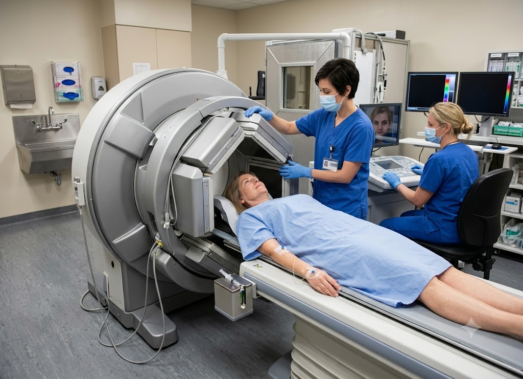
Nuclear medicine relies on tiny amounts of radioactive material, known as radiotracers, to show how organs and tissues are actually functioning inside the body. While most imaging tests capture structure, nuclear medicine captures activity blood flow, metabolism, and the presence of disease. Depending on the test, the tracer might be injected, swallowed, or inhaled. Once inside the body, a specialized camera picks up the energy it releases and turns that signal into an image doctors can interpret.
Physicians often turn to nuclear medicine when they need answers that other scans can’t give them. These tests can flag heart disease, bone problems, thyroid disorders, kidney issues, gallbladder disease, infection, inflammation, and various cancers. They can also show whether a treatment is actually working or whether a disease has come back. Below are seven of the most common nuclear medicine tests, what each one looks for, and what patients can expect along the way.
What is Nuclear Medicine and How Does It Work?
Nuclear medicine is a branch of radiology that uses trace amounts of radioactive substances to visualize both the internal workings and the structure of the body for diagnosis and, in some cases, treatment.
Core Mechanics of Functional Imaging
Nuclear medicine introduces small quantities of radioactive material into the body to capture physiological function and cellular activity in progress. Standard imaging tools X-rays, CT, and MRI mostly show anatomy. They work like a detailed photograph of an organ, picking up on its shape, density, or physical condition.
Nuclear medicine works differently: it’s a form of molecular imaging that shows how tissues and organs are behaving in real time. Instead of waiting for a disease to physically reshape an organ, this kind of imaging can pick up cellular changes, shifts in blood flow, or altered chemical processes at the molecular level. That’s often what makes it possible to catch problems tumors, restricted blood flow to the heart, or neurological disease well before they’d show up on a CT scan or X-ray.
Production and Administration of Radiopharmaceuticals
Every nuclear medicine scan depends on engineered compounds called radiopharmaceuticals, or radiotracers, which are built from two parts:
The carrier molecule: chosen because it naturally gravitates toward a particular organ, tissue, or metabolic process. The thyroid, for example, draws in iodine to make hormones, and fast-growing bone cells readily take up certain phosphate compounds. The carrier uses these existing biological routes to deliver the tracer where it’s needed.
The radioactive tag: an unstable isotope attached to the carrier. As it moves toward a more stable state, it decays and gives off energy gamma rays or positrons. Because this energy can pass through tissue, it can be picked up by a camera outside the body.
Routes of Clinical Administration
How a radiotracer enters the body depends on what’s being examined:
Intravenous injection: the most typical route. The tracer goes straight into a vein and circulates through the bloodstream to reach organs such as the heart, bones, or brain.
Oral ingestion: used to check the digestive tract, how quickly the stomach empties, or for certain thyroid procedures. The patient drinks or swallows the tracer.
Inhalation: used to examine how air moves through the lungs and to check for blood clots. The patient breathes in the tracer as a gas or fine mist.
Once the tracer reaches its target tissue, it gives off gamma photons, which are picked up by external detectors a gamma camera or a PET scanner. A computer then converts the pattern and intensity of these signals into 2D or 3D images that reflect what’s actually happening in the tissue.
Patient Safety and Radiobiological Half-Lives
Radiation safety is a common worry, but these procedures are tightly regulated and considered safe. The radiation dose from a typical scan is small often comparable to or lower than what a person gets from a routine CT scan.
Medical staff follow the ALARA principle (As Low As Reasonably Achievable), using only the minimum amount of radioactive material needed to get a clear image.
Two things keep the radiation exposure low:
Physical half-life. Isotopes used for imaging are chosen because they decay quickly. Common agents like Technetium-99m or Fluorine-18 lose most of their radioactivity within a matter of hours.
Biological clearance. At the same time, the kidneys and liver filter the tracer out of the bloodstream, and it leaves the body through urine or stool within a day or two. Patients are usually told to drink extra water after a scan to help this process along.
Because these tracers are used in such small amounts, allergic reactions are rare far less common than the reactions sometimes triggered by iodine-based contrast dye used in CT scans.
What Diseases Can 7 Common Nuclear Medicine Tests Detect?
Together, these seven tests can identify a wide range of conditions cancers, coronary artery disease, neurological disorders like Alzheimer’s, bone fractures and infections, thyroid dysfunction, kidney blockages, and gallbladder inflammation. Because they show how the body is functioning rather than just how it looks, these scans give doctors information that helps with staging disease, tracking how well treatment is working, and guiding overall care. Each scan uses a tracer matched to a specific organ or process, which keeps the results targeted and sensitive.
Positron Emission Tomography (PET) Scan
A PET scan tracks how tissue absorbs a radiotracer to measure cellular metabolism. The tracer most often used is FDG (fluorodeoxyglucose) essentially a sugar molecule labeled with radioactive Fluorine-18. Since metabolic changes tend to show up before structural ones, PET is especially good at catching disease early.
Oncology (cancer detection):
Cancer cells burn through glucose much faster than normal cells. FDG builds up heavily in these areas, lighting up as “hot spots” on the scan. This lets doctors tell benign growths apart from malignant ones, map tumor edges for biopsy or surgery, find hidden metastases for accurate staging, and check whether chemotherapy is working.
Cardiology (heart conditions):
PET can assess how well heart muscle is functioning by measuring both blood flow and glucose use. After a heart attack, it can tell the difference between tissue that’s permanently scarred and tissue that’s just “stunned” but still alive. Knowing which areas are still viable helps cardiologists decide whether procedures like bypass surgery or angioplasty are worth pursuing.
Neurology (brain disorders):
Since the brain runs almost entirely on glucose, PET can track shifts in how the brain uses it, which helps with:
- Telling dementias apart: It can spot the specific drop in metabolism seen in Alzheimer’s disease and distinguish it from frontotemporal or vascular dementia.
- Locating seizure origins: In patients whose epilepsy doesn’t respond to medication, a PET scan taken between seizures can pinpoint the exact area where they start, guiding potential surgery.
Single-Photon Emission Computed Tomography (SPECT) Scan
SPECT works much like PET but uses different tracers Technetium-99m, Thallium-201, or Iodine-123 which release single gamma photons instead of positrons. A gamma camera rotates fully around the patient, and a computer combines the readings into 3D cross-sectional images showing blood flow through tissue.
Cardiology (coronary artery disease):
SPECT is the backbone of nuclear stress testing. A tracer such as Tc-99m sestamibi is given both at rest and during exertion (typically on a treadmill), and comparing the two shows areas where blood flow is restricted due to narrowed or blocked arteries.
Neurology (cerebral blood flow):
SPECT can map blood flow in the brain, which reflects brain activity. It’s useful for spotting dead tissue after a stroke, finding hidden damage from a traumatic brain injury, and locating the source of seizures for surgical planning.
Oncology and orthopedics (bone imaging):
Paired with CT (SPECT/CT), this scan can detect small bone lesions stress fractures invisible on X-rays, deep bone infections, and early metastases from cancers like breast or prostate cancer.
The Skeletal Bone Scan
A bone scan screens the entire skeleton for metabolic changes using a technetium-labeled tracer that mimics natural bone-building material and attaches to areas where bone is actively repairing or breaking down.
Cancer metastasis screening:
Cancers such as breast, prostate, lung, and kidney cancer often spread to bone. As tumor cells invade, the body responds by remodeling the area, and the tracer collects there showing up as hot spots months before an X-ray would reveal anything.
Stress and hidden fracture detection:
For athletes with ongoing pain, a bone scan can catch tiny stress fractures or hidden breaks that don’t show up on X-rays because the bone’s shape hasn’t visibly changed. The scan instead picks up the heightened activity of bone cells working to repair the damage.
Finding osteomyelitis:
Deep bone infections can be hard to diagnose. A bone scan detects the surge in blood flow and bone turnover that comes with the body’s inflammatory response, allowing treatment to start before the infection causes lasting damage.
Thyroid Scan and Uptake Test
This test evaluates both the shape and the function of the thyroid gland, taking advantage of the gland’s natural tendency to absorb iodine. Either Iodine-123 or Technetium-99m is used.
The Radioactive Iodine Uptake (RAIU) Test
This portion measures how well the thyroid is functioning overall. After swallowing a measured dose of radioactive iodine, a probe placed over the neck checks the uptake at set intervals (around 4–6 hours and again at 24 hours).
- High uptake points to hyperthyroidism an overactive gland, often linked to Graves’ disease.
- Low uptake suggests hypothyroidism (an underactive gland) or thyroiditis (inflammation blocking iodine absorption).
The Thyroid Scintigraphy Scan
While the uptake test measures overall function, this scan creates a visual image of the gland, letting doctors classify nodules by how active they are:
- Hot nodules: Overactive tissue that absorbs the tracer heavily and produces excess hormone. These are rarely cancerous.
- Cold nodules: Inactive tissue that shows up as a blank spot. Because cold nodules carry a higher cancer risk, they usually need a follow-up biopsy.
Myocardial Perfusion Imaging (MPI)
Also known as a nuclear stress test, MPI maps blood flow to the heart both at rest and under stress.
Step 1 resting images:
The patient is given an injection of a technetium-based tracer (such as sestamibi) while at rest. It settles into heart muscle in proportion to blood flow, and a gamma camera captures the baseline picture.
Step 2 stress images:
The patient is then stressed physically, either by exercise or with a medication that dilates the coronary arteries. At peak stress, another dose of tracer is given, and images are taken of the heart working at full capacity.
Step 3 comparing the two:
- Reversible defect (ischemia): Blood flow looks normal at rest but drops noticeably under stress a sign of a significant blockage that could benefit from stenting or bypass.
- Fixed defect (infarction): Poor tracer uptake in both rest and stress images points to permanent scarring from a past heart attack tissue that won’t be helped by revascularization.
Renal (Kidney) Scintigraphy
Also called a renogram, this scan tracks blood flow, filtering ability, and drainage through the urinary system, usually with tracers like Tc-99m MAG3 or Tc-99m DTPA.
Checking for renal artery stenosis:
Narrowed arteries feeding the kidneys can cause hard-to-control high blood pressure. To check for this, the scan is paired with an ACE inhibitor. A sharp drop in filtration in one kidney after the medication confirms narrowing in that artery.
Finding urinary tract blockages:
Kidney stones, tumors, or structural issues can block the flow of urine, causing painful swelling (hydronephrosis). The scan follows the tracer as it moves through the kidney and drains into the bladder. If it pools and won’t clear even after a diuretic is given that confirms a blockage needing surgical attention.
Monitoring kidney transplants:
After a transplant, this scan offers a non-invasive way to check blood flow to the new kidney, how well it’s producing urine, and whether there are early signs of rejection or a urine leak.
Gallbladder HIDA Scan
A HIDA scan (hepatobiliary iminodiacetic acid scan) follows the path of bile from the liver, through the bile ducts, into the gallbladder, and on to the small intestine. It uses a technetium-labeled tracer that behaves like bilirubin, allowing doctors to watch digestion happen in real time.
Diagnosing acute cholecystitis:
This is sudden, painful gallbladder inflammation, usually from a gallstone blocking the cystic duct. Normally, the tracer moves from the liver into the bile ducts and gallbladder. If the gallbladder never fills even after an hour, while the liver and intestine light up normally that confirms cholecystitis.
Measuring biliary dyskinesia:
For patients with ongoing abdominal pain but no visible gallstones, this scan can measure how well the gallbladder empties. A hormone called CCK is given to trigger contraction; if too little bile is expelled, it points to a functional disorder that may be resolved by removing the gallbladder.
Spotting post-surgical bile leaks:
After gallbladder removal or a liver injury, bile can sometimes leak into the abdomen. A HIDA scan is highly sensitive at catching this, tracking tracer as it escapes normal channels and pools elsewhere helping surgeons locate and repair the leak.
What Else Should You Know About Nuclear Medicine Procedures?
Beyond diagnosing specific conditions, it helps to understand how to prepare for these scans, how they compare with other imaging methods, how precisely their tracers are targeted, and where the technology is heading. Together, these details show why nuclear medicine plays such a distinct role in modern medicine focusing on how the body functions rather than simply how it looks.
This is made possible by carefully chosen radiopharmaceuticals and imaging systems that keep advancing, offering more precision and new clinical uses, especially in oncology and neurology.
Clinical Patient Preparation Protocols
Following preparation instructions matters since these scans measure real physiological activity, skipping the guidelines can cause healthy tissue to absorb the tracer the wrong way and throw off the results.
Fasting and hydration: For heart or digestive scans and especially FDG-PET scans for cancer patients usually need to fast for 4 to 12 hours beforehand. Fasting keeps blood sugar and insulin low. Eating beforehand can cause insulin to spike, pushing the radioactive sugar into muscle instead of letting it concentrate in tumor cells. Drinking plenty of water is encouraged, since it keeps blood flow steady and helps the kidneys clear the tracer afterward.
Managing medications and stimulants:
- Before a cardiac stress test: avoid caffeine, nicotine, chocolate, and decaf drinks for at least 24 hours caffeine can block the receptors that certain stress medications rely on.
- Other medication adjustments: beta-blockers may need to be paused since they slow the heart rate, and iodine-containing medications or cough syrups should be avoided before a thyroid scan so they don’t interfere with iodine uptake.
Comfort and safety: Loose clothing without metal (zippers, underwires, snaps) is recommended, since metal can block gamma rays and create artifacts on the image. Jewelry and piercings should be removed. It’s also essential that patients who are pregnant, think they might be, or are breastfeeding tell their care team right away while doses are low, fetal tissue is sensitive to radiation, and breastfeeding mothers may need to pause nursing for a period depending on the tracer used.
Modality Matrix: Nuclear Medicine vs. CT vs. MRI
Choosing between nuclear medicine and structural imaging like CT or MRI usually comes down to one question: do you need to know what an organ looks like, or how it’s actually working?
CT and MRI focus on anatomy they produce detailed images of physical structures, borders, and densities. Nuclear medicine instead maps physiology tracking metabolism, chemical activity, and blood flow, often catching problems before they’ve changed an organ’s shape.
| Imaging Modality | How It Works | What It Shows Best | Best Used For |
|---|---|---|---|
| CT | Rotating X-ray beams measure tissue density | Detailed cross-sections of bone, vessels, organs | Emergency triage for bleeding, fractures, pulmonary embolism |
| MRI | Magnetic fields and radio pulses align hydrogen atoms | Detailed contrast in soft tissue | Ligament tears, spinal issues, brain tumors, subtle nerve injuries |
| Nuclear Medicine (SPECT/PET) | Detects gamma photons from tracers inside the body | Metabolism, blood flow, and cellular activity in real time | Early cancer detection, heart tissue viability, dementia diagnosis |
Targeted Biological Architecture of Radiopharmaceuticals
Precision in nuclear medicine comes from pairing the right radioactive tag with the right chemical carrier, so the tracer heads exactly where it’s needed.
FDG (fluorodeoxyglucose):
targets glucose metabolism. Cancer cells and active neurons use glucose quickly, and because FDG resembles regular glucose, cells absorb it then get trapped inside once altered by the cell. This creates the bright spots seen on PET scans over tumors or overactive seizure zones.
Technetium-99m medronate:
targets calcium structures in bone. When bone is injured, infected, or affected by cancer, bone-building cells rush to repair it, and the tracer settles into these active zones revealing fractures or metastases.
Radioactive iodine:
targets thyroid cells. Since the thyroid is the only organ that actively concentrates iodine to make hormones, and the body can’t distinguish stable iodine from its radioactive form, doctors use low-dose Iodine-123 to map nodules, or high-dose Iodine-131 to destroy thyroid cancer cells directly, with minimal effect elsewhere.
Next-Generation Technological Evolutions
Nuclear medicine is evolving quickly, driven by hybrid imaging systems, artificial intelligence, and targeted molecular treatments.
Hybrid imaging (PET/CT and PET/MRI):
Nuclear scans alone show metabolic activity but lack sharp anatomical detail. Combining scanners solves this:
- PET/CT: overlays a functional PET scan directly onto an anatomical CT scan taken in the same session, letting radiologists pinpoint the exact location of a hot spot relative to nearby structures.
- PET/MRI: replaces the CT with MRI, useful for brain and soft-tissue pelvic imaging, combining metabolic data with excellent soft-tissue detail while reducing radiation exposure.
Theranostics:
This approach pairs diagnosis and treatment using the same targeting molecule:
- Find the target: A diagnostic tracer, such as Gallium-68 PSMA, binds to receptors on prostate cancer cells, and a PET scan confirms the tumor expresses this target.
- Swap the isotope: The same targeting molecule is paired with a stronger, treatment-grade isotope, such as Lutetium-177, instead of the diagnostic one.
- Deliver treatment: The new radiopharmaceutical is infused and binds to the same cancer receptors identified earlier, delivering focused radiation that destroys tumor cells while sparing surrounding healthy tissue.
AI integration: Machine learning is being used to filter noise from raw scan data, which can allow lower tracer doses and shorter scan times without sacrificing image quality. AI is also being used to scan large image datasets for subtle patterns that might be easy to miss, helping catch conditions like Alzheimer’s earlier and tailoring cancer treatment more precisely.
Conclusion
Nuclear medicine tests reveal how the body is functioning, not just what it looks like. They can help catch disease earlier, evaluate how organs are working, track whether treatment is succeeding, and uncover problems that standard imaging might miss. Radiation exposure is generally low and carefully managed, though it’s still worth discussing the risks and benefits with a healthcare provider particularly during pregnancy or breastfeeding. Understanding why a particular scan has been recommended can make the whole process feel far less intimidating.
Frequently Asked Questions
1. What is nuclear medicine?
Nuclear medicine is a field of medical imaging that uses small amounts of radioactive tracers to study how organs and tissues function. The tracer moves through the body and gathers in specific areas depending on the test being done. A special camera picks up its signal and creates images doctors use to spot disease or unusual activity. Some techniques in nuclear medicine are also used for treatment, not just diagnosis.
2. What are common nuclear medicine tests?
Common tests include bone scans, thyroid scans, PET scans, cardiac stress tests, kidney scans, gallbladder scans, and lung ventilation-perfusion scans. Each relies on a different tracer to examine a specific organ or disease process for instance, a bone scan can detect cancer that’s spread to bone, while a thyroid scan evaluates nodules or an overactive gland. The choice of test depends on symptoms, history, and what the doctor is trying to investigate.
3. What can nuclear medicine detect?
It can detect a wide range of issues heart disease, cancer, bone infections, fractures, thyroid disorders, kidney problems, gallbladder dysfunction, and abnormal blood flow in the lungs. It can also confirm whether organs are functioning properly. Because these tests measure function rather than structure, they can reveal disease before changes are visible on other scans. Results are typically reviewed alongside symptoms, blood work, and other imaging.
4. Is nuclear medicine safe?
Generally, yes. The amount of radioactive tracer used is small and clears from the body over time through natural decay or excretion. Side effects are uncommon, though mild discomfort from the injection or a rare allergic reaction can occur. Patients who are pregnant or breastfeeding should inform their provider beforehand, since extra precautions may be needed.
5. How should I prepare for a nuclear medicine test?
Preparation depends on the specific scan. Some require fasting, avoiding caffeine, pausing certain medications, or drinking extra fluids before or after. It’s important to tell your care team about pregnancy, breastfeeding, allergies, kidney disease, and any medications or supplements you’re taking. Following these instructions helps ensure accurate, high-quality results.
6. How long does a nuclear medicine scan take?
This varies by test. Some are done in under an hour, while others take several hours or require delayed imaging later the same day, since the tracer sometimes needs time to reach its target organ. Your imaging center will walk you through the schedule so you know when the tracer is given and when the images will be taken.