10 Causes of Low Amniotic Fluid During Pregnancy
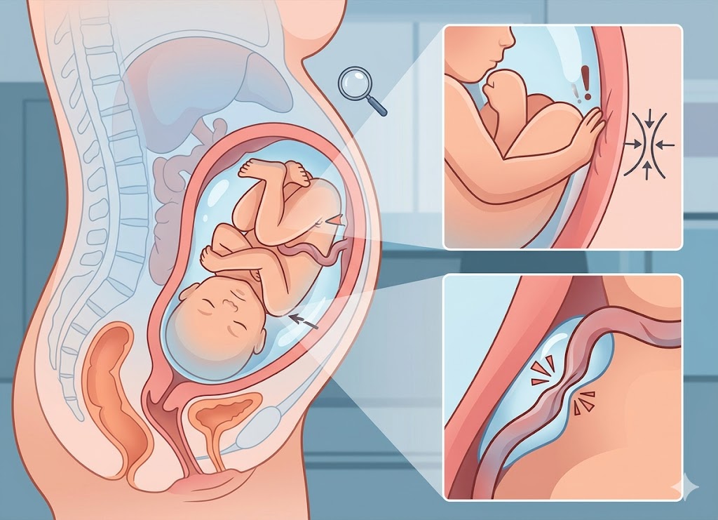
Amniotic fluid plays an essential role in supporting a healthy pregnancy. It surrounds the developing baby, cushions it from physical impact, helps maintain a stable temperature, supports lung development, and gives the fetus enough room to move. When the amount of fluid becomes lower than expected for the stage of pregnancy, it is known as oligohydramnios, or low amniotic fluid.
Understanding why amniotic fluid may become low is important because the condition can sometimes affect fetal growth and increase the risk of pregnancy complications. Some causes are temporary and manageable, while others may be associated with maternal health problems, placental issues, or fetal abnormalities.
In this article, we’ll look at 10 common causes of low amniotic fluid during pregnancy, along with its possible effects, symptoms, diagnostic methods, and potential risks.
What Is Oligohydramnios?
Oligohydramnios is a pregnancy condition in which there is less amniotic fluid surrounding the fetus than expected. Amniotic fluid, sometimes simply called the “waters,” is a clear to slightly yellow fluid that performs several important functions throughout pregnancy.
It cushions the baby from physical pressure and helps protect against injury. It also helps prevent the umbilical cord from becoming compressed, allowing oxygen and nutrients to continue reaching the fetus. In addition, the fluid provides space for movement and plays an important role in normal lung development.
The amount of amniotic fluid is constantly changing. The fetus swallows the fluid, processes it through the developing kidneys, and later produces urine, which becomes the main source of amniotic fluid during the second half of pregnancy.
When this balance is disrupted, it may indicate a problem involving the mother, placenta, or fetus. This is why low fluid levels generally require medical evaluation and monitoring.
The Normal Amount of Amniotic Fluid During Pregnancy?
The amount of amniotic fluid naturally changes throughout pregnancy. It generally increases as the pregnancy progresses and may reach approximately 800 to 1,000 milliliters around 34 to 36 weeks. After that point, the amount normally begins to decrease as delivery approaches.
During early pregnancy, much of the fluid comes from water and electrolytes that pass from the mother’s bloodstream through the membranes surrounding the fetus. As the fetus develops, its kidneys begin producing urine, which becomes the primary contributor to amniotic fluid.
For example, there may be only around 30 milliliters of fluid at 10 weeks. By approximately 16 weeks, the amount may reach about 200 milliliters and continue increasing through the later stages of pregnancy.
After about 36 weeks, a gradual decline is normal. By 40 weeks, the amount may be around 600 milliliters.
A reduction that occurs too early or becomes more significant than expected may indicate oligohydramnios. The appropriate fluid level is always considered in relation to the exact stage of pregnancy.
How Is Low Amniotic Fluid Level Medically Defined?
Doctors usually diagnose low amniotic fluid through specific measurements taken during an ultrasound examination. The two main measurements are the Amniotic Fluid Index (AFI) and the Maximum Vertical Pocket (MVP).
Oligohydramnios is commonly diagnosed when the AFI is below 5 centimeters or the MVP is below 2 centimeters. These measurements help healthcare providers determine whether the amount of fluid is below the expected range.
The method used may depend on the clinical situation, healthcare provider preference, and whether the pregnancy involves one baby or multiples.
The Amniotic Fluid Index (AFI)
The Amniotic Fluid Index is calculated by dividing the uterus into four sections. During the ultrasound, the deepest unobstructed pocket of fluid in each section is measured.
The four measurements are then added together to produce the AFI.
An AFI below 5 centimeters is generally considered diagnostic of oligohydramnios. An AFI between approximately 5.1 and 8 centimeters may be considered borderline low and can lead to closer monitoring.
The Maximum Vertical Pocket (MVP)
The Maximum Vertical Pocket, also called the Single Deepest Pocket (SDP), measures the deepest unobstructed pocket of amniotic fluid in the uterus.
Unlike the AFI method, the uterus is not divided into four sections. Instead, the sonographer looks for the largest clear pocket that does not contain fetal body parts or the umbilical cord.
An MVP of less than 2 centimeters is generally considered consistent with oligohydramnios.
This approach can be especially useful in twin or multiple pregnancies because it reduces the chance of overlapping measurements. Both AFI and MVP can help clinicians determine whether additional evaluation and fetal monitoring are needed.
10 Main Causes of Low Amniotic Fluid in Pregnancy
Low amniotic fluid can develop for many different reasons involving the membranes, placenta, mother, or fetus. Identifying the underlying cause is important because treatment and monitoring depend on what is causing the fluid level to fall.
Some causes, such as dehydration, may be temporary and reversible. Others, including certain fetal abnormalities or serious placental problems, may require more intensive medical care.
Rupture of the Amniotic Membranes
A tear in the amniotic sac is one of the most direct causes of low amniotic fluid. The fluid may escape as a noticeable gush or as a slow, ongoing leak that can sometimes be mistaken for urine or normal pregnancy discharge.
When the membranes rupture at term before labor begins, it is called Premature Rupture of Membranes (PROM). If the rupture happens before 37 weeks, it is known as Preterm Premature Rupture of Membranes (PPROM).
Even a very small tear can result in a continuous leak. Over time, the fluid may leave the amniotic sac faster than the fetus can replace it.
Doctors may use several tests to determine whether the membranes have ruptured, including:
- Visual Pooling: A sterile speculum examination is used to look for fluid collecting in the vagina.
- Nitrazine Paper Test: This test checks the pH of the fluid. Amniotic fluid is more alkaline than normal vaginal secretions.
- Fern Test: A fluid sample is dried and examined under a microscope for a characteristic fern-like pattern.
Placental Insufficiency and Vascular Dysfunction
Placental problems can contribute to low amniotic fluid because the placenta is responsible for supplying the fetus with oxygen and nutrients.
When placental blood flow becomes reduced, the fetus may receive less oxygen and nutrients. The fetus can respond by redirecting blood toward essential organs such as the brain and heart.
This adaptation, sometimes called the “brain-sparing” response, can reduce blood flow to the kidneys. Because fetal urine becomes the main source of amniotic fluid during the second half of pregnancy, reduced kidney blood flow can lead to lower urine production and, consequently, less amniotic fluid.
Post-Term Pregnancy Progression
A pregnancy that continues beyond the due date can increase the risk of oligohydramnios. As pregnancy progresses well beyond the expected delivery date, the placenta may become less efficient at supporting the fetus.
Reduced placental function can affect blood and oxygen delivery to the baby. In response, fetal blood flow may again be redirected toward essential organs, including the brain and heart.
Reduced kidney blood flow can lower fetal urine production, causing amniotic fluid levels to decline.
For this reason, pregnancies that continue beyond the expected delivery date are often monitored closely. Healthcare providers may use fetal testing and, depending on the circumstances, consider delivery.
Underlying Maternal Health Conditions
Certain maternal health conditions can affect blood flow to the placenta and contribute to low amniotic fluid.
Hypertension and Preeclampsia: High blood pressure and preeclampsia can affect blood vessels and reduce blood flow through the uterine arteries. This may decrease placental function and ultimately reduce fetal urine production.
Pre-existing Diabetes: Long-term, poorly controlled diabetes can damage blood vessels and affect placental function. Although gestational diabetes is more commonly associated with excessive amniotic fluid, advanced pre-existing diabetes may contribute to reduced fluid levels in some circumstances.
Congenital Fetal Abnormalities
When low amniotic fluid develops early in the second trimester, fetal abnormalities involving the kidneys or urinary tract may be considered.
Because fetal urine becomes the main source of amniotic fluid during the second half of pregnancy, problems that prevent the kidneys from producing urine or stop urine from reaching the amniotic space can cause a significant reduction in fluid.
Some abnormalities that may be identified during detailed fetal imaging include:
- Renal Agenesis: One or both kidneys fail to develop. When both kidneys are absent, there may be an extremely severe reduction or near absence of amniotic fluid.
- Obstructive Uropathy: A blockage in the urinary tract can prevent urine from leaving the bladder and entering the amniotic space.
- Polycystic Kidney Disease: This genetic condition can interfere with normal kidney tissue and reduce the kidneys’ ability to function properly.
Adverse Medication Side Effects
Certain medications can affect fetal kidney function and may contribute to low amniotic fluid. Medication use during pregnancy should always be reviewed with a healthcare provider.
Nonsteroidal Anti-Inflammatory Drugs (NSAIDs): These medications are generally discouraged during the third trimester because they can affect fetal circulation and kidney function, potentially reducing urine production.
ACE Inhibitors and ARBs: These blood pressure medications can interfere with fetal kidney development and function, particularly when used later in pregnancy. This can contribute to severe reductions in amniotic fluid.
Twin-to-Twin Transfusion Syndrome (TTTS)
Twin-to-Twin Transfusion Syndrome can occur in identical twin pregnancies that share one placenta.
The condition develops because abnormal blood vessel connections in the shared placenta allow blood to move unevenly between the twins.
The twin who acts as the donor may lose a significant amount of blood volume. As a result, the donor twin’s kidneys may produce less urine, leading to low amniotic fluid in that baby’s sac.
At the same time, the recipient twin may receive too much blood. This can increase blood volume, place stress on the cardiovascular system, and cause increased urine production and excessive amniotic fluid.
Intrauterine Growth Restriction (IUGR)
Intrauterine Growth Restriction occurs when a fetus does not reach its expected growth potential. Poor placental function is one of the major factors associated with IUGR.
When the placenta is unable to provide enough oxygen and nutrients, the fetus may redistribute blood flow toward the brain and heart. This can reduce blood flow to the kidneys and lower urine production.
When ultrasound shows both restricted fetal growth and low amniotic fluid, it can be an important sign of placental dysfunction and may require closer monitoring.
Acute Maternal Dehydration
Dehydration can temporarily contribute to low amniotic fluid levels. It may occur when a pregnant person does not drink enough fluids, experiences significant vomiting, or becomes severely dehydrated due to heat or illness.
Reduced maternal blood volume can affect blood flow to the uterus and placenta. This may temporarily reduce fetal urine production and lower the amount of amniotic fluid.
In cases where dehydration is the main cause, improving hydration may help restore fluid levels. Depending on the severity, this may involve drinking fluids or receiving intravenous fluids under medical supervision.
Idiopathic Oligohydramnios (Unknown Origin)
Sometimes, amniotic fluid levels become low even though doctors cannot identify a specific underlying cause. This is known as idiopathic oligohydramnios.
It is considered a diagnosis of exclusion, meaning healthcare providers first evaluate other possible explanations. They may check for membrane rupture, fetal kidney or urinary tract abnormalities, placental problems, maternal health conditions, and medication exposure.
When low fluid occurs near term without another identifiable cause, doctors may recommend closer fetal monitoring. This can include Non-Stress Tests (NSTs), Biophysical Profiles (BPPs), and repeat ultrasound examinations.
If the fluid level becomes critically low or the fetus shows signs of distress, delivery may be recommended depending on the stage of pregnancy and overall clinical situation.
Symptoms and Risks Associated with Oligohydramnios
Low amniotic fluid does not always cause noticeable symptoms. Some people only learn they have oligohydramnios during a routine prenatal ultrasound.
Possible clues include a uterus measuring smaller than expected for the stage of pregnancy or leakage of amniotic fluid.
The risks depend on several factors, including how low the fluid level is, why it became low, and how early in pregnancy the condition develops. Oligohydramnios that occurs earlier in pregnancy can present greater developmental concerns than low fluid detected close to delivery.
Signs of Low Amniotic Fluid Might a Pregnant Person Notice
Although many people do not experience obvious symptoms, certain changes may provide clues that further evaluation is needed.
Fluid Leakage
One of the clearest possible signs is fluid leaking from the vagina. It may occur as a sudden gush or as a slow, continuous trickle.
Because a small leak can resemble urine leakage or increased vaginal discharge, unusual or uncontrollable fluid loss should be reported to a healthcare provider for evaluation.
Reduced Fetal Movement
Some people may notice reduced fetal movement when amniotic fluid becomes significantly low.
Amniotic fluid gives the baby space to move, stretch, and kick. A significant reduction in fluid can restrict this space, potentially changing the frequency or strength of movements.
Any noticeable decrease in fetal movement should be discussed with a healthcare professional promptly.
Small Fundal Height
Fundal height is measured during routine prenatal visits by checking the distance from the pubic bone to the top of the uterus.
The measurement generally corresponds with the number of weeks of pregnancy. If the uterus consistently measures smaller than expected, it may indicate restricted fetal growth, low fluid, or another issue and may lead to an ultrasound for further evaluation.
Potential Health Risks for the Baby
Low amniotic fluid can create several potential risks for the baby. The severity depends greatly on when the condition develops, how severe it becomes, and how long it lasts.
Umbilical Cord Compression
Amniotic fluid provides cushioning around the umbilical cord and helps prevent it from being compressed between the fetus and the uterine wall.
When fluid levels become very low, the cord may be more easily compressed. This can temporarily reduce the flow of oxygen and nutrients to the fetus and may result in abnormal fetal heart rate patterns or fetal distress.
Pulmonary Hypoplasia (Underdeveloped Lungs)
Severe oligohydramnios that develops early in pregnancy can interfere with normal lung development.
The fetus makes breathing movements that help the lungs develop while surrounded by amniotic fluid. When fluid levels remain severely low during critical stages of lung development, the lungs may not develop normally.
This condition, called pulmonary hypoplasia, can cause serious breathing problems after birth.
Musculoskeletal and Facial Deformities
Long-lasting severe oligohydramnios can restrict the baby’s movement and leave very little cushioning space around the fetus.
Prolonged pressure may contribute to musculoskeletal problems such as joint contractures or clubfoot. In severe cases, the baby’s facial features may also be affected by prolonged compression.
Labor and Delivery Complications
Low amniotic fluid can increase the risk of umbilical cord compression during contractions. This may lead to temporary drops in the baby’s heart rate during labor.
Fetal stress may also increase the chance of meconium being passed before birth. If complications develop during labor, medical intervention, including cesarean delivery, may become necessary.
The Diagnostic Nuances and Related Amniotic Fluid Conditions
Diagnosing oligohydramnios depends largely on ultrasound measurements such as the Amniotic Fluid Index and Maximum Vertical Pocket.
These measurements help healthcare providers determine whether the fluid level is low and whether the change appears temporary or may be related to an underlying condition.
Comparing oligohydramnios with polyhydramnios, which means excessive amniotic fluid, also helps explain the importance of maintaining an appropriate fluid balance during pregnancy.
The Difference Between an AFI and MVP Measurement
Two main ultrasound techniques are used to estimate amniotic fluid levels. Although both evaluate the available fluid, they measure it in different ways.
Amniotic Fluid Index (AFI)
For an AFI measurement, the uterus is divided into four sections. The sonographer measures the deepest clear vertical pocket of fluid in each section, making sure the pocket does not contain fetal body parts or the umbilical cord.
The four measurements are then added together.
An AFI below 5 centimeters is generally considered diagnostic of oligohydramnios, while an AFI between 5.1 and 8 centimeters may be considered borderline low.
Maximum Vertical Pocket (MVP)
The Maximum Vertical Pocket, also called the Single Deepest Pocket, uses a simpler approach.
Instead of dividing the uterus into four sections, the sonographer looks throughout the uterus for the deepest unobstructed pocket of fluid.
If the deepest pocket measures less than 2 centimeters, oligohydramnios is diagnosed.
Clinical Comparison and Application
- AFI Profile: The AFI evaluates fluid from four separate areas and provides an overall measurement. However, because it combines several measurements, it may sometimes identify low fluid in situations where the clinical significance is uncertain.
- MVP Profile: MVP focuses on the single deepest pocket and can reduce some measurement variability. It is commonly used in multiple pregnancies and may be useful when assessing certain pregnancy risks.
How Does Oligohydramnios Compare to Polyhydramnios?
Oligohydramnios and polyhydramnios represent opposite problems involving amniotic fluid levels.
Oligohydramnios means there is too little fluid, while polyhydramnios means there is too much.
Low fluid may result from reduced fetal urine production or leakage from the amniotic membranes. Excessive fluid can develop when fluid is produced in greater amounts or when the fetus cannot swallow and process fluid normally.
| Clinical Feature | Oligohydramnios (Low Fluid) | Polyhydramnios (Excess Fluid) |
|---|---|---|
| Primary Mechanisms | Reduced fetal urine production due to kidney problems or placental dysfunction, or fluid loss caused by membrane rupture. | Increased fluid production or reduced fetal swallowing, including situations associated with maternal diabetes or certain fetal abnormalities. |
| Primary Risks to the Fetus | Umbilical cord compression, impaired lung development, and musculoskeletal problems. | Cord prolapse, abnormal fetal position, and fetal distress. |
| Primary Risks to the Mother | Greater likelihood of induction and possible operative delivery. | Increased risk of preterm labor, placental complications, and postpartum hemorrhage. |
Can Borderline Low Amniotic Fluid Resolve on Its Own?
Borderline low fluid levels, often described as an AFI between 5 and 8 centimeters, may improve when the underlying cause is temporary.
Reversible Factors
Dehydration can temporarily reduce amniotic fluid. When maternal fluid levels fall, blood volume may decrease, which can affect placental blood flow and fetal urine production.
Improving hydration may help restore maternal blood volume and increase placental blood flow. In some cases, fluid levels may return toward normal after appropriate hydration.
Persistent Pathological Obstacles
Hydration will not correct low fluid when the underlying cause is a more persistent problem.
Conditions such as placental insufficiency, an undetected membrane leak, fetal growth restriction, or congenital kidney abnormalities may cause fluid levels to remain low or continue declining.
Even when the fluid reduction is only borderline, continued monitoring is important. Doctors may recommend repeat ultrasounds, fetal growth assessments, and non-stress tests to monitor the baby’s condition.
The Long-Term Outlook for the Baby
The long-term outlook following a pregnancy complicated by low amniotic fluid can vary considerably.
Several factors influence the outcome, including the cause of the low fluid, when it developed, and how severe or prolonged the condition became.
1. The Root Cause of the Deficit
When low fluid results from a temporary problem such as maternal dehydration, the outlook may be favorable once the underlying issue is addressed.
In contrast, severe congenital abnormalities affecting both kidneys can have a much more serious prognosis.
2. Gestational Age at Onset
The stage of pregnancy when oligohydramnios develops is particularly important.
Severe low fluid early in the second trimester can interfere with lung development and increase the risk of pulmonary hypoplasia. When the condition develops later in pregnancy, the baby’s lungs have had more time to mature, which may reduce some developmental risks.
3. Severity and Duration of the Deficit
The longer and more severe the fluid deficiency, the greater the potential for complications.
Chronic low fluid can restrict fetal movement and contribute to musculoskeletal problems. When placental dysfunction is also present, fetal growth may be affected.
Close prenatal monitoring and appropriate medical care can help identify complications early and guide decisions about treatment and delivery.
FAQs
What is low amniotic fluid?
Low amniotic fluid, also known as oligohydramnios, occurs when the amount of fluid surrounding the baby is lower than expected for the stage of pregnancy.
What are the most common causes of low amniotic fluid?
Possible causes include dehydration, placental problems, fetal kidney abnormalities, ruptured membranes, certain medications, and pregnancies that continue beyond the expected due date.
How is low amniotic fluid diagnosed?
Oligohydramnios is generally diagnosed using ultrasound measurements such as the Amniotic Fluid Index (AFI) or the deepest vertical pocket, also called the Maximum Vertical Pocket.
Can dehydration cause low amniotic fluid?
Yes. Maternal dehydration can sometimes contribute to reduced amniotic fluid, particularly during the second and third trimesters.
Is low amniotic fluid dangerous for the baby?
It can be, particularly when the fluid level is significantly reduced or the condition develops early in pregnancy. Potential complications include restricted fetal growth, umbilical cord compression, and problems with lung development.
Can low amniotic fluid be treated?
Treatment depends on the underlying cause and how far along the pregnancy is. Management may include hydration, closer fetal monitoring, amnioinfusion during labor in certain situations, or delivery when medically appropriate.
What symptoms may suggest low amniotic fluid?
Many people do not have noticeable symptoms. Possible signs include fluid leakage, reduced fetal movement, or a uterus that measures smaller than expected.
Can low amniotic fluid improve on its own?
In some cases, particularly when dehydration or another temporary factor is responsible, fluid levels may improve after the underlying issue is addressed.
Who is at higher risk for low amniotic fluid?
Risk may be higher in pregnancies affected by high blood pressure, diabetes, placental problems, multiple pregnancy, or pregnancy that continues beyond the due date.
When should I contact my healthcare provider?
Contact your healthcare provider if you notice fluid leaking from the vagina, reduced fetal movement, contractions, or other concerning pregnancy symptoms. Prompt evaluation can help determine whether the symptoms are related to low amniotic fluid or another pregnancy complication.
Conclusion
Understanding the possible causes of low amniotic fluid can help expectant parents recognize why monitoring may be necessary. Oligohydramnios can result from several different factors, including dehydration, membrane rupture, placental problems, maternal health conditions, medications, and fetal abnormalities.
Not every case has the same level of risk. The potential effects depend on the underlying cause, how low the fluid becomes, and when during pregnancy the condition develops.
Regular prenatal care and ultrasound monitoring can help detect changes in amniotic fluid before serious complications develop. Once low fluid is identified, healthcare providers can investigate the cause and determine the appropriate monitoring or treatment plan.
If you are diagnosed with oligohydramnios, staying in close contact with your healthcare team and following the recommended care plan can help support the health and well-being of both you and your baby.