3D Mammography (Tomosynthesis): How It Helps Detect Breast Cancer
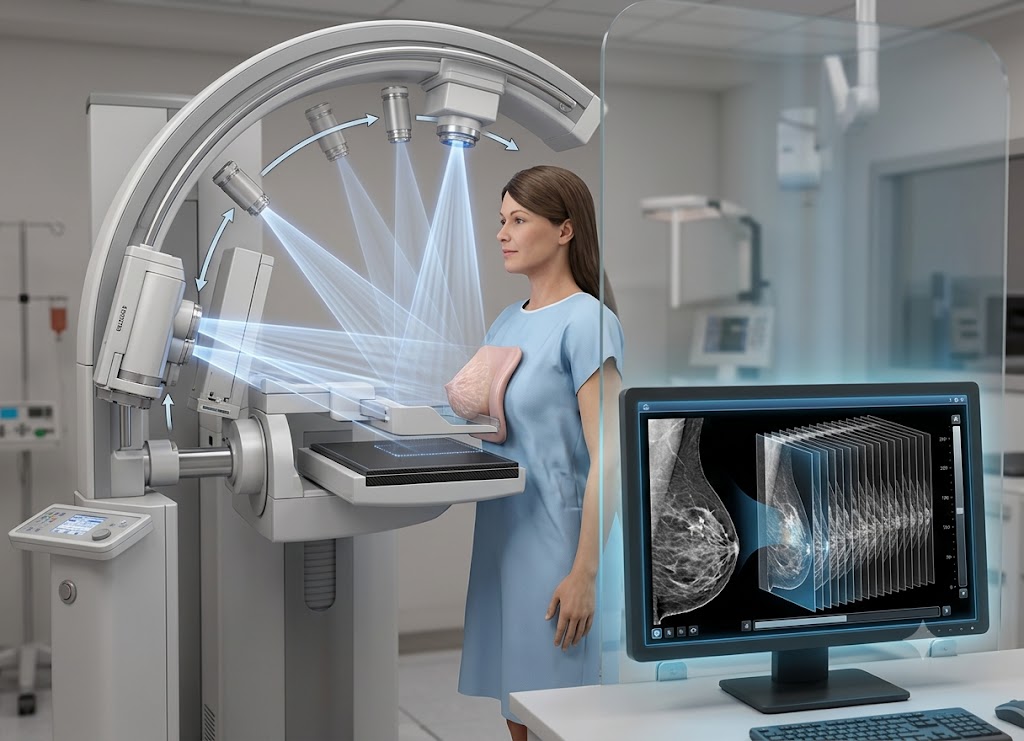
Tomosynthesis is an advanced breast imaging method that uses multiple low-dose X-ray images to create a three-dimensional view of breast tissue.
Also known as 3D mammography, it allows radiologists to examine the breast layer by layer instead of relying on a single flat image. By separating overlapping tissue, tomosynthesis can make certain abnormalities easier to see, particularly in people with dense breast tissue.
Breast cancer screening has become more detailed as imaging technology continues to improve. Tomosynthesis may help identify small breast cancers and reduce the chance that overlapping tissue hides an abnormal area.
It is often performed with digital mammography and may also reduce the need for additional imaging after unclear findings. This article explains how tomosynthesis works, how it compares with traditional mammography, what it can detect, and what patients can expect during a 3D mammogram.
What Is Tomosynthesis?
Tomosynthesis is an advanced breast imaging technique that produces a three-dimensional view of breast tissue using multiple low-dose X-ray images taken from different angles. Instead of creating only two flat images like a traditional mammogram, tomosynthesis collects several images and combines them into thin sections that can be reviewed individually.
Also called 3D mammography, tomosynthesis is used for both breast cancer screening and diagnostic evaluation. It can be particularly useful when breast tissue is dense because overlapping structures can make abnormalities harder to identify on a standard mammogram.
During the test, the breast is gently compressed while the X-ray tube moves through a small arc and captures several images. The examination is quick and uses a low level of radiation while providing radiologists with additional information about the breast.
How Tomosynthesis Works: How does 3D mammography create a clearer picture of breast tissue?
To understand what tomosynthesis is, it helps to look at how it addresses one of the main challenges of traditional mammography, overlapping breast tissue. Digital breast tomosynthesis is the medical term for what is commonly called a 3D mammogram.
Instead of viewing all breast tissue as one flat image, this technology allows radiologists to review the tissue in separate layers.
The Physics and Mechanics of Image Acquisition
The main difference between a traditional 2D mammogram and tomosynthesis is the way the X-ray images are collected.
In a conventional 2D mammogram, the X-ray tube remains in a fixed position while images are taken from standard views of the compressed breast.
During a tomosynthesis exam, the breast is compressed in a similar way, but the X-ray tube moves through a controlled arc. It captures multiple low-dose images from different angles within a few seconds.
Specialized computer software then processes these images and reconstructs them into a three-dimensional dataset. The radiologist can review the breast as a series of thin layers rather than a single flat picture.
Eliminating the Masking Effect: The Power of Slices
One of the major advantages of viewing the breast in separate slices is that it can reduce the masking caused by overlapping tissue.
On a traditional 2D mammogram, different structures in the breast are superimposed in the same image. Fat, ducts, glands, and other tissues can overlap and sometimes create shadows that look suspicious.
At the same time, a small abnormality can sometimes be hidden behind normal tissue.
Tomosynthesis helps address this problem by allowing the radiologist to scroll through individual layers. A helpful way to imagine this is to picture a forest. Looking at the forest from one angle may make it difficult to see what is behind the first row of trees. Viewing the area row by row provides a much clearer picture.
In the same way, reviewing breast tissue in slices can help radiologists determine whether an area is a true abnormality or simply overlapping normal tissue.
Improving Tumor Characterization and Identifying Distortion
By displaying breast tissue in layers, mammogram tomosynthesis can provide additional information about the shape and structure of suspicious areas.
Assessing Borders and Margins
The appearance of a mass can help doctors determine whether it is more likely to be benign or suspicious.
Benign findings such as some cysts and fibroadenomas may have smooth, well-defined borders. Some breast cancers can have irregular or spiculated edges that extend into surrounding tissue.
On a 2D image, overlapping tissue may make these borders harder to evaluate. A 3D view can separate the mass from nearby structures and provide a clearer look at its margins.
Spotting Architectural Distortion
Breast cancer can sometimes cause architectural distortion. This means the normal arrangement of breast tissue appears pulled, twisted, or disrupted, even when there is no obvious lump.
This subtle change can be difficult to recognize on a flat mammogram because surrounding tissue may overlap the affected area. Reviewing the breast layer by layer can make these structural changes easier to identify.
Precise Spatial Localization
When a suspicious area requires additional testing or a biopsy, knowing its location is important.
Because tomosynthesis collects images from multiple angles, it provides information about the depth and position of an abnormality. This can help doctors plan additional imaging or guide a biopsy when necessary.
Comparing Imaging Capabilities
The two imaging methods differ in how they collect and display breast tissue:
| Imaging Feature | Traditional 2D Mammogram | Digital Breast Tomosynthesis (3D) |
|---|---|---|
| X-Ray Tube Motion | Remains in a fixed position during each image. | Moves through a controlled arc around the breast. |
| Images Captured | Produces standard flat images of each breast. | Captures multiple low-dose images that are reconstructed into thin slices. |
| Impact of Tissue Density | Overlapping dense tissue can make abnormalities harder to see. | Separate tissue layers can make some abnormalities easier to identify. |
| False-Alarm Recalls | Overlapping tissue can sometimes create findings that require additional imaging. | Reviewing individual slices may help clarify whether a finding is real or due to overlapping tissue. |
| Distortion Detection | Some subtle structural changes may be difficult to see. | Layered images can provide a clearer view of tissue architecture. |
Benefits of Tomosynthesis: Is 3D mammography more effective at finding breast cancer than 2D mammography?
The potential benefits of a tomosynthesis mammogram have been studied in large breast cancer screening populations. Research suggests that 3D mammography can improve detection of certain breast cancers while also reducing some unnecessary callbacks in particular screening settings.
Clinical Data: Impact on Cancer Detection Rates
Studies have found that mammogram tomosynthesis can detect more invasive breast cancers than traditional 2D mammography in some populations.
Increasing Invasive Cancer Detection
Research has shown that adding tomosynthesis to breast cancer screening can increase the detection of invasive cancers compared with 2D mammography alone. The exact improvement varies between studies and can depend on factors such as age, breast density, screening practices, and the population being studied.
Why Invasive Tracking Matters
Invasive breast cancers have grown beyond the milk ducts or lobules into surrounding breast tissue and may have the potential to spread to other parts of the body.
Finding breast cancer when it is smaller and more localized can give doctors more treatment options. Depending on the cancer type and stage, treatment may include surgery, radiation, hormone therapy, chemotherapy, targeted therapy, or other approaches.
Reducing False Positives and Patient Recall Rates
Another potential benefit of tomosynthesis mammography is that it may reduce the number of patients called back for additional imaging.
Eliminating the “Summation Artifact”
On a traditional 2D mammogram, normal breast structures can overlap and create a dense area or shadow. This is sometimes called a summation effect or summation artifact.
The finding may look suspicious even though it is simply normal tissue positioned on top of itself.
With tomosynthesis, radiologists can review the breast one layer at a time. This may help determine whether a suspicious-looking area is a true mass or simply overlapping tissue.
The Impact on Patients
Fewer unnecessary callbacks can reduce anxiety and the need for additional tests in some patients. A clearer initial screening result may also reduce the time and expense associated with follow-up imaging.
However, callbacks are sometimes necessary and do not automatically mean that breast cancer is present.
High-Density Breast Tissue: The “Polar Bear in a Snowstorm”
Tomosynthesis may be particularly helpful for people with dense breast tissue.
The Camouflage Challenge
On mammograms, fatty tissue generally appears darker, while dense glandular and fibrous tissue appears white. Many breast abnormalities also appear white.
When a suspicious area is surrounded by dense tissue, it can therefore be harder to see on a traditional 2D mammogram. This is one reason breast density can affect mammography performance.
Dense breast tissue is also associated with a higher risk of developing breast cancer, making appropriate screening particularly important.
Overcoming the Barrier
Digital breast tomosynthesis can reduce the impact of overlapping dense tissue by displaying the breast in separate layers.
This layered view may help radiologists identify masses, distortions, and other changes that could be difficult to distinguish on a traditional flat image.
High-Risk Candidates and the Shift in Standards of Care
Breast density is not the only factor that may influence the use of 3D mammography. Personal and family history can also affect screening decisions.
- Personal History of Breast Cancer: People who have previously had breast cancer may require closer surveillance. Surgical changes, scarring, or other changes in the breast can sometimes make imaging more challenging, and additional imaging may be useful depending on the situation.
- Genetic Factors and Family History: People with certain inherited genetic changes, such as BRCA1 or BRCA2, or those with a strong family history of breast cancer may have a higher lifetime risk. Their healthcare team may recommend a more personalized screening plan.
The New Standard of Care
Tomosynthesis has become widely available and is commonly used for breast cancer screening. Some professional organizations support its use as an option for routine screening, although recommendations can vary based on age, risk level, breast density, and local guidelines.
For people at increased risk, healthcare providers may also recommend additional screening methods based on individual circumstances.
Other important considerations for a tomosynthesis exam
A tomosynthesis appointment is similar to a traditional mammogram. Knowing what happens during the exam, as well as understanding radiation exposure and possible insurance issues, can help patients feel more prepared.
The Appointment Experience and What to Expect
If you are wondering what is a 3d mammogram like, the process is generally very similar to having a standard mammogram.
Pre-Exam Preparation
Patients are often asked to avoid deodorant, antiperspirant, powder, or lotion on the breasts and underarms on the day of the exam.
Some of these products can contain substances that appear as small marks on an X-ray image and may interfere with image interpretation.
It is also important to tell the imaging staff if you are pregnant or could be pregnant.
During the Scan
A technologist positions the breast on the imaging platform and gently compresses it with a plastic paddle. Compression helps spread the breast tissue, reduces movement, and improves image quality.
During tomosynthesis, the X-ray tube moves through a small arc while capturing multiple low-dose images. You will need to remain still and may be asked to hold your breath briefly.
The compression may feel uncomfortable for some people, but the actual imaging process is usually completed within a few seconds for each view. A tomosynthesis bilateral exam evaluates both breasts.
Radiation Safety, Machine Availability, and Insurance
Understanding the tomosynthesis meaning also involves considering its radiation exposure, availability, and cost.
Managing Radiation Exposure
Tomosynthesis uses X-rays, so it involves exposure to a small amount of radiation. Depending on the equipment and whether a separate 2D image is also obtained, the radiation dose can be higher than that of a standard digital mammogram.
Many modern systems can create a synthetic 2D image from the tomosynthesis data. This can reduce the need for a separate 2D exposure in some settings.
The imaging team uses the lowest practical radiation dose needed to obtain useful diagnostic images.
Logistical and Insurance Factors
Although digital breast tomosynthesis is increasingly available, patients may still encounter practical differences depending on where they receive care.
- Regional Availability: Not every imaging center has 3D mammography equipment, particularly in some smaller or rural areas.
- Reading and Interpretation Time: Tomosynthesis produces substantially more image data than traditional mammography, so radiologists may need additional time to review the images.
- Insurance Coverage: Coverage for 3D mammography can vary depending on the healthcare system, insurance plan, location, and whether the exam is being used for screening or diagnostic purposes. Checking with your insurance provider before the appointment can help avoid unexpected costs.
Understanding Your Results and the BI-RADS Report
Your mammogram with tomosynthesis report may use the Breast Imaging Reporting and Data System, commonly called BI-RADS. This standardized system helps radiologists describe breast imaging findings and recommend appropriate follow-up.
The categories range from 0 to 6:
- BI-RADS 0: Incomplete; additional imaging or comparison with previous images is needed.
- BI-RADS 1 or 2: Negative or benign findings.
- BI-RADS 3: Probably benign; short-term follow-up is usually recommended.
- BI-RADS 4 or 5: Suspicious findings that may require a biopsy.
- BI-RADS 6: Cancer that has already been confirmed by biopsy.
The use of tomosynthesis does not change the BI-RADS system itself. Instead, it provides radiologists with additional image information that may help them evaluate certain findings more clearly.
A synthetic 2D image may also be available for comparison with previous mammograms, depending on the equipment and imaging center.
Structural vs. Functional Imaging: Tomosynthesis vs. CEM
People researching advanced breast imaging may also wonder how tomosynthesis compares with Contrast-Enhanced Mammography, or CEM.
The key difference is what each test is designed to show. Tomosynthesis mainly evaluates the structure and appearance of breast tissue, while CEM uses contrast to provide information about how breast tissue takes up the contrast agent.
Tomosynthesis (Structural Imaging)
Tomosynthesis focuses on the physical structure of the breast. By separating overlapping tissue into thin layers, it can help radiologists identify masses, calcifications, architectural distortion, and other structural changes.
It is commonly used for breast cancer screening and for evaluating certain abnormal or unclear mammogram findings.
Contrast-Enhanced Mammography (Functional Imaging)
CEM involves injecting an iodine-based contrast material into a vein before breast imaging. The contrast can highlight areas with increased blood flow, which may occur in some cancers.
Because CEM requires an injection and specialized imaging, it is generally used for selected diagnostic or high-risk situations rather than as a routine screening test for everyone.
Doctors may use CEM to further evaluate certain unclear findings, assess the extent of a known breast cancer, or help with treatment planning.
Conclusion
Tomosynthesis is an important development in breast imaging that provides a more detailed, layered view of breast tissue than traditional 2D mammography. By collecting images from multiple angles, it can reduce the effect of overlapping tissue and help radiologists identify certain abnormalities more clearly.
Although no breast cancer screening test can detect every cancer, 3D mammography can be a useful part of screening and diagnostic care. Your age, breast density, personal history, family history, and other risk factors can all influence which screening approach is appropriate. Talk with your healthcare provider about your individual screening needs and whether tomosynthesis is a suitable option for you.
Frequently Asked Questions
1. What is tomosynthesis?
Tomosynthesis is a breast imaging technique that uses multiple low-dose X-ray images taken from different angles to create a three-dimensional view of breast tissue. The images are reconstructed into thin layers that radiologists can examine individually. This can reduce the effect of overlapping tissue and make certain abnormalities easier to see.
2. How does tomosynthesis differ from a regular mammogram?
A traditional mammogram produces two-dimensional images, while tomosynthesis uses multiple X-ray views to create a layered 3D image. The additional views can help radiologists examine breast tissue more clearly, particularly when overlapping or dense tissue makes a standard mammogram harder to interpret.
3. Can tomosynthesis detect breast cancer earlier?
Tomosynthesis can improve the detection of some breast cancers compared with traditional 2D mammography alone. Research has found that it may detect more invasive cancers in certain screening populations. However, no imaging method can identify every breast cancer, so regular screening and appropriate follow-up remain important.
4. Is tomosynthesis safe?
Tomosynthesis is widely used for breast imaging and involves a low dose of X-ray radiation. The exact radiation exposure depends on the equipment and imaging method used. Patients who are pregnant or may be pregnant should inform their healthcare provider or imaging technologist before the exam.
5. What happens during a tomosynthesis exam?
The breast is placed on an imaging platform and gently compressed, similar to a traditional mammogram. The X-ray tube then moves through a small arc and takes multiple images from different angles. Each view usually takes only a few seconds, although the overall appointment may take longer because of positioning and preparation.
6. Who may benefit from tomosynthesis?
Tomosynthesis may be useful for many people undergoing breast cancer screening and can be particularly helpful when breast tissue is dense or previous mammogram findings are unclear. It may also be used to investigate symptoms or suspicious findings. The most appropriate screening method depends on individual risk factors and recommendations from a healthcare provider.