Ruptured Spleen Warning Signs: 8+ Symptoms You Should Never Ignore
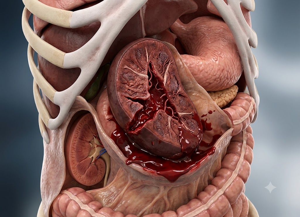
A ruptured spleen is a medical emergency in which the organ tears open and blood escapes into the abdominal cavity. Sitting on the upper left side of the abdomen, the spleen filters blood, helps fight infection, and recycles aging blood cells. Because it holds such a rich blood supply, a rupture can trigger heavy internal bleeding that turns life-threatening within a short window. Recognizing the warning signs quickly and getting emergency help is what makes the difference.
Most ruptured spleens follow blunt trauma to the abdomen a car crash, a sports collision, or a hard fall though infections, blood disorders, or a spleen that’s already enlarged can also set the stage for a tear. Symptoms sometimes show up right after the injury, and other times they build slowly over hours or even days. Watch for severe abdominal pain, dizziness, fainting, a racing pulse, and other signs the body is heading into shock. Below is a closer look at more than eight warning signs of a splenic rupture, what typically causes one, and how it’s diagnosed and treated.
What Is a Ruptured Spleen?
A ruptured spleen means the organ’s outer capsule has torn, letting blood spill into the abdominal cavity. Tucked under the rib cage on the upper left side of the abdomen, the spleen filters worn-out blood cells, stores platelets, and supports the immune system. Its capsule surrounds a dense network of blood vessels, so once it tears, bleeding can escalate fast making this one of the more dangerous abdominal injuries a person can have.
Blunt trauma a vehicle collision, a sports hit, a fall accounts for most cases. But a spleen can also give way on its own if it’s already swollen from something like mononucleosis, a blood cancer, or an inflammatory condition. Because symptoms can surface immediately or creep in over the following days, prompt evaluation matters: left unaddressed, the blood loss can progress to shock or worse.
Signs of a Ruptured Spleen: What are the 8+ warning signs that indicate a splenic rupture?
The signs clinicians watch for most closely are pain and tenderness in the upper left abdomen, pain that radiates to the left shoulder (called Kehr’s sign), confusion, dizziness or lightheadedness, blurry vision, fainting, and the pale skin and rapid heartbeat typical of shock. One important caveat: none of these have to appear right away some can take hours or days to surface.
It helps to think of them in three groups pain felt right at the site, pain that’s referred elsewhere in the body, and the broader signs of internal blood loss. The sections below break down each category.
Pathophysiology of Splenic Trauma
The spleen sits in the upper left abdomen, tucked behind the 9th, 10th, and 11th ribs, and serves as the body’s main blood filter and reservoir. It draws a heavy volume of blood straight from the aorta through the splenic artery. Inside, it’s made of soft, spongy tissue red and white pulp wrapped in a thin capsule that offers little structural support.
Because that inner tissue is so vascular and fragile, a blunt hit to the lower left torso can squeeze the organ against the ribs hard enough to tear the capsule or fracture the tissue beneath it.
Once torn, blood pours into the space around the abdominal organs turning what started as a localized injury into a body-wide emergency.
Clinical Progression of Localized Abdominal Signs
Symptoms in the abdomen tend to unfold in a fairly predictable order. How fast and how badly depends on whether the capsule holds (leading to a hematoma that ruptures later) or gives way right away.
Pain in the upper left abdomen: While blood is still trapped under an intact capsule, the person may only feel a dull ache. Once the capsule tears, the pain sharpens and becomes constant worse with movement, coughing, or deep breaths, since the diaphragm pushes down against the injured area as the lungs expand.
Peritoneal irritation: Blood pooling in the abdomen irritates the lining of the cavity, which causes the abdominal muscles to tense up involuntarily as a protective reflex.
More advanced signs:
- Rebound tenderness a sharp jump in pain when pressure on the abdomen is suddenly released, pointing to widespread irritation of the abdominal lining.
- Ballance’s sign a fixed area of dullness when tapping the left flank, along with a dullness on the right side that shifts with position, both hinting at a large pool of clotted blood near the spleen.
Neurological Mechanisms of Referred Pain (Kehr’s Sign)
One telltale symptom of a splenic rupture is Kehr’s sign a sudden, sharp pain at the tip of the left shoulder. It’s a case of referred pain: the brain misreads where the signal is actually coming from because certain nerve pathways overlap.
When someone lies flat or tilts head-down, blood pooling in the abdomen drifts upward and collects under the left side of the diaphragm. The diaphragm is controlled by the phrenic nerve, which branches from the C3 through C5 spinal nerve roots in the neck.
Those same nerve roots also carry sensation from the skin at the tip of the shoulder. So when the pooled blood irritates the phrenic nerve, the pain signals mix with shoulder-sensation signals at that same spinal level.
Since the brain is far more used to reading signals from skin than from internal organs, it assumes the pain is coming from the shoulder rather than from deep inside the diaphragm.
Systemic Cascade of Hemorrhagic Shock
As blood loss continues, the body’s total blood volume drops, and the patient moves through the recognizable stages of hemorrhagic shock.
Compensatory stage (fast heart rate):
As blood volume falls, pressure sensors in the major arteries trigger a surge of adrenaline. This pushes the heart rate above 100 beats a minute to keep blood moving. Blood pressure can still look normal at this point because the heart is working harder to compensate.
Decompensation (blood gets rerouted):
To protect the brain and heart, the body narrows blood vessels near the skin and limbs, redirecting blood to the core. That’s what produces the pale, cool, clammy skin associated with shock.
Late stage (the brain starts losing blood flow):
If bleeding isn’t controlled, the heart eventually can’t keep up with the brain’s oxygen needs.
- Early signs: lightheadedness, dizziness, tunnel vision or blurred vision.
- Later signs: anxiety, restlessness, confusion.
- Collapse: blood pressure drops, and the person faints as circulation to the brain fails.
Diagnostic Grading & Trauma Protocols
Trauma teams rely on fast imaging and standardized scoring systems to size up the injury and decide next steps.
FAST exam (Focused Assessment with Sonography for Trauma): A quick bedside ultrasound, done within seconds of arrival, aimed at the space between the spleen and left kidney. A dark pocket on the scan signals active bleeding.
Contrast CT scan: The gold-standard test for patients who are stable enough to undergo it it maps exactly how deep the tears run and checks for ongoing bleeding.
AAST Splenic Injury Grading
| Grade | Tissue Damage | What Imaging Shows | Typical Next Step |
|---|---|---|---|
| I | Small tear or minor hematoma under the capsule | Under 10% of surface affected; tear less than 1 cm deep | Watchful waiting with bed rest and repeat blood tests |
| II | Moderate hematoma or laceration | 10–50% of surface affected; tear 1–3 cm deep, vessels untouched | Close ICU monitoring with frequent vital checks |
| III | Larger hematoma or deeper tear | Over 50% of surface affected; tear deeper than 3 cm or reaching internal vessels | ICU care, often paired with angioembolization to block the bleeding vessel |
| IV | Major tissue damage, blood supply compromised | Significant vessel injury cutting off flow to more than a quarter of the spleen | Surgery if the patient becomes unstable, otherwise aggressive embolization |
| V | Spleen shattered or its main vessel torn away | Total loss of structure and blood supply | Emergency splenectomy to stop fatal bleeding |
Complications of a Ruptured Spleen: What is a ruptured spleen and why is it a life-threatening emergency?
A ruptured spleen happens when the organ’s capsule tears and blood leaks into the abdominal cavity. It’s dangerous precisely because the spleen holds so much blood a rupture can cause rapid, massive blood loss and push the body into hemorrhagic shock, which can be deadly without fast treatment.
To see why this matters so much, it helps to understand what the spleen does and what happens when it fails. The immediate danger is blood loss, but there’s also a longer-term infection risk to manage once the organ is gone.
Vascular Hemodynamics of Splenic Hemorrhage
The spleen bleeds so dangerously because of how its internal blood vessels are arranged. It handles roughly 5% of the body’s cardiac output about 250 to 300 milliliters of blood pass through it every minute.
Inside, blood moves from small arterioles into an open network of tissue called red pulp, rather than staying contained in thick-walled vessels. That open, sponge-like structure is efficient for filtering blood, but it also means there’s little to slow the flow once something goes wrong.
The organ’s thin capsule offers minimal resistance. Once it tears, there’s nothing holding the blood back it drains straight into the abdominal cavity, often faster than it’s noticed.
Pathophysiology of Post-Splenectomy Complications
When surgeons remove the spleen (a splenectomy) to control bleeding, the patient permanently loses the organ’s filtering and immune functions, leaving a lasting gap in their defenses.
Loss of immune filtering: The spleen’s white pulp is packed with immune cells that specialize in hunting down bacteria with a slippery outer coating like Streptococcus pneumoniae, Haemophilus influenzae, and Neisseria meningitidis. Without the spleen catching these organisms, they can multiply unchecked in the bloodstream.
The OPSI risk: That unchecked growth can trigger Overwhelming Post-Splenectomy Infection (OPSI) a fast-moving form of sepsis that can go from mild, flu-like symptoms to full septic shock and organ failure in under a day. Because that risk never fully goes away, lifelong preventive care is essential for anyone without a spleen.
Post-Splenectomy Patient Management Timeline
Losing the spleen means a much higher lifetime infection risk, so care teams follow a structured schedule of vaccines and medication.
- Days 1–14 (early recovery): Blood counts are watched closely. Platelet counts often spike since the spleen normally stores platelets. Compression stockings or blood thinners may be used to lower clotting risk.
- Around week 2 (first vaccines): For emergency surgeries, key vaccines are given roughly two weeks out, once the immune system has had time to recover from the operation and can respond well. This covers the three major encapsulated bacteria: pneumococcus, meningococcus, and Hib.
- Years 1–5+ (daily antibiotics): Low-dose antibiotics such as penicillin or amoxicillin are often prescribed daily, especially in the early years when OPSI risk is highest.
- Ongoing, for life: Booster vaccines every five years keep antibody levels up. Patients are also advised to keep emergency antibiotics on hand and seek care right away at the first sign of fever.
Causes of a Ruptured Spleen: What events or conditions can cause the spleen to rupture?
Splenic ruptures fall into two categories: trauma, which is by far the more common cause, and spontaneous (non-traumatic) rupture, which usually points to an underlying illness that’s already weakened the spleen. Traumatic ruptures come from a direct hit to the abdomen; spontaneous ones happen when an enlarged, fragile spleen gives way with little or no outside force.
Knowing the difference matters both for preventing rupture in people already at risk and for helping doctors quickly figure out what’s going on in an emergency.
Traumatic Biomechanics of Splenic Rupture
Trauma is the leading cause of splenic rupture, usually from a strong, sudden force striking the left upper abdomen or lower chest, squeezing the spleen against the spine or rib cage.
Rapid deceleration and seatbelt injuries: In a car crash, the sudden stop can drive the seatbelt or steering wheel hard into the left side of the torso. That compression spikes the pressure inside the spleen’s blood-filled tissue, bursting the capsule.
Direct, focused impact:
- Contact sports: A hard hit in football, rugby, lacrosse, or hockey a helmet strike or a stick to the flank can concentrate enough force on the spleen to tear it.
- Falls from height: A fall from a ladder, roof, or stairs can channel the full impact of the fall into a single point on the body.
Rib fractures piercing the spleen: The lower left ribs normally shield the spleen, but under enough force they can break. The jagged edges of a fractured rib can then puncture the capsule and cut into the tissue beneath it.
Pathophysiology of Atraumatic (Spontaneous) Rupture
A spontaneous rupture happens without any real outside force, almost always because an underlying condition has caused splenomegaly an enlarged spleen.
A healthy spleen is compact and well-protected by the ribs. Disease can make it swell, leaving the tissue heavy and fragile while the capsule stretches thin.
In that stretched state, the capsule loses its give. Everyday pressure changes in the abdomen a hard cough, a sneeze, vomiting, even rolling over can be enough to tear it.
Clinical Etiology Matrix of Underlying Conditions
A range of illnesses can enlarge the spleen and raise the odds of a spontaneous tear.
| Category | What’s Happening | Examples | Managing the Risk |
|---|---|---|---|
| Blood cancers | Abnormal white cells flood the spleen, causing it to swell | Chronic lymphocytic leukemia, acute myeloid leukemia, Hodgkin and non-Hodgkin lymphoma | Regular ultrasounds to track spleen size; avoid heavy lifting or straining |
| Infections and inflammation | An overactive immune response engorges the spleen | Mononucleosis (Epstein-Barr virus), malaria, typhoid fever, endocarditis | No contact sports for 3–4 weeks with mono, until an ultrasound shows normal size |
| Vascular congestion | High pressure in liver veins backs blood up into the spleen | Cirrhosis, portal hypertension, splenic artery aneurysms | Beta-blockers to ease portal pressure; surgical shunts when needed |
| Metabolic storage disorders | Abnormal proteins or fats build up inside the spleen | Gaucher disease, amyloidosis, sarcoidosis | Enzyme replacement therapy to slow the buildup |
Diagnostic Timeline for Suspected Splenic Injury
When someone arrives at the ER after left-sided trauma, or with unexplained pain in the upper left abdomen, doctors move through a set sequence.
- Minutes 1–5 initial check: Vital signs are checked for early shock a heart rate above 100 or dropping blood pressure and large IV lines are placed in case fluids or a transfusion are needed.
- Minutes 5–10 FAST scan: A bedside ultrasound scans the space near the spleen and left kidney. A dark fluid pocket points to active bleeding and helps guide immediate decisions.
- Minutes 15–45 CT scan: If vitals are stable, a contrast CT scan gives a detailed picture, letting doctors see how deep the tear goes, whether the capsule is intact, and assign an official injury grade.
Emergency Care for a Ruptured Spleen: What immediate steps should be taken for a suspected ruptured spleen?
The single most important step is calling 911 (or your local emergency number) right away this is a time-critical, life-threatening situation that needs advanced medical care fast. After the call, keep the person as calm and still as possible until paramedics arrive. Driving to the hospital yourself is strongly discouraged.
Should you call 911 for a suspected ruptured spleen?
Yes without hesitation. This is a genuine emergency where minutes matter, since a ruptured spleen can cause massive bleeding and shock in a short span of time. Driving the person yourself carries real risks.
Paramedics are trained specifically for trauma and shock. They can start IV fluids, give oxygen, and track vital signs the whole way to the hospital care that can make the difference between life and death. The motion of getting into a car and the bumps of the drive can also worsen internal bleeding, while EMS crews know how to move and stabilize a patient safely. On top of that, paramedics can radio ahead so the trauma team is ready and waiting the moment the patient arrives. And if the person’s condition suddenly worsens en route losing consciousness, for instance an ambulance is equipped to respond immediately, unlike a personal car. All of this is why calling 911 is the right first move, every time.
What can you do while waiting for medical help to arrive?
While you wait, there are a few simple things that can help keep the person stable and avoid making things worse.
Have them lie down on their back, and if it doesn’t cause more pain, raise their feet about 12 inches. This “shock position” uses gravity to help keep blood flowing toward the brain and heart. Cover them with a blanket blood loss can bring on hypothermia quickly, and shivering burns energy and oxygen the body can’t spare right now.
Keep the person calm and still stress and movement can raise heart rate and blood pressure, which may speed up internal bleeding. Speak calmly and reassuringly. Don’t give them anything to eat or drink, since an empty stomach lowers the risk of complications if emergency surgery is needed. And don’t press on the abdomen or apply heat or ice unlike external bleeding, pressure won’t stop internal bleeding, and it could actually dislodge a clot or worsen the injury. Your job is simply to keep them steady until trained help takes over.
What are the diagnostic procedures, recovery outlook, and special cases for a splenic rupture?
Diagnosis combines a physical exam with imaging, and recovery depends heavily on how severe the injury is including tricky cases like delayed rupture, which need careful follow-up.
Clinical Diagnostic Protocols in the Emergency Room
When someone arrives with suspected trauma to the upper left abdomen, or signs of internal bleeding, the ER team runs through a set process.
Physical exam: Doctors check for signs of circulatory collapse a heart rate above 100, blood pressure under 90 mmHg, pale and clammy skin. They also press on the abdomen to check for rigidity, guarding, and rebound tenderness. A positive Kehr’s sign sharp pain at the left shoulder tip is a strong clue that blood is pooling under the diaphragm.
FAST ultrasound: For patients whose vital signs are unstable, this rapid bedside scan is the first move, done in under five minutes. It targets the space between the spleen and left kidney; a dark, fluid-filled pocket confirms bleeding. If the scan is positive in an unstable patient, they go straight to surgery no further testing needed.
Contrast CT scan: For patients who are stable, a CT scan is the definitive test. The contrast dye highlights blood flow, letting radiologists pinpoint the tear, measure any hematomas, spot ongoing bleeding, and assign an official grade all of which shapes whether surgery is needed.
Solid Organ Injury Comparison Matrix
The spleen is the organ most often injured in blunt abdominal trauma, but the liver and kidneys carry similar risks, each with its own pattern of symptoms.
| Organ | Where the Pain Shows Up | Key Clue | Special Risk | Long-Term Outlook |
|---|---|---|---|---|
| Spleen | Upper left abdomen, sometimes radiating to the left shoulder | Kehr’s sign | Lifelong OPSI risk if removed | Immune filtering is permanently lost if removed |
| Liver | Upper right abdomen, sometimes radiating to the right shoulder | Fractured lower right ribs, abdominal swelling | Bile leaking into the abdomen can cause severe inflammation | Excellent the liver regenerates well |
| Kidney | Flank or lower back pain | Blood in the urine | Urine leaking into surrounding tissue can cause localized infection | Excellent one healthy kidney can usually compensate |
Treatment and Recovery Pathways
Treatment depends on how severe the injury is and how stable the patient’s vital signs remain.
Watching and waiting: For milder to moderate injuries (Grades I–III) in stable patients, doctors often try to preserve the spleen. That means ICU monitoring, strict bed rest, and blood tests every four to six hours to catch any drop in hemoglobin. Interventional radiologists may also perform angioembolization threading a catheter into the splenic artery and sealing off the bleeding vessel with coils or gel foam, stopping the bleed while keeping the rest of the organ intact. Full recovery can take weeks, and heavy lifting or contact sports are off-limits for months afterward to avoid re-injury.
Surgery: If vital signs collapse or the spleen is severely damaged, surgery becomes necessary right away.
- Splenorrhaphy repairing the tear with sutures, clotting agents, or a mesh wrap, used whenever the spleen can realistically be saved.
- Splenectomy full removal of the spleen, reserved for cases where the bleeding can’t otherwise be controlled.
Long-term care after splenectomy: Without a spleen, the body loses its ability to clear certain bacteria, so patients follow a lifelong prevention plan vaccines against pneumococcus, meningococcus, and Hib around two weeks after surgery, boosters every five years, and often daily low-dose antibiotics for the first several years, when infection risk is highest.
The Anatomy of a Delayed Splenic Rupture
A delayed rupture is a rare pattern where the classic signs of internal bleeding don’t show up until hours, days, or even weeks after the original injury.
- Day 0 the initial injury: Blunt trauma tears the internal tissue of the spleen, but the outer capsule holds, trapping the bleeding in a hematoma beneath it.
- Days 1–10 a false sense of stability: The pressure of the trapped blood clot acts like a plug, temporarily stopping further bleeding. The person’s pain may fade, making the injury seem minor.
- Day 14 and beyond sudden rupture: As the clot breaks down, or with something as small as a cough or a lift, pressure spikes and the stretched capsule finally gives way. Blood floods the abdominal cavity, causing sudden rigidity, a drop in blood pressure, and shock.
A word of caution: This delayed pattern is exactly why anyone who takes a hard hit to the lower left ribs should get checked out, even if they feel completely fine right after it happens.
Conclusion
A ruptured spleen is a life-threatening condition that calls for immediate medical attention. Severe pain in the upper left abdomen, pain radiating to the left shoulder, dizziness, fainting, a racing heart, confusion, pale skin, or any signs of shock should never be brushed off especially after an injury. Emergency care may involve IV fluids, blood transfusions, close monitoring, or surgery to repair or remove the spleen, depending on how severe the bleeding is. Getting help quickly gives the best chance at a full recovery and lowers the risk of serious complications.
Frequently Asked Questions
1. What is a ruptured spleen?
A ruptured spleen happens when the organ’s outer capsule tears, letting blood leak into the abdominal cavity. Because the spleen is so richly supplied with blood vessels, even a small tear can cause significant bleeding. It’s treated as a medical emergency, and without prompt care, the blood loss can become life-threatening.
2. What are the warning signs of a ruptured spleen?
Typical signs include severe pain in the upper left abdomen, pain radiating to the left shoulder, dizziness, fainting, weakness, a fast heartbeat, low blood pressure, pale skin, and confusion. Some people also notice abdominal swelling or tenderness. These signs can appear right after an injury or build up gradually if the bleeding is slower. Anyone with a suspected ruptured spleen should be seen in an emergency department right away.
3. What causes a ruptured spleen?
Blunt trauma from car accidents, contact sports, falls, or physical assault is the most common cause. Less often, infections like mononucleosis, blood cancers, inflammatory diseases, or an already enlarged spleen can make rupture more likely, sometimes with little or no outside force involved. What caused the rupture often shapes how it’s treated.
4. How is a ruptured spleen diagnosed?
Diagnosis combines a physical exam, blood tests, and imaging. Ultrasound can quickly detect internal bleeding in an emergency, while a contrast CT scan is the usual choice for assessing the injury in stable patients. Blood tests help gauge how much blood has been lost. Because treatment decisions hinge on the severity of bleeding, fast diagnosis is essential.
5. Can a ruptured spleen heal without surgery?
In some cases, yes if the patient is stable and the bleeding is limited, treatment may involve hospitalization, bed rest, IV fluids, repeat imaging, and close monitoring rather than surgery. But if bleeding is severe or vital signs are unstable, emergency surgery or a bleeding-control procedure is usually needed. The right approach depends on the extent of the injury and the person’s overall condition.
6. When should I go to the emergency room for a ruptured spleen?
Seek emergency care right away if you notice symptoms of a ruptured spleen, especially following an abdominal injury. Severe abdominal pain, left shoulder pain, dizziness, fainting, trouble breathing, confusion, a racing heartbeat, or any signs of shock all call for urgent evaluation. Don’t drive yourself if you feel faint or unstable calling emergency services is the safer choice.