6 Open-Angle Glaucoma Symptoms to Protect Your Vision
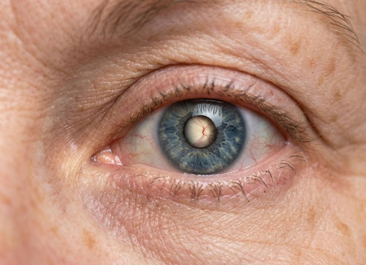
Open-angle glaucoma is often called “the sneak thief of sight,” and the name fits. It is the most common type of glaucoma, making up about 80% to 90% of cases. Over time, the eye’s drainage channels clog, and fluid can no longer leave the eye as it should.
Pressure then builds inside the eye and strains the optic nerve, the cable that carries images from the retina to the brain. Damage to this nerve is permanent.
The biggest problem is that the early disease gives no warning. There is no pain, no redness, no discomfort, and no blurriness. Your brain covers for small blind spots by leaning on your better eye and filling in the gaps. By the time you notice a shadow or a loss of clarity, up to 40% of your optic nerve fibers may already be gone.
Regular comprehensive eye exams are therefore essential, and paying attention to subtle changes in your vision adds another layer of protection. Below, you’ll find the 6 symptoms to watch for, how rising eye pressure erodes your field of vision, and which treatments can stop the disease from getting worse.
What is Open-Angle Glaucoma?
Open-angle glaucoma is a long-term optic nerve disease. It is usually linked to raised pressure inside the eye (intraocular pressure, or IOP) and causes slow, permanent vision loss.
Your eye constantly makes a clear fluid called aqueous humor, which feeds the tissues at the front of the eye. This fluid should drain through the trabecular meshwork, a spongy filter where the iris meets the cornea. In a healthy eye, the amount produced matches the amount drained, so pressure stays steady.
In open-angle glaucoma, the drainage angle looks normal and stays open, but the meshwork slowly becomes clogged and resists outflow. Fluid is made faster than it drains, so pressure creeps up without pain. That pressure strains the optic nerve, which holds over a million nerve fibers, and reduces its blood supply. Over months and years, the fibers die off and vision fades.
Why is Open-Angle Glaucoma Called the Silent Thief of Sight?
It earns the name because it causes no pain or obvious symptoms in the early and middle stages. Vision slips away so slowly that people often don’t realize it until real, permanent damage is done.
Most conditions warn you with pain or sudden changes. This one works quietly in the background. The first loss almost always happens in the side (peripheral) vision, and the brain fills in the missing areas using the surrounding scene, so the picture still looks complete.
Someone with early glaucoma can still read an eye chart perfectly and see clearly straight ahead. Trouble only becomes noticeable when the side vision loss grows into “tunnel vision” or begins to reach the center of vision. By then, 40% or more of the optic nerve fibers may be lost. Nerve fibers don’t grow back, so lost vision is gone for good.
That is why routine comprehensive eye exams matter so much. An eye doctor can spot early nerve changes or high pressure long before you feel anything.
The Eye’s Drainage System Malfunction in Open-Angle Glaucoma
The trabecular meshwork gradually becomes more resistant to flow, so it can’t filter fluid out of the eye efficiently.
Think of a slow-draining sink. The ciliary body is the faucet, steadily producing aqueous humor. The fluid fills the basin (the front chamber of the eye) and leaves through the drain (the trabecular meshwork). When the drain is clear, the water level, and therefore the eye pressure, stays steady.
In open-angle glaucoma, tiny debris, cell changes, and stiffening of the tissue slowly block the drain over many years. The faucet keeps running at the same speed, but the drain can’t keep up. The water level rises, and so does the pressure on the walls of the basin. In the eye, this is the slow, painless rise in IOP.
The term “open-angle” comes from a test called gonioscopy, which shows that the drain’s entrance is not blocked or narrowed, unlike in angle-closure glaucoma. The trouble lies deeper, inside the meshwork itself. This steady pressure presses on the optic nerve head and causes the “cupping” and nerve fiber loss that define glaucoma damage.
6 Silent Symptoms of Open-Angle Glaucoma
The six subtle signs are:
- Changes in peripheral vision
- Frequent changes in eyeglass prescription
- Halos around lights
- Trouble adjusting to low light
- Occasional blurred vision or mild eye redness
- Mild eye pain or headaches
These signs are faint and come and go, and they’re easy to blame on age, tiredness, or dry eyes. That’s why so many people miss them. But minor issues can point to something serious, so they’re worth taking seriously.
None of them proves you have glaucoma. Still, they should not be ignored, especially if several show up together or you have known risk factors. Unlike the dramatic pain of an acute attack, the warnings of open-angle glaucoma are whispers. Let’s look at each one.
Initial Changes in Peripheral (Side) Vision
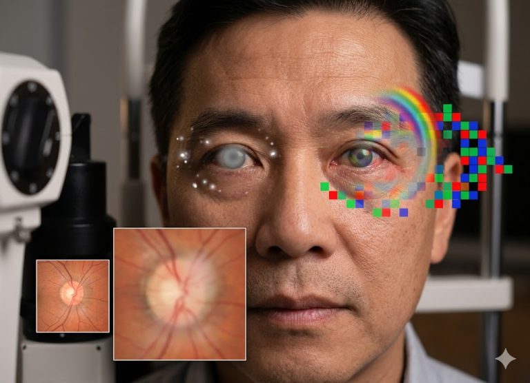
The classic first change is a small, patchy blind spot called a scotoma, usually in the mid-periphery of your vision. These often follow an arc that matches the curve of the nerve fibers across the retina. A common starting point is just off-center on the nose side.
A single small scotoma is almost impossible to notice. Your other eye covers the gap, and your brain fills it in. You won’t see a black spot; you just don’t receive information from that area. You might only catch it if you cover your good eye and an object briefly vanishes as it passes through the blind spot.
As the disease advances, these spots slowly enlarge and join together. The loss usually respects the horizontal midline, hitting the upper or lower half first. Only when the loss becomes tunnel vision does it become impossible to ignore, and at that point the disease is advanced and the damage is irreversible.
Frequent Changes in Eyeglass Prescription
Needing new glasses again and again, especially when the new pair still doesn’t look quite sharp, can be a subtle clue. Most prescription changes come from normal aging (like presbyopia) or changes in the cornea or lens. With glaucoma, the issue isn’t focusing power but the health of the optic nerve.
Nerve damage lowers visual quality in a way lenses can’t fix. You might lose contrast sensitivity, making similar shades or cluttered scenes harder to make out, or notice a general drop in sharpness. An optometrist may find only a small prescription change, and yet even with the right lenses your vision still feels “off.” That lingering dissatisfaction is a red flag.
You’re feeling the effect of nerve damage but reading it as a focusing problem. If you need a new prescription every year or more often and still struggle with vision quality, ask for a comprehensive eye exam that checks the optic nerve.
Halos Around Lights
Halos are bright, sometimes rainbow-colored rings around lights such as headlights and streetlamps. In glaucoma, they can happen when high eye pressure makes the cornea swell (corneal edema).
Halos are more often linked to cataracts or certain eye surgeries, but when other glaucoma risk factors are present they deserve prompt attention. The cornea must stay relatively dry to remain clear, and a layer of cells on its inner surface, the endothelium, pumps fluid out. When IOP rises sharply, it can overwhelm these cells, and fluid seeps into the cornea and makes it swell slightly.
The swollen cornea scatters light instead of focusing it cleanly, and your brain reads this as halos or starbursts, especially at night. Because eye pressure fluctuates in open-angle glaucoma, halos may appear only during pressure spikes. You might notice them one evening and not the next, and blame tired eyes. But halos that come and go, particularly with mild blurriness, are a warning sign worth checking with an eye doctor soon.
Difficulty Adjusting to Low Light
Struggling to see in dim places, or taking much longer to adjust when going from bright to dark, may be a sign. This poor dark adaptation can be one of the earliest functional losses in glaucoma.
Seeing in low light depends on the whole visual pathway, from the rod cells in the retina to the optic nerve fibers that carry their signals. Glaucoma destroys those fibers, so the nerve can’t deliver complete, accurate information. The weakness shows most in tough conditions like dim light, where signals are already faint.
You might find it harder to walk through a dark theater, drive at dusk, or find something in a poorly lit room, and your eyes may need much longer to adjust after coming in from bright sun. Night vision also declines with normal aging and cataracts, but glaucoma can make the change more pronounced or faster. If you or a loved one has a clear, worsening struggle in low light, get a comprehensive exam that includes an optic nerve check.
Blurred Vision or Eye Redness
Open-angle glaucoma can cause occasional blurriness when pressure swings affect the cornea. Mild, lingering redness may also signal that the eye is under stress from raised pressure. Both are easy to overlook because dry eye, allergies, and screen fatigue cause the same things. People often treat them with over-the-counter drops, which can delay a proper diagnosis.
The blurriness is usually brief. When IOP spikes, the cornea swells slightly and vision turns hazy. As pressure drops again, sight clears. This on-and-off pattern makes people assume they’re just tired.
Constant low-grade redness can also be a subtle clue. It isn’t a direct sign of nerve damage, but an eye under chronically high pressure may stay slightly irritated, with dilated surface blood vessels that give it a pink or bloodshot look. What matters is persistence without a clear cause. If your eyes are often briefly blurry, or stay red with no allergy or irritant to explain it, glaucoma could be involved.
Glaucoma
A dull, aching eye or a mild headache around the eyes or forehead can also be a faint sign, often as pressure fluctuates. This is very different from the severe, excruciating pain of an acute angle-closure attack, which is a medical emergency. In open-angle glaucoma, pressure rises so slowly that the body adapts and gives few pain signals.
Still, more sensitive people may feel a vague deep ache, a sense of pressure behind the eye, or a feeling of fullness rather than sharp pain. Low-grade frontal headaches are possible too. Since these are mild and irregular, they’re easily blamed on sinus problems, tension, or computer strain, and a painkiller that helps seems to confirm it.
The pattern is the clue. If you get repeated dull headaches in or around your eyes with no clear cause, consider that eye pressure could be involved. Tell your eye doctor, especially if you have other risk factors. It could be a useful piece of the puzzle.
What are the Main Causes and Risk Factors for Developing Open-Angle Glaucoma?
The main risk factors are raised intraocular pressure, older age, a family history of glaucoma, certain ethnic backgrounds, and some medical conditions. The exact reason the drainage system fails isn’t fully understood. High IOP is the most important risk factor and the only one you can change through treatment.
Glaucoma has many contributing factors. High pressure doesn’t guarantee you’ll get it, and some people develop it with normal pressure (normal-tension glaucoma). It helps to split risks into two groups.
Non-modifiable risks are things you can’t change. Risk rises sharply after age 60. People of African descent face a much higher risk, often at a younger age and with faster progression, and people of Hispanic and Asian descent also have increased risk. Family history is powerful: a parent or sibling with glaucoma can raise your risk up to nine times.
Modifiable or secondary risks include diabetes, high blood pressure, and heart disease, which affect blood flow to the optic nerve and make it more vulnerable. Eye-related factors such as high nearsightedness or a thin central cornea also raise risk, and a serious eye injury can lead to secondary glaucoma.
Who is Most at Risk for Developing Open-Angle Glaucoma?
The highest-risk groups are people over 60, people of African, Hispanic, or Asian descent, and anyone with a close relative who has the disease. Risk climbs steeply with age, which is why regular screening is standard in senior eye care.
Ethnicity is a strong predictor. People of African ancestry are six to eight times more likely than Caucasians to develop open-angle glaucoma. It also tends to start about ten years earlier and progress faster, often causing more severe vision loss. Likely reasons include genetics, thinner central corneas, and larger optic nerve cupping.
Hispanic individuals also face higher risk, especially at older ages, and glaucoma is a leading cause of blindness in this community. Some types vary by population too; angle-closure glaucoma, for example, is more common in people of Asian descent.
Other groups at risk include people with diabetes or high blood pressure, which can harm the blood supply to the optic nerve. Long-term steroid use, whether as eye drops, inhalers, or pills, is a well-known cause of raised eye pressure. Knowing which categories apply to you helps you and your doctor set the right exam schedule.
Open-Angle Glaucoma
Open-angle glaucoma has a very strong genetic component, and family history is one of its biggest non-modifiable risk factors. Researchers have linked several genes to a higher risk. These genes can affect how the trabecular meshwork develops and works, or how sturdy the optic nerve is, making it easier for eye pressure to cause harm.
The risk is highest if a first-degree relative (parent, sibling, or child) has the condition. Having a sibling with glaucoma can raise your risk as much as nine times compared with the general population. Because the link is so well established, a family history should prompt earlier and more frequent screening.
If you’re diagnosed, tell your close relatives. It lets them start regular comprehensive exams, including an optic nerve evaluation, often earlier than usually advised. Catching the disease early in these relatives is the best way to avoid the irreversible vision loss earlier generations may have suffered. Inheritance is complex, and not everyone who carries the genes will develop glaucoma, but the genetic makeup lowers the threshold for damage.
How to Protect Vision from the Effects of Open-Angle Glaucoma
You protect your vision mainly through early detection with regular comprehensive eye exams, plus lifelong, consistent use of the treatments that lower your eye pressure. Because glaucoma damage can’t be reversed, everything is aimed at preventing loss and slowing progression.
The foundation is finding the disease early, before nerve damage builds and before you notice anything. Only a routine exam by an eye care professional can pick up those early signs.
Once diagnosed, the goal is to bring IOP down to a “target pressure” that is safe for your optic nerve. That can be done with eye drops, laser treatment, or surgery.
Protecting your sight is a lifelong partnership with your ophthalmologist. You need to use medications exactly as prescribed and attend every follow-up so the disease can be monitored. There is no cure, but with early diagnosis and steady management, most people keep their functional vision for life.
Essential Treatments for Managing Eye Pressure
Treatment falls into four groups: eye drops, oral medications, laser therapy, and surgery. All aim to lower IOP to a safe level and protect the optic nerve.
Eye drops are nearly always the first step. Some reduce how much fluid the eye makes (beta-blockers and alpha-adrenergic agonists). Others improve drainage through the meshwork or another route (prostaglandins, the most commonly prescribed class).
Oral medications, such as carbonic anhydrase inhibitors, may be added if drops aren’t enough or cause bad side effects. They’re usually used short-term because of a higher chance of body-wide side effects.
Laser therapy is often next. Selective Laser Trabeculoplasty (SLT) is a common, minimally invasive office procedure. A low-energy laser stimulates the trabecular meshwork so it drains better and lowers IOP.
Surgery is used for advanced or stubborn cases. A trabeculectomy builds a new drainage channel that bypasses the clogged meshwork. Other options include Micro-Invasive Glaucoma Surgery (MIGS) devices and drainage implants such as valves or tube shunts. The best choice depends on how severe the glaucoma is, your target pressure, and your overall health and lifestyle.
Regular Comprehensive Eye Exams
Regular comprehensive exams are the most effective way to prevent major vision loss, because they find the disease long before you feel symptoms. A basic vision screening or glasses check is not enough. A comprehensive dilated exam for glaucoma includes several key tests:
- Tonometry measures eye pressure. It’s important, but pressure alone can’t confirm glaucoma.
- Dilated ophthalmoscopy lets the doctor look directly at the optic nerve for its shape, color, and health, and for signs like cupping or a thinning rim.
- Perimetry (visual field test) maps your side vision to find early blind spots you wouldn’t notice yourself.
- Optical Coherence Tomography (OCT) produces a detailed cross-section of the optic nerve and nerve fiber layer, measuring thickness and spotting thinning, often years before a visual field test shows any loss.
Together, these results let an ophthalmologist diagnose glaucoma at its earliest stage and start lowering pressure, which can stop or greatly slow the disease and preserve sight.
Open-Angle Glaucoma Diagnosis
Diagnosis needs several tests that together show the full state of the eye.
- Tonometry is usually first and measures IOP. High pressure is a major risk factor but not the only deciding one: some people have high pressure without nerve damage (ocular hypertension), and others develop glaucoma at normal pressure.
- Ophthalmoscopy checks the optic nerve for damage. The doctor looks through the pupil at the back of the eye to examine the nerve head’s shape, color, and health. Cupping or thinning is a red flag.
- Perimetry maps peripheral vision to find blind spots, a hallmark of glaucoma damage.
- Gonioscopy uses a special mirrored lens to inspect the drainage angle and confirm it is open and unblocked.
- Pachymetry measures corneal thickness, which can affect how accurate pressure readings are.
The Difference Between Open-Angle and Angle-Closure Glaucoma
The key difference is the anatomy of the drainage angle and how fast the disease develops.
In primary open-angle glaucoma (POAG), the angle where the iris meets the cornea is open, so nothing physically blocks access to the trabecular meshwork. The problem is rising resistance inside the meshwork, which makes fluid drain too slowly. Pressure climbs gradually and painlessly, silently damaging the optic nerve over years.
In angle-closure glaucoma, the drainage angle becomes physically blocked, often when the iris pushes forward and seals off the meshwork. This can happen suddenly (acute) or on and off (chronic). An acute attack is a medical emergency with a rapid pressure spike, severe eye pain, headache, nausea, blurred vision, and halos around lights. Without immediate treatment, it can cause permanent vision loss within hours.
Lifestyle Changes to Manage Glaucoma
Diet and exercise can’t cure or prevent glaucoma, but they can support your eye health alongside medical treatment.
Diet: Antioxidant-rich foods are believed to help protect the optic nerve. Vitamins A, C, and E, zinc, and the carotenoids lutein and zeaxanthin support nerve tissue. Good sources are leafy greens like spinach and kale, colorful fruits and vegetables such as berries, bell peppers, and carrots, and omega-3-rich fatty fish. These nutrients help fight oxidative stress, which is linked to nerve damage in glaucoma.
Exercise: Regular, moderate aerobic activity such as brisk walking, jogging, or swimming can lower IOP in some people. The effect is usually temporary and lasts only while you keep exercising, but it also improves cardiovascular health, which helps nourish the optic nerve.
What to avoid: Not every habit helps. Head-down or inverted positions, such as headstands or downward-facing dog in yoga, can raise IOP significantly and may be harmful. Also check with your doctor before taking high-dose vitamin supplements, since they can interfere with other medicines or conditions.
Lifestyle changes only complement treatment. Using your prescribed drops and medications and attending regular check-ups remain the most important steps.
Minimally Invasive Glaucoma Surgeries (MIGS)
MIGS is a newer group of procedures that lower IOP with a much better safety record than traditional glaucoma surgery. They use microscopic devices and tiny incisions to boost the eye’s natural drainage or create new pathways.
They offer a gentler alternative to trabeculectomy or tube shunts. Those operations lower pressure very well but carry higher risks, including infection, dangerously low eye pressure (hypotony), and cataract formation.
MIGS is generally reserved for mild to moderate open-angle glaucoma and is often done during cataract surgery, so both problems are treated in one session. Examples include the iStent, Hydrus Microstent, and Xen Gel Stent, each of which bypasses the trabecular meshwork in a slightly different way to improve fluid outflow.
FAQs
1. What is the difference between open and closed glaucoma?
It comes down to the drainage system’s structure and the speed of the disease. In open-angle glaucoma, the angle between the iris and cornea stays wide open, but the sponge-like drainage meshwork gradually clogs, like a slow sink. Pressure rises slowly, painlessly, and without symptoms. In closed-angle glaucoma, the iris bows forward and seals the drainage angle, trapping fluid. Pressure spikes quickly, and it is a painful emergency that can lead to blindness within days if untreated.
2. How serious is open-angle glaucoma?
Very serious. It is a chronic condition that develops silently and is a leading cause of permanent blindness worldwide. The optic nerve damage can’t be reversed. But it is very manageable when caught early through routine eye exams.
3. How long can you live with open-angle glaucoma?
A normal, full lifespan. The disease affects only the eye and optic nerve, not life expectancy or general health. With regular monitoring and steady treatment, most people keep their independence and vision throughout life.
4. Can you live with glaucoma without going blind?
Yes. Most people with glaucoma do not go blind. Modern eye drops, laser treatments like SLT, and advanced surgeries let doctors lower eye pressure and control the disease. Blindness usually happens only when glaucoma goes undiagnosed for decades or a patient stops following their treatment plan.
5. What’s the worst type of glaucoma to have?
Acute angle-closure and neovascular glaucoma are considered the most dangerous. Acute angle-closure is a sudden crisis with severe pain and nausea that can destroy sight within hours if pressure isn’t relieved. Neovascular glaucoma is a secondary form, often triggered by advanced diabetes, in which fragile new blood vessels grow over the drainage channels. It is aggressive, resists standard treatment, and is very hard to manage.
6. At what age does glaucoma start?
It can appear at any age, but primary open-angle glaucoma most often begins after 40, and risk rises with each decade. Since early stages have no symptoms, ophthalmologists recommend a comprehensive baseline eye exam for everyone over 40.
Conclusion
Open-angle glaucoma shows how some of the biggest health threats work in the shadows. By the time it shows itself through blurred text or shrinking side vision, a large part of your optic nerve has already been lost for good.
Waiting for obvious symptoms is a gamble your eyesight can’t afford. The damage can’t be undone, but with early detection and treatment, further loss can usually be stopped or greatly slowed. Knowing these 6 subtle warning signs, keeping up with regular comprehensive eye exams, and following your specialist’s treatment plan are the best ways to keep seeing clearly for decades.