7 Early Symptoms of Atelectasis Disease You Shouldn’t Ignore
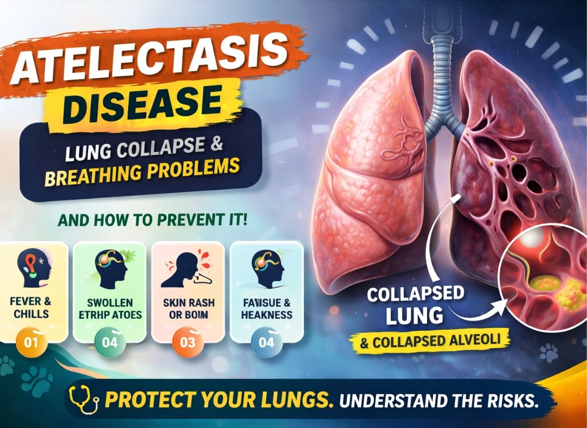
Atelectasis disease is a condition in which part or, in some cases, an entire section of the lung does not fully expand. When this happens, the affected area cannot fill with air normally, which may make breathing harder and can reduce oxygen levels. Atelectasis may be temporary and mild, particularly after surgery or periods of shallow breathing, but it can also develop because of an airway blockage or another lung problem.
One challenge with atelectasis is that its early signs may be mild and easy to dismiss. Symptoms such as slight shortness of breath, tiredness, or an ongoing cough may not immediately seem serious. However, they can sometimes indicate that part of the lungs is not working normally. In this article, “7 Early Symptoms of Atelectasis Disease You Shouldn’t Ignore,” we’ll discuss common early symptoms, possible causes, how doctors diagnose the condition, and when medical attention is important.
The Definition of a Collapsed Lung (Atelectasis) and Its Causes
The atelectasis medical definition refers to a condition in which some of the lung’s tiny air sacs, known as alveoli, lose air and partially or completely collapse. To understand the atelectasis meaning, it can help to imagine the lungs as a soft sponge filled with countless small air pockets. When some of those pockets lose their air, the affected lung tissue becomes denser and cannot participate normally in gas exchange.
When this happens over a significant area, the amount of oxygen reaching the bloodstream may decrease. The severity depends on how much lung is affected and what caused the collapse.
The Mechanics of Obstruction and Resorption
One common form of atelectasis lung collapse occurs when an airway becomes blocked. This is known as obstructive or resorptive atelectasis.
A blockage may be caused by thick mucus, an inhaled foreign object, or a growth inside the airway. Air that is already trapped beyond the blockage is gradually absorbed into the bloodstream. Because fresh air cannot replace it, the pressure inside the alveoli decreases and they eventually collapse.
This type of atelectasis is common after surgery. Anesthesia, pain, reduced movement, and shallow breathing can make it harder to cough effectively and clear mucus from the airways.
Compression and External Pressure
Compression atelectasis occurs when something outside the lung places pressure on it and prevents normal expansion. The airway itself may remain open, but the lung cannot expand because there is not enough room.
This can happen with atelectasis and pleural effusion, where fluid collects in the space surrounding the lung. A large mass or an enlarged structure in the chest can also place pressure on nearby lung tissue and produce subsegmental atelectasis, affecting only a small portion of a lobe.
The Role of Surfactant and Scarring
The lungs also depend on a substance called surfactant to reduce surface tension inside the alveoli. Without enough surfactant, the tiny air sacs can become more likely to collapse.
This is particularly important in premature infants because their lungs may not yet produce enough surfactant.
Atelectasis and scarring, also known as cicatrization, can occur when damaged lung tissue becomes stiff and fibrous. Conditions such as chronic inflammation or previous radiation therapy may cause scarring that contracts the surrounding tissue and contributes to permanent or difficult-to-reverse collapse.
Defining the Radiological Landscape
When doctors examine an atelectasis CXR, they look for areas of increased density that suggest lost air volume. Collapsed lung tissue is denser than normal air-filled lung tissue, so it may appear whiter on an X-ray.
Depending on its shape and location, the finding may be described as plate-like atelectasis or linear atelectasis. These forms often appear as thin streaks.
If changes are present near the bases of both lungs, the report may use terms such as bibasilar atelectasis or atelectatic changes at the lung bases. These descriptions help doctors determine where the reduced lung expansion is occurring.
The Most Common Causes of a Collapsed Lung
The atelectasis medical definition describes an area of lung that has lost normal air volume. Although people sometimes call atelectasis a “collapsed lung,” it differs from a pneumothorax. Atelectasis generally involves collapse of lung tissue or alveoli, while pneumothorax occurs when air enters the pleural space and causes the lung to pull away from the chest wall.
Atelectasis is generally divided into obstructive and non-obstructive forms.
Obstructive (Resorptive) Atelectasis: The Internal Blockage
Obstructive atelectasis occurs when something blocks an airway and prevents air from reaching the alveoli beyond the blockage. The trapped air is gradually absorbed, causing the affected area to lose volume.
The Mucus Plug: Thick mucus is a common cause of postoperative subsegmental atelectasis. Anesthesia can reduce the cough reflex, while pain may cause a person to breathe shallowly. As a result, mucus can collect in the smaller airways.
Foreign Bodies and Tumors: In children, inhaled objects such as food or small toys can block an airway and cause sudden collapse. In adults, a tumor or other growth may gradually narrow an airway and eventually prevent air from reaching part of the lung.
Atelectasis and Pneumonia: Lung infections can produce thick mucus and inflammatory material that obstructs an airway. As a result, pneumonia and atelectasis may occur together.
Non-Obstructive Atelectasis: External and Structural Pressures
Non-obstructive atelectasis occurs when there is no direct blockage inside the airway but the lung still cannot expand properly.
Compression Atelectasis: Fluid, air, masses, or other material surrounding the lung can put pressure on it. With atelectasis and pleural effusion, for example, fluid in the pleural space can compress nearby lung tissue.
Bibasilar and Dependent Atelectasis: Remaining in bed or in one position for an extended period can contribute to dependent atelectasis. Gravity and shallow breathing can cause the lower portions of the lungs to expand less effectively, sometimes producing bibasilar atelectasis.
Cicatrization (Atelectasis and Scarring): Previous lung damage, radiation, or chronic inflammatory disease can cause scar tissue to form. As the scar tissue contracts, it can pull surrounding lung tissue inward and contribute to persistent atelectasis.
Radiological Signatures: Plate-like and Round Atelectasis
The shape of atelectasis on a chest X-ray or CT scan can provide clues about its cause.
Plate-like atelectasis, also called linear atelectasis, often appears as a thin horizontal line and may be associated with shallow breathing.
Round atelectasis is less common. It can sometimes resemble a mass on imaging and is often associated with changes in the pleura. The pleural tissue folds or thickens around an area of lung, creating a rounded appearance.
7 Early Symptoms of a Collapsed Lung
The atelectasis medical definition describes a loss of normal lung expansion, but the symptoms a person experiences depend on how much lung is affected and how quickly the collapse develops.
Small areas of atelectasis may cause few or no symptoms. Larger areas can interfere with breathing and oxygen exchange, leading to more noticeable respiratory symptoms.
The Respiratory Struggle: Dyspnea and Tachypnea
Shortness of breath, or dyspnea, is one of the main symptoms that can occur when a significant area of the lung is affected.
As functional lung volume decreases, the body may compensate by breathing more quickly. This rapid breathing is called tachypnea.
If atelectasis occurs alongside pleural effusion, taking a deep breath may also be uncomfortable or difficult. The severity of shortness of breath depends on the extent and underlying cause of the collapse.
The Sensory Warning: Pleuritic Pain and Persistent Cough
Atelectasis may sometimes cause chest discomfort, particularly when the pleura becomes irritated. Pleural pain is often sharp and can become worse when taking a deep breath or coughing.
A cough may also develop. When mucus is blocking an airway, coughing is the body’s attempt to clear the obstruction. With compression atelectasis, the cough may be dry because there is no mucus plug to remove.
Systemic Alarm: Tachycardia and Cyanosis
When breathing is significantly impaired, the heart may beat faster to help circulate oxygen throughout the body. This increased heart rate is known as tachycardia.
Severe or widespread atelectasis can cause low blood oxygen levels. In serious cases, cyanosis may develop, causing the lips or nail beds to appear bluish or gray. This is a warning sign that requires urgent medical attention.
Clinical Findings: The Silent Side
During a physical examination, a doctor may notice reduced movement of one side of the chest or decreased breath sounds over an affected area.
A chest X-ray can provide additional information. Small areas may appear as linear or plate-like atelectasis, while larger areas of collapse may produce more obvious changes in lung density and volume.
How Does Atelectasis Differ From a Pneumothorax?
Although both atelectasis and pneumothorax can be described as forms of “lung collapse,” their causes are different.
Atelectasis usually involves loss of air within part of the lung, while pneumothorax occurs when air enters the pleural space and disrupts the normal pressure that keeps the lung expanded.
Atelectasis: The “Inward” Deflation
With atelectasis disease, the affected lung tissue loses volume because the alveoli are not adequately filled with air.
For example, a mucus plug can cause subsegmental atelectasis by blocking a small airway, while external pressure can produce compression atelectasis.
On an atelectasis CXR, the affected tissue may appear denser or whiter because it contains less air. There may also be signs of volume loss.
Pneumothorax: The “Outward” Pressure
A pneumothorax develops when air enters the pleural space between the lung and chest wall. This disrupts the normal negative pressure that helps keep the lung expanded.
The lung then partially or completely collapses toward the center of the chest.
On a chest X-ray, a pneumothorax typically appears as an area without normal lung markings, with the edge of the collapsed lung visible. This appearance differs from the increased density commonly seen with atelectasis.
Diagnostic and Treatment Differences
Because the causes are different, treatment also varies.
Treatment of Atelectasis: Treatment focuses on improving lung expansion and addressing the underlying cause. Depending on the situation, this may include deep-breathing exercises, incentive spirometry, clearing mucus, treating infection, or removing an airway blockage.
Treatment of Pneumothorax: Treatment is directed at removing air from the pleural space and allowing the lung to expand again. Depending on its size and severity, a pneumothorax may require observation, needle aspiration, or a chest tube.
Compression Atelectasis Link: A large pneumothorax can also compress the lung from the outside and produce a form of compression atelectasis.
When Should You Seek Medical Care for Collapsed Lung Symptoms?
The need for medical attention depends on the severity of symptoms and the underlying cause of the atelectasis.
Small areas discovered incidentally may require monitoring or treatment of the underlying problem, while sudden or severe breathing difficulty can represent a medical emergency.
Identifying a Medical Emergency: The “911” Threshold
Seek emergency medical care if you develop severe or rapidly worsening shortness of breath, especially if you are struggling to speak normally because of breathlessness.
Other emergency warning signs include:
- Cyanosis: Blue or gray lips, face, or fingernails may indicate dangerously low oxygen levels.
- Mental Alteration: New confusion, unusual drowsiness, fainting, or difficulty staying awake can occur when the brain is not receiving enough oxygen.
- Severe Chest Symptoms: Sudden or intense chest pain, especially when combined with difficulty breathing, should be evaluated immediately.
When to Schedule a Prompt Appointment
Not every case of atelectasis requires emergency treatment, but persistent symptoms should still be evaluated.
Contact a healthcare professional promptly if you experience:
- Persistent Productive Cough: Ongoing coughing with thick or discolored mucus may suggest infection or an airway obstruction.
- New-Onset Exercise Intolerance: Becoming unusually short of breath during activities that were previously easy can indicate a change in respiratory function.
- Pleuritic Aches: Chest discomfort that becomes worse when you breathe deeply, cough, or yawn should be discussed with a medical professional.
Preparing for the Diagnostic Deep-Dive
Providing detailed information about your symptoms can help your doctor determine whether atelectasis, pneumonia, pleural effusion, scarring, or another condition is responsible.
The Symptom Roadmap
- The “When” and “How”: Explain whether the symptoms appeared suddenly or developed gradually. The timing can help doctors narrow down possible causes.
- The Nature of the Pain: Describe the location, severity, and type of pain and whether it becomes worse with breathing or coughing.
- Post-Event Context: Tell your doctor if you recently had surgery, were hospitalized, or spent a long period in bed, as these factors can increase the risk of atelectasis.
Vital History for the Pulmonologist
- Respiratory Baseline: Mention conditions such as COPD, asthma, or a history of smoking, as these may affect airway function.
- Environmental Exposure: Tell your doctor about significant exposure to substances such as asbestos or other lung irritants, particularly if imaging has shown pleural thickening or round atelectasis.
How is a collapsed lung diagnosed by a doctor?
Doctors diagnose atelectasis by combining a physical examination with imaging and, when needed, additional tests.
Because atelectasis involves a change in lung volume and air content, imaging is particularly useful for confirming its location and possible cause.
The Physical Exam: Listening for Silence
The first step is usually a physical examination. Using a stethoscope, a doctor listens to the lungs for changes in breath sounds.
Breath sounds may be reduced over an area affected by atelectasis. A doctor may also use percussion, or gentle tapping on the chest, to identify changes in the sound produced by the underlying tissue.
These findings can suggest that part of the lung is not filling with air normally.
Diagnostic Imaging: The Visual Proof
Imaging helps confirm the presence and location of atelectasis.
Atelectasis CXR (Chest X-ray): A chest X-ray is commonly used to evaluate suspected atelectasis. Areas of collapsed lung may appear more opaque or white than normal air-filled lung. Doctors may also look for signs of volume loss, including linear or plate-like atelectasis.
CT Scan: A CT scan provides more detailed images and may be used when an X-ray does not clearly explain the problem. It can help identify causes such as tumors, foreign bodies, scarring, or pleural fluid.
Physiological Monitoring: Oxygen and Blood Gases
Doctors may also evaluate how atelectasis is affecting oxygen levels.
- Pulse Oximetry: A small device placed on the finger measures blood oxygen saturation.
- Arterial Blood Gas (ABG): In patients with significant respiratory problems, an arterial blood sample can measure oxygen and carbon dioxide levels more precisely.
Specialized Procedures: Bronchoscopy
If an airway blockage is suspected, a bronchoscopy may be recommended.
During this procedure, a thin flexible tube with a camera is passed through the airways. It allows the doctor to identify problems such as mucus plugs or foreign objects and, in some cases, remove the obstruction.
Can a Collapsed Lung be Prevented?
Some forms of atelectasis can be prevented, particularly those associated with surgery, prolonged bed rest, or shallow breathing.
The goal is to encourage regular lung expansion, keep airways clear, and address conditions that increase the risk of collapse.
Post-Surgical Pulmonary Hygiene
Atelectasis commonly occurs after surgery because anesthesia, pain, and reduced activity can cause people to take shallow breaths.
Deep Breathing and Coughing: Deep-breathing exercises can help expand the lower portions of the lungs. Controlled coughing can also help clear mucus from the airways.
Incentive Spirometry: An incentive spirometer encourages slow, deep inhalation. It is often used after surgery to promote lung expansion and reduce the risk of postoperative atelectasis.
Mobilization and Positioning
Remaining in bed for long periods can increase the risk of dependent atelectasis.
Early Mobilization: Getting out of bed and walking as soon as medically appropriate can encourage deeper breathing and improve overall lung function.
Frequent Repositioning: For people who cannot walk, changing positions regularly can help different areas of the lungs expand and may reduce prolonged compression of the lower lung regions.
Lifestyle and Chronic Management
Long-term lung health also plays an important role in reducing certain risks of atelectasis.
Smoking Cessation: Smoking damages the airways and interferes with the normal movement of mucus. Quitting smoking can improve airway function and reduce respiratory complications.
Hydration: Adequate fluid intake can help keep mucus thinner and easier to clear, although hydration needs vary depending on a person’s health and medical conditions.
Managing Underlying Conditions: Proper management of asthma, COPD, and other respiratory disorders can help maintain open airways and reduce the risk of mucus buildup and related complications.
Conclusion
Atelectasis disease can range from a small, temporary area of reduced lung expansion to a more extensive problem that affects breathing and oxygen levels. Symptoms such as shortness of breath, chest discomfort, rapid breathing, or a persistent cough should not be ignored when they are new, worsening, or unexplained.
Recognizing symptoms early can help identify the underlying cause and allow appropriate treatment. Depending on the situation, management may involve breathing exercises, physical activity, treatment of infection, removal of an airway blockage, or management of another condition causing the collapse.
If breathing difficulty becomes severe or develops suddenly, urgent medical attention is important. For persistent or recurring symptoms, a healthcare professional can determine whether atelectasis or another lung condition is responsible.
Frequently Asked Questions (FAQ) About Atelectasis Disease
What is atelectasis disease?
Atelectasis disease occurs when part or all of a lung does not fully expand. This usually happens because some of the alveoli lose air or because an external force prevents normal expansion. Small areas may cause few symptoms, while larger areas can interfere with breathing and oxygen exchange.
What causes atelectasis disease?
Atelectasis can develop for several reasons. Common causes include mucus blocking an airway, inhaled foreign objects, tumors, shallow breathing after surgery, prolonged bed rest, pneumonia, pleural fluid, and pressure from surrounding structures. Identifying the underlying cause is important for choosing the appropriate treatment.
What are the early symptoms of atelectasis disease?
Early symptoms can include shortness of breath, shallow breathing, chest discomfort, coughing, and reduced exercise tolerance. Some people may have very few symptoms when only a small area of the lung is affected. More extensive atelectasis can cause more noticeable breathing difficulties.
Is atelectasis disease dangerous?
The seriousness of atelectasis depends on how much lung is affected and why the collapse occurred. Small areas may cause little concern, while extensive atelectasis can reduce oxygen levels or increase the risk of complications such as pneumonia. Severe breathing difficulty requires prompt medical evaluation.
How is atelectasis disease treated?
Treatment depends on the underlying cause. Doctors may recommend deep-breathing exercises, incentive spirometry, coughing techniques, physical activity, or treatment for an underlying infection. If an airway is blocked by mucus or another object, a procedure such as bronchoscopy may sometimes be necessary.
Can atelectasis disease be prevented?
Some types of atelectasis can be reduced through regular movement, deep-breathing exercises, proper postoperative care, and avoiding smoking. People recovering from surgery may be encouraged to use an incentive spirometer and get out of bed as soon as medically appropriate. Managing chronic lung conditions can also help reduce respiratory complications.