Understanding Bronchiectasis: 7 Early Signs and Treatment Options
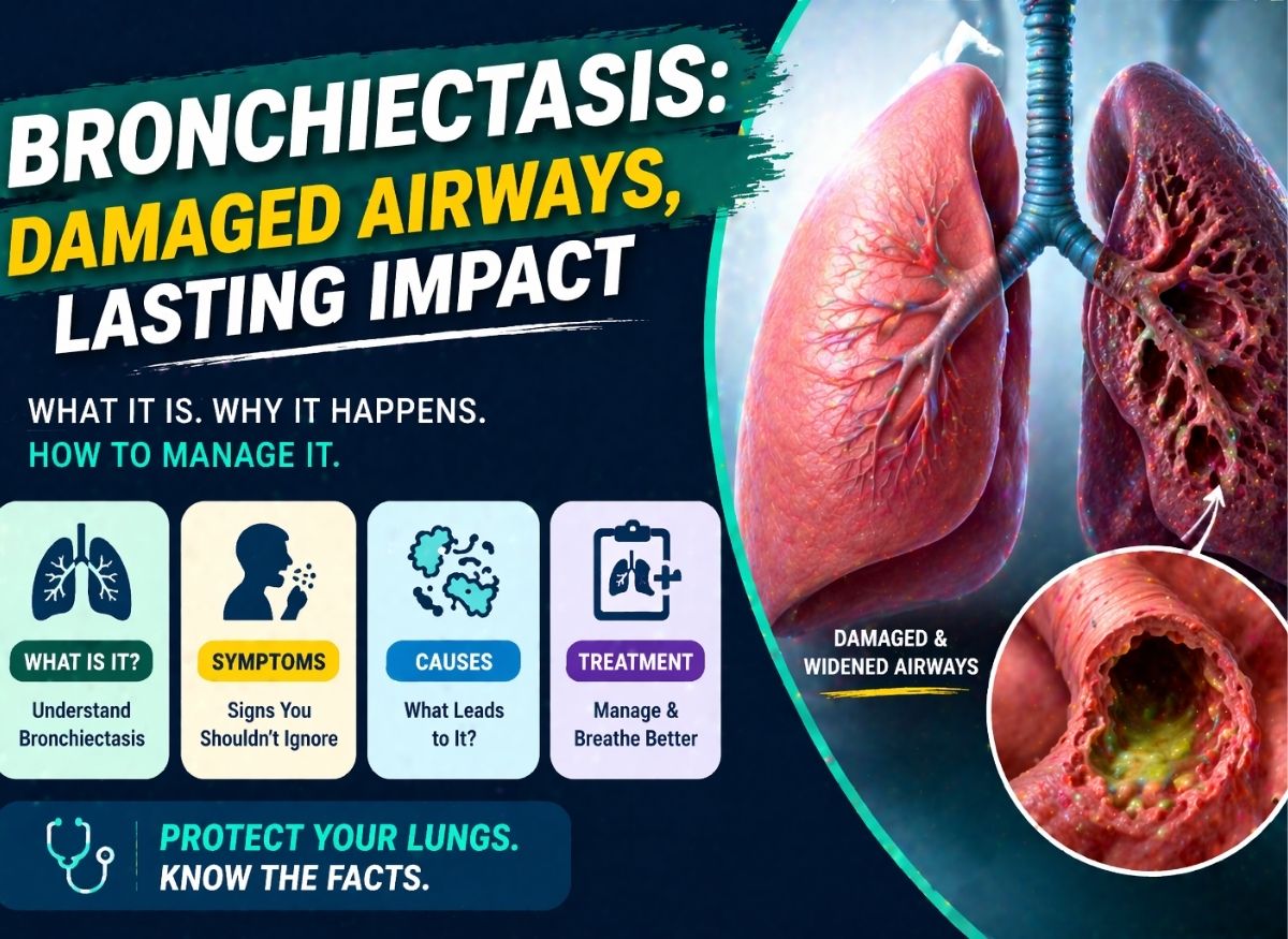
Bronchiectasis is a chronic lung condition in which the airways become permanently widened and damaged, making it hard to clear mucus from the lungs. The mucus that builds up creates a place where bacteria can grow, leading to repeated infections and inflammation. Over time, it can affect breathing, reduce lung function, and lower quality of life if it isn’t properly managed.
One challenge is that the early symptoms are often mild and easy to overlook. Many people mistake a persistent cough or fatigue for a minor respiratory issue. But recognizing the early signs is essential for getting the right diagnosis and starting treatment early. Below, we explore the warning signs, underlying causes, and the most effective ways to manage the condition and protect your lung health.
What Is Bronchiectasis?
Bronchiectasis is best understood as a structural change to the lungs rather than a temporary illness. It’s a chronic, irreversible condition in which the large airways (the bronchi) become abnormally and permanently dilated. This happens when the elastic and muscular parts of the bronchial walls are destroyed, usually through a persistent cycle of inflammation and infection. The airways lose their tone and “springiness,” becoming flaccid and distorted, and can no longer move air and clear waste efficiently.
How Airway Clearance Fails
At the core of the condition is the breakdown of the “mucociliary escalator.” In a healthy lung, mucus traps debris and bacteria, and microscopic cilia sweep it upward. In bronchiectasis, the widened airways act like stagnant pools instead of flowing rivers. Mucus collects in the dilated pockets and becomes a nutrient-rich breeding ground for germs. This creates a self-perpetuating “vicious cycle”: bacteria trigger an immune response, and the resulting inflammation weakens the airway walls further, causing more dilation.
Classifications You May Encounter
- Bronchiolectasis refers to a similar dilation in the much smaller, terminal branches of the lungs (the bronchioles).
- Focal bronchiectasis means damage is confined to a single lobe or segment, often from a localized obstruction or a previous severe pneumonia.
- Bilateral bronchiectasis means the widening is present in both lungs, which is more common in systemic conditions like cystic fibrosis or primary ciliary dyskinesia.
How It Appears on Imaging
Diagnosis is typically confirmed with High-Resolution CT (HRCT), which can detect even mild changes before they show up on a standard X-ray. When a radiologist notes mild bronchiectasis, they’re seeing an airway whose diameter slightly exceeds that of its accompanying blood vessel, a classic finding known as the “signet ring sign.” These early structural shifts mark the start of the lung’s remodeling. Because the damage is permanent, catching it at this stage is crucial for starting therapies that keep it from progressing to a severe, debilitating state.
7 Early Warning Signs
Recognizing bronchiectasis early is the most effective way to halt the cycle of airway destruction. Because the condition is a permanent change in lung structure, the body shows a specific cluster of symptoms reflecting its inability to clear secretions.
1. A persistent daily cough. The most definitive early sign is a chronic, daily productive cough. Unlike a cold, it lasts for months and is usually worst in the morning, since mucus pools in the dilated airways during sleep. If damage reaches the smallest passages (bronchiolectasis), the cough may bring up smaller amounts of very thick, plug-like sputum.
2. Recurrent chest infections. People often first suspect a problem when they catch “one cold after another.” With focal bronchiectasis, infections may keep settling in the same spot in the chest. Because stagnant mucus in widened airways is a reservoir for bacteria, even a minor viral sniffle can escalate into a bacterial infection, requiring repeated antibiotics that only offer temporary relief.
3. Shortness of breath. Breathlessness often begins subtly with exertion. It’s common when damage is concentrated at the bottom of both lungs (bibasilar bronchiectasis), areas vital for gas exchange, so even mild changes can make it feel like you can’t take a satisfying deep breath. Over time, as airways become more distorted, it can start limiting activities like climbing stairs.
4. Coughing up blood (hemoptysis). Though alarming, blood-streaked mucus is a common early sign of the inflammation involved. Chronic irritation and repeated infection make the small blood vessels lining the airways fragile, and even a strong cough can make them leak. It’s often minor, but it signals significant stress on the airway lining and needs formal evaluation.
5. Wheezing. A whistling sound while breathing is often mistaken for asthma but can reflect mild cylindrical bronchiectasis. The widened tubes are more prone to kinking or blockage by thick mucus plugs, and air forced through partially obstructed or inflamed passages creates the high-pitched wheeze of restricted airflow.
6. Chest pain. Early bronchiectasis can cause a dull, heavy ache or a sharp, stabbing pain called pleurisy, which occurs when inflammation spreads to the pleura, the thin membrane surrounding the lungs. The strain of chronic coughing can also fatigue the muscles between the ribs, causing persistent soreness that makes breathing feel like a chore.
7. Persistent fatigue. Profound fatigue is often overlooked. The body burns enormous energy fighting constant low-grade infection and systemic inflammation, and reduced oxygen efficiency adds to it, producing a deep, “bone-weary” tiredness. It’s a sign the lungs aren’t delivering the oxygen the body needs while their condition drains its resources.
When to See a Doctor
Seeing a physician at the first sign of these changes is the most important step in preventing permanent lung disability. The longer airways stay filled with stagnant mucus, the more the surrounding tissue is remodeled and scarred.
The Three-Week Rule and Recurring Infections
If you have a productive cough lasting more than 21 days without improvement, it’s no longer a simple viral infection. Two or more “chest colds” or bouts of pneumonia within a year is also a major red flag, since it suggests the lungs are failing to clear bacteria, leading to chronic colonization that needs more than standard antibiotics.
Red Flags Needing Urgent Evaluation
- Coughing blood: any amount suggests inflammation has eroded a blood vessel and calls for urgent imaging to make sure a major bleed isn’t imminent
- Changes in sputum: a sudden shift from clear or white to dark green or rust-colored, or becoming thicker and foul-smelling, indicates a high bacterial load
- Sharp chest pain when breathing deeply: often suggests infection has reached the lining of the lung
The Diagnostic Path
A doctor may hear “crackles” or “rhonchi” through a stethoscope, but a standard X-ray often misses mild cylindrical bronchiectasis. HRCT is the gold standard, letting radiologists look for the signet ring sign. A sputum culture may also identify specific bacteria like Pseudomonas, so treatment can be targeted rather than generic.
If you’ve already had a scan, you may see these terms on your report:
- Mild cylindrical bronchiectasis: the most common form, with bronchi uniformly dilated like a tube
- Mild traction bronchiectasis: lung scarring (fibrosis) physically pulls the airways open
- Mild bibasilar bronchiectasis: damage at the base of both lungs, often related to chronic aspiration or systemic issues
Goals of Treatment
Treatment centers on active management rather than a cure. Since the architectural change is permanent, the focus is on containment: addressing the mechanical failure of the airways to halt the cycle where stagnant mucus invites infection and infection causes further damage.
Effective Airway Clearance
The most critical goal is restoring the lungs’ ability to clean themselves. Physical and mechanical methods such as oscillating positive expiratory pressure (OPEP) devices, chest physiotherapy, and specialized breathing exercises shake mucus loose from dilated pockets and move it to larger airways where it can be coughed out. This shrinks the bacterial reservoir, reduces the daily cough, and helps prevent the thick, plug-like mucus of bronchiolectasis.
Preventing Flare-Ups and Infections
A major goal is reducing exacerbations, periods when symptoms suddenly worsen, usually from a bacterial surge. Each infection risks new scarring that pulls the airways wider. To prevent them, doctors may prescribe:
- Mucolytics to thin mucus and make it easier to cough up
- Prophylactic antibiotics, such as long-term, low-dose inhaled or oral macrolides, for people with frequent infections, to keep bacterial colonies in check
- Vaccinations, keeping flu and pneumonia shots current, a primary defense against the severe infections that accelerate damage
Managing Symptoms
Though the damage is structural, the inflammation is manageable. If wheezing is present, bronchodilators can open smaller passages and improve airflow. Calming chronic inflammation also makes blood vessels less fragile, addressing hemoptysis, and lessens chest pain and the “rattle” or whistle sometimes heard.
Preserving Lung Function and Quality of Life
The ultimate goal is preventing mild changes from progressing to severe respiratory failure. Pulmonary rehabilitation, a program of exercise and education, builds stamina and reduces fatigue. With a rigorous daily clearance routine and quick response to early signs of respiratory infection, people can preserve healthy lung tissue, stay active, and avoid needing supplemental oxygen for as long as possible.
Airway Clearance and Lifestyle Habits
Management is essentially a daily commitment to mechanical hygiene, designed to work around the body’s broken natural defenses. Applied consistently, these techniques can help turn a progressive illness into a stable, manageable one.
Airway Clearance Techniques (ACTs)
The airways have lost their recoil, like overstretched balloons that can’t push air or fluid out. ACTs act as an external pump.
- Active Cycle of Breathing (ACBT): a repeatable cycle beginning with “breathing control” (relaxed diaphragmatic breathing) to prevent airway collapse, followed by “thoracic expansion” (deep breaths with a 3-second hold) to get air behind the mucus, and finally the “huff” (forced exhalation) to shear mucus off the airway walls
- OPEP devices such as the Acapella or Aerobika create a vibrating pressure that travels deep into the lungs, shaking thick, stubborn mucus loose so it can move into larger airways
- Gravity-assisted drainage: for bibasilar disease, “postural drainage” (chest lower than hips) lets gravity pull fluid toward the throat, and combined with chest percussion (clapping) it’s highly effective at clearing the lung bases
Lifestyle Support
- Hydration. Mucus is largely water, and dehydration makes sputum like “industrial glue.” Drinking 2 to 3 liters of water daily (unless contraindicated) keeps mucus slippery so your device and huffing work well.
- Avoiding irritants. Because the immune response is already hyperactive, external irritants fuel the fire. Avoiding tobacco smoke and vaping is essential, as the chemicals paralyze the few remaining functional cilia. Watching air quality and avoiding wood-burning stoves can also prevent flare-ups.
- Pulmonary rehabilitation. More than just exercise, it’s a medically supervised program that teaches your body to use oxygen more efficiently, strengthens the accessory breathing muscles to reduce fatigue, and teaches energy-conservation techniques for daily tasks.
How It’s Diagnosed in Detail
Diagnosis moves from observing symptoms to confirming irreversible structural change, so a physical exam alone is rarely enough. Doctors need to look deep into the bronchial tree to identify patterns that shape long-term management and urgency.
Confirming the Structural Shift
While a stethoscope may reveal crackles or wheezing, HRCT is the gold standard. In healthy lungs, airways taper toward the edges; in bronchiectasis, that tapering disappears. Radiologists look for two classic markers:
- Signet ring sign: the dilated airway appears much larger than its accompanying blood vessel, like a ring with a jewel
- Tram track opacities: parallel lines representing thickened, scarred bronchial walls that can no longer contract
Identifying the Driver
Once damage is confirmed, the focus shifts to finding the cause. A sputum culture shows which bacteria, such as Pseudomonas aeruginosa, are colonizing the stagnant mucus. Blood tests check for immune deficiencies or genetic causes like cystic fibrosis. These help determine whether the disease is focal (localized) or bilateral, which often points to a systemic or genetic origin.
Long-Term Complications
Leaving bronchiectasis unmanaged allows a slow demolition of the lungs. The cycle of inflammation causes more than a cough and can eventually lead to systemic failure.
- Cor pulmonale. Damaged lungs can’t process oxygen efficiently, so lung blood vessels constrict, forcing the right side of the heart to pump against high pressure, which can eventually cause right-sided heart failure.
- Respiratory failure. Chronic scarring can leave the body unable to clear carbon dioxide, requiring permanent supplemental oxygen.
- Massive hemoptysis. Chronic inflammation erodes the bronchial arteries, and if a major vessel ruptures, a person can cough up life-threatening amounts of blood.
- Pneumothorax. Scarred, weakened lung areas, especially with traction bronchiectasis, can rupture and leak air into the chest cavity, collapsing the lung.
Who Is at Higher Risk
Bronchiectasis is often the end point of various underlying health challenges. Anyone can develop it after a catastrophic lung event, but some people have biological or genetic vulnerabilities that make it far more likely.
Genetic conditions. In cystic fibrosis, a genetic mutation causes thick, glue-like mucus the lungs can’t move, leading to bilateral bronchiectasis that begins in childhood and worsens with age. In primary ciliary dyskinesia, a structural defect in the cilia stops them beating effectively, so mucus stagnates and causes bronchiolectasis and widespread dilation. Signs often appear much earlier in life in these groups.
Immune system problems. People with common variable immunodeficiency (CVID) lack antibodies needed to neutralize common respiratory bacteria, so even minor infections linger and damage the airways. Autoimmune diseases like rheumatoid arthritis or Sjögren’s syndrome can lead the body to attack its own lung tissue, and the resulting scarring pulls airways open, causing traction bronchiectasis.
Post-infectious and structural damage. Many cases stem from a single severe insult, such as childhood whooping cough, measles, or severe tuberculosis, leaving focal damage where the infection was most intense. Allergic bronchopulmonary aspergillosis (ABPA), an overreaction to the Aspergillus fungus, can cause a specific central form of cylindrical bronchiectasis. In these cases, the damage is the scar left by a battle fought years or even decades earlier.
Frequently Asked Questions
What is bronchiectasis?
A chronic condition in which the lung airways become widened, scarred, and damaged, making it harder to clear mucus and leading to repeated infection and inflammation. Over time this cycle can worsen lung function and breathing, so early diagnosis and treatment matter for slowing progression and managing symptoms.
What causes it?
Severe or repeated lung infections like pneumonia or tuberculosis, and underlying conditions like cystic fibrosis, immune disorders, or autoimmune disease. Sometimes the cause is unknown, called idiopathic bronchiectasis. Identifying the cause helps guide treatment.
What are the early signs?
A persistent mucus-producing cough, fatigue, and shortness of breath, sometimes with frequent chest infections or wheezing. These develop gradually and can be mistaken for other respiratory conditions, so ongoing or worsening symptoms are worth attention.
How is it diagnosed?
Usually with imaging such as a CT scan, along with lung function tests, sputum tests to identify infection, and blood tests to check for underlying conditions. A thorough evaluation confirms the diagnosis and severity.
What treatments are available?
Treatment focuses on managing symptoms, preventing infections, and improving lung function, using airway clearance techniques, antibiotics for infections, and inhaled medications to reduce inflammation, sometimes with physical therapy and breathing exercises. Long-term management is key to fewer flare-ups and better quality of life.
Can it be cured?
No, but it can be managed effectively. With consistent care, many people control their symptoms and lead active lives. The goal is to prevent further lung damage and reduce infections, with regular follow-ups to monitor the condition.
The Bottom Line
Bronchiectasis is a long-term condition, but with early detection and proper care it can be managed well. Recognizing symptoms like a chronic cough, frequent lung infections, and shortness of breath helps you seek medical attention before the condition worsens, and early diagnosis allows better symptom control and less further lung damage.
There’s no cure, but airway clearance techniques, medications, and lifestyle adjustments can greatly improve quality of life. Staying consistent with treatment and monitoring symptoms are key to reducing flare-ups and maintaining lung function. If you have persistent respiratory symptoms, seeing a healthcare provider is an important step toward managing bronchiectasis and supporting long-term respiratory health.