Patent Foramen Ovale: Symptoms, Risks, and Treatment Options
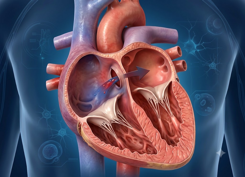
A patent foramen ovale (PFO) is a small opening between the heart’s upper chambers that remains open after birth. Before birth, this opening is a normal part of fetal circulation because it allows blood to bypass the developing lungs.
In most people, it closes naturally soon after birth. However, in some people, it remains open into adulthood. Many people with a PFO have no symptoms and may only discover it during tests for another medical condition.
Although a PFO is usually harmless, it can be linked to certain health concerns in some people. One important concern is that a blood clot may occasionally pass through the opening and travel to the brain, potentially causing an ischemic stroke.
Doctors may investigate a PFO after an unexplained stroke, particularly in younger adults who do not have another obvious cause. This article covers PFO symptoms, potential risks, diagnosis, and treatment options, including when monitoring, medication, or closure may be considered.
What is a Patent Foramen Ovale (PFO)?
A Patent Foramen Ovale (PFO) is a small, tunnel-like opening between the right and left atria, the two upper chambers of the heart. It is a normal part of fetal circulation but may fail to close completely after birth.
A PFO often causes no problems and may never be discovered. In certain situations, however, it can allow blood to move between the heart chambers in an unusual way. A PFO is different from an atrial septal defect (ASD). An ASD is an actual hole in the tissue separating the atria, while a PFO is more like a flap or tunnel that has not fully sealed.
To understand why a PFO can remain open, it helps to look at how the foramen ovale works before birth.
Fetal Circulation and the Role of the Foramen Ovale
Before birth, a baby’s lungs are filled with fluid and do not yet exchange oxygen. The placenta provides oxygen and nutrients instead. Because the fetal lungs do not need to handle normal blood flow, the fetal circulation uses special pathways called shunts to redirect blood.
The Oxygenation Pathway
Oxygen-rich blood comes from the placenta through the umbilical vein. It partly bypasses the liver through a vessel called the ductus venosus before entering the inferior vena cava, where it mixes with blood returning from the lower part of the body.
Shunting Inside the Heart
The oxygen-rich blood enters the right atrium of the fetal heart. A small tissue flap called the Eustachian valve helps direct blood toward the foramen ovale.
The foramen ovale allows blood to move from the right atrium into the left atrium without passing through the lungs. From there, the blood enters the left ventricle and is pumped through the aorta so the developing brain and other important organs receive oxygen-rich blood.
Post-Natal Transition: How the Flap Seals
When a newborn takes the first breath, major changes occur in the pressure inside the heart.
As the lungs fill with air, blood vessels in the lungs open and resistance in the pulmonary circulation decreases. More blood begins flowing through the lungs, picks up oxygen, and returns to the left atrium. This raises pressure on the left side of the heart.
At the same time, clamping the umbilical cord reduces blood returning to the right atrium. The change in pressure pushes the septum primum against the septum secundum, covering the opening much like a flap.
Over the first year of life, these tissue layers usually fuse together completely in many people. When they do not fully join, the opening can remain patent, or open.
What is a PFO? Anatomical Variant vs. Congenital Defect
A PFO occurs when the tissue layers that normally close after birth do not completely fuse. As a result, the tunnel-like opening remains open.
What is a PFO in Medical Terms?
Medically, a patent foramen ovale is generally considered an anatomical variant rather than a traditional congenital heart defect. The main difference is whether actual heart tissue is missing.
| Structural Feature | Patent Foramen Ovale (PFO) | Atrial Septal Defect (ASD) |
|---|---|---|
| Anatomical Structure | A flap or tunnel formed by overlapping tissue layers that did not completely fuse. | A true opening in the atrial wall caused by missing septal tissue. |
| Blood Flow Dynamics | Blood flow through the opening is usually intermittent and may occur during temporary pressure changes. | Blood can flow continuously through the opening because the tissue is actually missing. |
| Clinical Classification | A common anatomical variant found in a significant portion of healthy adults. | A congenital heart defect that may require medical or surgical management. |
Hemodynamics of a PFO Heart Condition
In everyday conditions, a PFO usually remains closed and does not interfere with normal circulation. Pressure in the left atrium is normally slightly higher than pressure in the right atrium, which keeps the tissue flap closed.
Certain activities can briefly change this pressure balance. Coughing, sneezing, vomiting, heavy lifting, or straining during a bowel movement can temporarily increase pressure in the right atrium. This is sometimes referred to as a Valsalva maneuver.
If right-sided pressure becomes higher than left-sided pressure, the flap can briefly open. This may allow some blood to move from the right atrium to the left atrium without passing through the lungs.
What are the Symptoms of a Patent Foramen Ovale?
Most people with a Patent Foramen Ovale have no symptoms and do not know they have it. In some cases, however, a PFO may be associated with conditions such as cryptogenic stroke, migraine with aura, or, rarely, low oxygen levels and shortness of breath.
A PFO itself does not usually cause pain or interfere with the heart’s electrical rhythm. It therefore does not typically cause symptoms such as chest pain, palpitations, or ongoing fatigue.
The medical importance of a PFO comes from what can potentially pass through the opening when it temporarily allows blood to move from the right side of the heart to the left.
The Right-to-Left Shunt: Bypassing the Pulmonary Filter
Normally, blood returning from the body enters the right side of the heart and travels to the lungs. The lungs help filter small clots and other particles from the venous circulation before blood returns to the left side of the heart.
With a PFO, a temporary rise in right-sided heart pressure can open the flap and create a right-to-left shunt.
If a small blood clot or another particle is present in the bloodstream at that moment, it may pass through the PFO into the left atrium. From there, it can enter the arterial circulation and potentially travel to the brain or another organ.
Clinical Conditions Scientifically Linked to a PFO
Because a PFO often causes no symptoms, it is usually discovered during testing for another medical concern rather than during a routine examination.
Cryptogenic Stroke and TIA
One of the most important concerns associated with a PFO is a cryptogenic stroke. This is an ischemic stroke that occurs without another clearly identified cause, such as significant artery disease or atrial fibrillation.
A possible mechanism is called a paradoxical embolism:
- A blood clot forms in a deep vein, often in the legs.
- The clot travels through the veins toward the right atrium.
- A sudden increase in right-sided heart pressure causes the PFO to open.
- The clot passes into the left atrium and then into the arterial circulation, where it can travel to the brain and block blood flow.
A temporary blockage may result in a Transient Ischemic Attack (TIA), sometimes called a mini-stroke.
Migraine with Aura
Research has found an association between PFO and migraine with aura, which can involve visual symptoms such as flashing lights, blind spots, or other changes before a headache.
Researchers continue to study the reason for this association. One theory is that certain substances or microscopic clots in venous blood may bypass the lungs through the PFO and reach the brain.
Decompression Sickness (“The Bends”)
A PFO can also be relevant to scuba divers. During a dive, nitrogen dissolves into body tissues because of increased underwater pressure. As a diver rises toward the surface, nitrogen can form small bubbles in the bloodstream.
Normally, these bubbles travel to the lungs, where they can be filtered out. With a PFO, a temporary pressure increase may allow some bubbles to cross into the arterial circulation instead.
If this happens, the bubbles may travel to the brain or spinal cord and contribute to neurological decompression sickness.
Rare Structural Manifestations: Platypnea-Orthodeoxia Syndrome
Although a PFO does not usually cause breathing problems, it can contribute to a rare condition called Platypnea-Orthodeoxia Syndrome (POS).
POS causes significant shortness of breath (platypnea) and a decrease in blood oxygen levels (orthodeoxia) when a person sits or stands. Symptoms may improve when the person lies down.
The syndrome generally occurs when a PFO is combined with another physical change, such as an enlarged aorta, a shifted diaphragm, or certain lung conditions.
When the person stands, changes in the position or shape of nearby structures can alter blood flow through the right atrium. This may direct more deoxygenated blood through the PFO into the left side of the heart, lowering oxygen levels and causing breathlessness or fatigue.
Advanced Cardiovascular Diagnostic Procedures
When doctors suspect a right-to-left shunt, they use specialized heart and blood-flow tests rather than relying only on a standard chest X-ray or EKG.
Transthoracic Echocardiogram (TTE) with Bubble Study: This is a non-invasive test that uses ultrasound to view the heart. Agitated sterile saline containing tiny bubbles is injected into a vein. Doctors watch the heart to determine whether the bubbles cross from the right atrium to the left atrium.
Transesophageal Echocardiogram (TEE): If a TTE does not provide enough information, a TEE may be used. The patient is sedated while a thin ultrasound probe is guided into the esophagus, which lies close to the heart. This provides detailed images of the atrial septum and PFO.
Transcranial Doppler (TCD): This test uses ultrasound to monitor blood flow in the brain’s arteries during a bubble study. Detecting bubbles in the brain can provide evidence of a right-to-left shunt.
Diagnostic Evaluation Checklist
When a patient has a cryptogenic stroke or unexplained neurological symptoms, evaluation for a PFO may include:
- Clinical History Review: Reviewing previous unexplained strokes, migraine with aura, or decompression sickness related to diving.
- Physical Valsalva Testing: Looking for changes associated with coughing or straining.
- Surface TTE with Saline Contrast: Using an ultrasound and bubble study to screen for a shunt.
- High-Resolution TEE: Examining the atrial septum and PFO structure in greater detail.
- Transcranial Doppler Tracking: Checking whether bubbles or other shunted material reach the brain’s circulation.
What is the Stroke Risk associated with a Patent Foramen Ovale?
A PFO is common and is usually harmless. However, it becomes more clinically important when a younger or middle-aged adult experiences an unexplained ischemic stroke.
A stroke without an identifiable cause may be classified as cryptogenic. In these cases, doctors may investigate whether a PFO could have allowed a clot to travel from the venous circulation into the arteries.
The Anatomy of a Paradoxical Embolism
To understand how a PFO could contribute to stroke, it helps to follow the possible path of a blood clot:
- Source: Venous System: A clot forms in a deep vein of the legs or pelvis.
- Route: To the Heart: The clot travels through the veins to the right atrium.
- The Paradoxical Shunt: A temporary increase in right-sided pressure opens the PFO, allowing the clot to pass into the left atrium.
- Destination: The Brain: The clot enters the arterial circulation and may travel to a blood vessel in the brain, causing an ischemic stroke.
Normally, venous clots travel to the right side of the heart and then to the lungs, where the pulmonary circulation can trap them. A PFO can provide another route if pressure conditions allow the clot to cross into the left side of the heart.
Anatomical Risk Factors: High-Risk PFO Features
Not all PFOs have the same characteristics. Cardiologists can use detailed imaging, including TEE, to examine the size, shape, and movement of the opening.
Certain anatomical features may be associated with a higher likelihood of a clot crossing through the PFO.
Atrial Septal Aneurysm (ASA)
An atrial septal aneurysm is a flexible and unusually mobile section of tissue in the wall between the heart’s upper chambers. When it moves significantly, it may be associated with a greater chance of blood crossing through a PFO.
Large Shunt Size
The size of the right-to-left shunt can be estimated during a bubble study by observing how many bubbles cross into the left atrium. A larger shunt may provide a greater pathway for material to cross between the chambers.
Long PFO Tunnel
The structure and length of the PFO tunnel can also be considered during evaluation. Doctors may assess its shape and size when determining the overall characteristics of the PFO.
Clinical and Hypercoagulable Risk Multipliers
The presence of a PFO is only one part of the overall risk assessment. The likelihood of a clot-related complication may also depend on how easily a person forms blood clots.
- Thrombophilia (Hypercoagulable States): Certain inherited or acquired conditions can make blood more likely to clot, including Factor V Leiden, prothrombin gene mutations, and antiphospholipid syndrome.
- A Personal History of DVT: A previous deep vein thrombosis provides evidence that a person has experienced venous clot formation.
- Situational Stasis: Long periods of limited movement, prolonged bed rest, long flights, or recovery from major surgery can slow blood flow and increase the risk of developing a clot.
Summary of High-Risk PFO Criteria
When doctors evaluate a PFO after a cryptogenic stroke, they may consider several clinical and anatomical factors:
| High-Risk Feature | Clinical Presentation / Imaging Finding | Why It May Increase Risk |
|---|---|---|
| Atrial Septal Aneurysm | Excessive movement or bulging of the atrial septal tissue. | May be associated with increased blood movement through the PFO. |
| Large Shunt Grade | A larger number of bubbles crossing during a bubble study. | Suggests a more significant right-to-left shunt. |
| Long PFO Tunnel | A longer tunnel identified during TEE. | Provides additional structural information for risk assessment. |
| Hypercoagulable Blood | Laboratory evidence of a clotting disorder. | May increase the likelihood of venous clot formation. |
| History of DVT/PE | Current or previous confirmed venous clots. | Indicates a known source of potential emboli. |
What are the Closure Options for a Patent Foramen Ovale?
The main treatment approaches for a PFO that is believed to be involved in a stroke include medication to reduce clot formation and a minimally invasive catheter procedure to close the opening.
The decision is individualized and may involve both a neurologist and a cardiologist. The goal is to reduce the risk of another stroke. Simply finding an asymptomatic PFO does not usually mean that it needs to be closed.
What are the primary treatments for a PFO after a stroke?
The main options after a presumed PFO-related cryptogenic stroke are long-term medical therapy with antiplatelet or anticoagulant medication and percutaneous PFO closure.
The choice depends on factors such as the patient’s age, stroke characteristics, PFO anatomy, other medical conditions, and personal circumstances.
- Medical Therapy: Medication does not close the PFO. Instead, it helps reduce the likelihood of blood clots forming.
- Antiplatelet Agents: Medications such as aspirin or clopidogrel reduce platelet activity and can help prevent certain blood clots.
- Anticoagulants: Medications such as warfarin or direct oral anticoagulants affect the body’s clotting process. They may be considered when a person has another reason to require anticoagulation, such as certain clotting disorders.
- Percutaneous PFO Closure: This is a minimally invasive procedure that places a device over the PFO to close the opening.
- The Procedure: A catheter is inserted into a large vein, commonly through the groin, and guided toward the heart. A collapsible closure device is positioned across the PFO, with one part on each side of the opening. Once released, the device holds the tissue together and blocks the passage.
- Recovery and Long-Term Outcome: Heart tissue gradually grows around the device over the following months. Many patients can return home the same day or after overnight observation and may need antiplatelet medication for a period after the procedure.
When is PFO closure recommended over medication?
PFO closure may be considered for carefully selected younger adults who have experienced a cryptogenic ischemic stroke that is believed to be related to the PFO. Certain anatomical features may also influence the decision.
The choice is not automatic. Doctors first need to determine whether the PFO is likely to have contributed to the stroke and whether closure is appropriate for the individual patient.
The comparison between closure and medication may involve several factors:
Patient Profile: A person who has experienced a confirmed ischemic stroke with no other identified cause may be evaluated for PFO closure. As people get older, other potential causes of stroke become more common, so doctors carefully assess the entire clinical picture.
High-Risk PFO Anatomy: Features such as a large right-to-left shunt or an atrial septal aneurysm may affect the treatment discussion. The RoPE (Risk of Paradoxical Embolism) score is another tool doctors may use when considering how likely a PFO is to have contributed to a stroke.
Evidence from Clinical Trials: Studies including RESPECT, REDUCE, and CLOSE have compared PFO closure with medical treatment in selected patients. These trials have helped establish the role of closure in preventing recurrent stroke for certain patients.
Risk-Benefit Discussion: Closure has potential procedural risks, including bleeding around the catheter site and complications such as atrial fibrillation. Medication also has potential risks, including bleeding. Doctors and patients generally discuss these factors together before choosing a treatment approach.
Key considerations for diagnosing and managing a PFO
Managing a patent foramen ovale requires consideration of heart anatomy, medical history, and other factors that may affect stroke or clot risk. Because most PFOs cause no symptoms, treatment is generally focused on preventing complications rather than treating everyday discomfort.
Definitive Diagnosis and Structural Distinction
Doctors use specialized imaging to determine whether a PFO is present and whether it is allowing a right-to-left shunt. These tests can also help distinguish a PFO from other conditions, including an atrial septal defect.
Transthoracic Echocardiogram (TTE) with Bubble Study
A TTE is a non-invasive ultrasound examination of the heart. During a bubble study, agitated sterile saline is injected into a vein, producing tiny bubbles.
The physician watches the bubbles as they move through the right atrium. If they cross into the left atrium, particularly during a Valsalva maneuver, this may indicate a right-to-left shunt.
Transesophageal Echocardiogram (TEE)
A TEE uses a thin ultrasound probe that is passed into the esophagus to obtain detailed images of the heart. Because the esophagus is located close to the heart, the test can provide a clearer view of the atrial septum.
TEE can also help doctors evaluate the structure of the PFO and identify features such as an atrial septal aneurysm.
Transcranial Doppler (TCD)
TCD uses ultrasound to monitor blood flow through arteries in the brain while a bubble study is performed. If bubbles are detected in the brain’s circulation, this can support the presence of a right-to-left shunt.
Distinguishing a PFO from an Atrial Septal Defect (ASD)
It is important to tell a PFO apart from an atrial septal defect because their structures are different:
- Patent Foramen Ovale: A flap-like tunnel formed by overlapping tissue layers that did not completely fuse after birth. It generally remains closed and may open temporarily when pressure changes.
- Atrial Septal Defect: A congenital defect in which part of the tissue separating the atria is missing, creating a permanent opening between the chambers.
Management Pathways: Medical Therapy vs. Transcatheter Closure
When a PFO is found in someone who has experienced an otherwise unexplained stroke, doctors may consider medication or a catheter-based closure procedure.
Medical Therapy
For some patients, particularly those without features that suggest a higher-risk PFO, medical treatment may be appropriate. The goal is to reduce the chance of blood clot formation.
- Antiplatelet Medications: Medicines such as low-dose aspirin or clopidogrel reduce platelet activity and can lower the risk of certain clot-related events.
- Anticoagulants: Medicines such as warfarin or direct oral anticoagulants may be used when there is an additional reason for anticoagulation, including certain clotting disorders.
Percutaneous Transcatheter PFO Closure
For selected patients, a catheter-based closure procedure may be considered.
A specialist guides a thin catheter through a vein in the groin to the heart. A small double-disc closure device is positioned across the PFO. The device holds the overlapping tissue together, and over time, the body’s tissue grows around it.
Post-Procedural Recovery Timeline
Recovery after PFO closure is generally focused on healing the catheter entry site, allowing the device to settle, and preventing clot formation during the healing period.
Days 1 to 2 (Immediate Post-Procedure): Patients may be observed for several hours or overnight before returning home. Care focuses on monitoring the catheter entry site and checking for early complications.
Weeks 1 to 2 (Activity Modification): Light daily activities may usually be resumed relatively soon, while strenuous exercise and heavy lifting may be restricted for a short period according to the doctor’s instructions.
Months 1 to 6 (Endothelialization & Medication): The body gradually grows tissue over the closure device. Antiplatelet medication may be prescribed during this period, and follow-up imaging may be used to check the device and make sure the PFO remains closed.
Year 1 and Beyond (Long-Term Maintenance): Additional follow-up may be performed during the first year. Long-term medication and monitoring depend on the individual’s medical history and the treatment plan.
Special Precautions and Scuba Diving Risks
People with an open PFO may need to take special precautions in high-pressure environments such as scuba diving.
The Mechanics of “The Bends” in Scuba Diving
During a dive, increased pressure causes nitrogen to dissolve into the body’s tissues. As the diver rises, nitrogen can form small bubbles in the bloodstream.
Normally, these bubbles travel to the lungs and are filtered out. With a PFO, a temporary increase in right-sided heart pressure can allow some bubbles to cross into the arterial circulation.
Once there, the bubbles may travel to the brain or spinal cord and contribute to neurological decompression sickness.
Safety Protocol for Divers with a Confirmed PFO
Divers with a confirmed PFO should discuss their individual situation with a cardiologist and a physician experienced in dive medicine. If they continue diving without closure, a conservative approach may be recommended to reduce the amount of nitrogen absorbed.
- Depth and Time Limits: Avoid deeper dives and limit underwater exposure.
- Ascent Profile Changes: Follow slow ascent procedures and appropriate safety stops.
- Specialist Consultation: Work with a cardiologist and dive-medicine specialist to determine the safest approach based on individual risk.
Conclusion
A patent foramen ovale is a common heart condition that causes no symptoms in many people and does not require treatment in most cases. However, in certain situations, it may be associated with complications such as stroke when a blood clot passes through the opening and reaches the brain.
Treatment decisions depend on factors such as previous stroke history, PFO anatomy, overall health, and other clotting or cardiovascular risks. Medical evaluation can help determine whether monitoring, medication, or PFO closure is appropriate.
Frequently Asked Questions
1. What is a patent foramen ovale?
A patent foramen ovale is a small flap-like opening between the right and left atria of the heart. It is normal before birth because it allows blood to bypass the developing lungs. After birth, it usually closes, but it remains open in some people. Most people with a PFO have no symptoms and do not need treatment.
2. What symptoms can a patent foramen ovale cause?
Most people with a PFO have no noticeable symptoms. It is often found during an echocardiogram performed for another reason. In some cases, it may be associated with an unexplained stroke, migraine with aura, or low oxygen levels. Doctors consider the person’s overall medical history when deciding whether the PFO may be contributing to a problem.
3. Can a patent foramen ovale cause a stroke?
A PFO may contribute to stroke in some people by allowing a blood clot from the veins to cross into the arterial circulation and travel to the brain. This is called a paradoxical embolism. However, many people with a PFO never experience a stroke, and doctors usually investigate other possible causes before determining that a PFO was responsible.
4. How is a patent foramen ovale diagnosed?
A PFO is commonly evaluated with an echocardiogram. A bubble study may be performed by injecting tiny bubbles into a vein and watching whether they cross from the right side of the heart to the left. A transesophageal echocardiogram may provide more detailed images when needed.
5. Does a patent foramen ovale need treatment?
Many people with a PFO do not need treatment, particularly when there are no symptoms or related complications. If a person has experienced an unexplained stroke that may be connected to the PFO, doctors may consider medications or a catheter-based closure procedure. The appropriate option depends on individual risk factors.
6. What happens during patent foramen ovale closure?
PFO closure is a minimally invasive procedure that uses a catheter, usually inserted through a blood vessel in the groin, to place a small closure device over the opening. The device blocks the passage while heart tissue gradually grows around it. Open-heart surgery is generally not required. Follow-up care and medication are commonly needed for a period after the procedure.