What Is Hematopoiesis? Understanding the Formation of Blood Cells
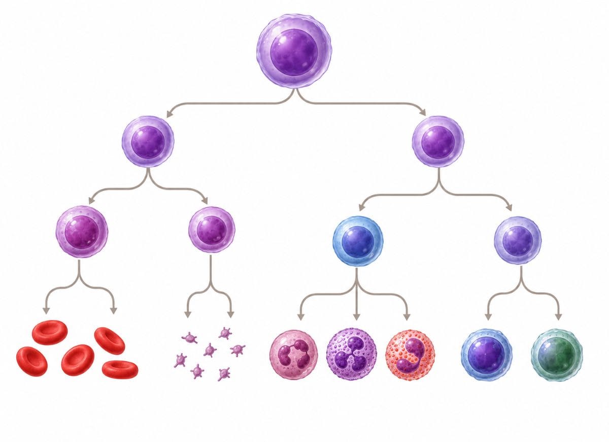
Your body never stops making blood. Right now, millions of fresh blood cells are being produced to take the place of ones that have worn out or finished their job. This never-ending renewal keeps oxygen moving, keeps infections in check, and keeps bleeding under control. Without it, the body would grind to a halt within days.
This ongoing manufacturing process is called hematopoiesis a word built from Greek roots meaning “blood” and “to make.” It describes how a small population of blood stem cells gradually turns into all the mature blood cells circulating in your veins: red blood cells, white blood cells, and platelets, each with its own job to do.
Most of this happens inside the bone marrow, the soft tissue packed inside certain bones. There, hematopoietic stem cells go through a tightly choreographed series of divisions and transformations. On a typical day, the body churns out around 2 million red blood cells every single second a staggering pace that shows just how much this system does behind the scenes.
Why does any of this matter? Because when blood cell production breaks down, the consequences show up as real disease anemia, leukemia, lymphoma, bone marrow failure, and various immune disorders all trace back to problems somewhere in this pipeline. Below, we’ll walk through what hematopoiesis actually is, where it takes place, how it unfolds step by step, and what can go wrong along the way.
What is Hematopoiesis?
At its core, hematopoiesis is the process that builds every blood cell in your body from a single type of ancestor cell the hematopoietic stem cell (HSC). It’s the engine behind your entire circulatory and immune system, keeping a steady output of red blood cells (erythrocytes) for carrying oxygen, white blood cells (leukocytes) for defending against illness, and platelets (thrombocytes) for stopping bleeding.
None of this happens by accident. A dense web of growth factors, cytokines, and transcription factors directs stem cells down specific developmental roads, and this signaling network can shift production up or down depending on what the body needs more red cells during low oxygen, more white cells during an infection, and so on.
It’s a constant balancing act: new cells are made at roughly the same rate old ones are cleared out, keeping the whole system in equilibrium. If that balance breaks, the fallout is serious the body loses its ability to carry oxygen, fight infection, or seal off injuries, which can cascade into failure across multiple organs.
What are Hematopoietic Stem Cells (HSCs)?
Hematopoietic stem cells are a small, specialized population living mainly in the bone marrow, and they’re the single source from which every blood and immune cell in the body eventually descends. What makes them special is two abilities working together: they can renew themselves, and they can turn into many different cell types.
Self-renewal means an HSC can divide and leave behind a copy of itself that stays an HSC this is what keeps the stem cell reservoir from running dry over a lifetime. Differentiation is the other half of the story: an HSC can also commit to a developmental path, spinning off progenitor cells with narrowing options until they finally become one specific, mature cell type from either the myeloid or lymphoid family.
On their own, HSCs don’t look or act like any mature blood cell they’re unspecialized but they carry the genetic instructions to become any of them. That flexibility is exactly why bone marrow and stem cell transplants work: give the body a fresh supply of HSCs, and it can rebuild its entire blood and immune system from scratch.
Where Does Hematopoiesis Occur Throughout Life?
The location where blood cells are made isn’t fixed it moves as the body develops, shifting to whatever site best fits the needs of that stage of life.
It all starts in the yolk sac, around the third week after conception, in what’s called primitive hematopoiesis. At this early point, the focus is almost entirely on making red blood cells so the rapidly growing embryo gets enough oxygen.
As development continues, the job passes to the aorta-gonad-mesonephros (AGM) region, then to the fetal liver by the second trimester, with some help from the spleen. This later stage “definitive” hematopoiesis is where the lasting stem cell population gets established and a much wider variety of blood cells starts being produced. By the third trimester, the process begins handing off to the bone marrow.
After birth, the bone marrow takes over completely and stays the primary site for life. In adults, active blood production is concentrated in the red marrow found in flat bones the sternum, pelvis, vertebrae, ribs and the ends of long bones like the femur and humerus. The remaining marrow space is yellow marrow, mostly fat tissue, though it can convert back into active red marrow if the body suddenly needs more blood cells, such as after severe blood loss.
The Process of Hematopoiesis
Once a hematopoietic stem cell activates, it faces its first major fork in the road: becoming part of the myeloid lineage or the lymphoid lineage. The myeloid side produces most innate immune cells along with red blood cells and platelets, while the lymphoid side is dedicated entirely to lymphocytes the cells driving adaptive immunity.
This choice isn’t random. It’s shaped by a mix of the cell’s internal genetic programming and outside signals from the bone marrow environment. An HSC first becomes a multipotent progenitor, which then locks in as either a common myeloid progenitor (CMP) or a common lymphoid progenitor (CLP) a decision that, once made, can’t be reversed.
From the CMP branch come red blood cells, megakaryocytes (which produce platelets), mast cells, and myeloblasts, which go on to form granulocytes (neutrophils, eosinophils, basophils) and monocytes. The CLP branch is narrower, giving rise only to B-lymphocytes, T-lymphocytes, and Natural Killer (NK) cells. This split keeps the two arms of the blood and immune system working in a coordinated, non-overlapping way.
What Blood Cells Are Produced by The Myeloid Lineage?
Starting from the common myeloid progenitor, this lineage produces the cells responsible for carrying oxygen, stopping bleeding, and mounting the body’s first response to invaders. It’s the busiest branch of the whole system, generating the largest share of blood cells day to day.
Erythrocytes (red blood cells) have no nucleus and a distinctive disc shape. Thanks to the iron-rich protein hemoglobin, they carry oxygen out to the tissues and bring carbon dioxide back.
Thrombocytes (platelets) aren’t full cells at all they’re tiny fragments pinched off from megakaryocytes. Their job is to plug damaged vessels and kick off the clotting cascade.
Mast cells live in tissues and drive allergic and inflammatory reactions, releasing histamine and heparin from their granules when triggered.
Myeloblasts are the precursor stage that later splits into granulocytes and monocytes.
Granulocytes neutrophils, eosinophils, and basophils each carry distinctive granules. Neutrophils swallow and destroy bacteria; eosinophils target parasites and help regulate allergic inflammation; basophils release histamine during allergic responses.
Monocytes are the largest white blood cells. They travel through the bloodstream before settling into tissues, where they turn into macrophages or dendritic cells both essential for cleaning up debris and kicking off the adaptive immune response.
What Blood Cells Are Produced By The Lymphoid Lineage?
Branching off from the common lymphoid progenitor, this lineage is responsible for lymphocytes the cells that give the immune system its precision and memory. Rather than reacting broadly like myeloid cells, lymphoid cells specialize in recognizing specific threats and remembering them for next time. Three cell types come from this branch: T-cells, B-cells, and NK cells.
T-lymphocytes drive cell-mediated immunity. They start in the bone marrow but finish maturing in the thymus, where they’re trained to tell self from non-self. Helper T-cells (CD4+) coordinate the wider immune response, while cytotoxic T-cells (CD8+) directly destroy infected or cancerous cells.
B-lymphocytes mature entirely in the bone marrow and are the backbone of antibody-based immunity. When a B-cell meets its matching antigen, it turns into a plasma cell that pumps out antibodies to neutralize or flag pathogens. Some B-cells become long-lived memory cells, giving faster protection the next time the same threat shows up.
Natural Killer cells, despite sharing a progenitor with T and B cells, act more like innate immune cells attacking infected or abnormal cells immediately, without needing to recognize a specific antigen first.
Steps of Hematopoiesis: How are red blood cells and platelets formed?
Red blood cells develop through erythropoiesis, controlled by the hormone erythropoietin (EPO). Platelets form through thrombopoiesis, driven by thrombopoietin (TPO) and built from oversized precursor cells called megakaryocytes. Both pathways branch off the same myeloid progenitor but end up producing very different, nucleus-free cells suited to very different jobs.
Erythropoiesis is all about packing in hemoglobin and shrinking the cell down into a flexible shape that can squeeze through tiny capillaries a process that ends with the cell ejecting its own nucleus.
Thrombopoiesis takes a completely different route: a single giant cell grows enormous, then breaks apart to release thousands of platelet fragments straight into the bloodstream. Together, these two pathways keep the blood able to carry oxygen and stop bleeding, and both are adjusted constantly to match the body’s needs.
What are The Stages of Erythropoiesis?
Turning a stem cell into a mature red blood cell takes about a week, moving through several stages marked by shrinking cell size, rising hemoglobin, and finally the loss of the nucleus.
Proerythroblast the first committed red-cell precursor. Large, with a big round nucleus and a cytoplasm that stains blue due to heavy ribosome activity.
Basophilic erythroblast a bit smaller, with the nucleus starting to tighten up while ribosomes work at full speed producing globin chains for hemoglobin.
Polychromatic erythroblast hemoglobin production ramps up here, giving the cytoplasm a mixed pink-and-blue look. The cell keeps shrinking, and this is the last point at which the cell can still divide.
Orthochromatic erythroblast (normoblast) hemoglobin synthesis is nearly finished, the cytoplasm turns mostly pink, and the nucleus becomes dense and inactive before finally being pushed out of the cell.
Reticulocyte now nucleus-free, but still carrying leftover ribosomal material. It moves out of the bone marrow into circulation and finishes maturing over the next day or two.
Mature erythrocyte once the last ribosomes and organelles are cleared out, the cell becomes a fully functional, biconcave red blood cell ready to transport oxygen.
How Does Thrombopoiesis Create Platelets?
Platelets don’t come from ordinary cell division they’re released when giant megakaryocyte cells break apart. Thrombopoietin (TPO) is the main hormone pushing this process forward.
It begins with a stem cell committing to the megakaryocyte path, first becoming a megakaryoblast. This cell copies its DNA repeatedly without actually splitting a process called endomitosis resulting in one enormous cell with a single nucleus containing many sets of chromosomes (up to 64 times the normal amount).
As it matures through the promegakaryocyte stage into a full megakaryocyte, it becomes one of the biggest cells in the bone marrow. It builds an internal membrane network and sends long branching arms called proplatelets out through the marrow’s blood vessel walls and into the bloodstream itself.
The force of blood flow then shears these proplatelet extensions into thousands of individual platelets. A single megakaryocyte can generate somewhere between 1,000 and 3,000 platelets before its remaining nucleus is cleared away by marrow macrophages.
Steps of Hematopoiesis: How are the different white blood cells formed?
White blood cells come from three separate pathways collectively known as leukopoiesis: granulopoiesis (neutrophils, eosinophils, basophils), monocytopoiesis (monocytes), and lymphopoiesis (B-cells, T-cells, NK cells). Each pathway starts with its own committed progenitor and follows a unique sequence of division and maturation before producing a finished, functional white blood cell.
Granulopoiesis is defined by the appearance of specific granules that distinguish the three granulocyte types. Monocytopoiesis is a shorter, more direct route to a large phagocytic cell that finishes developing once it reaches tissue. Lymphopoiesis stands apart because T-cells don’t finish maturing in the bone marrow at all they relocate to the thymus.
Together, these three pathways keep the body supplied with a varied, balanced set of white blood cells ready to respond to whatever threat comes along.
What is The Process of Granulopoiesis?
Granulopoiesis builds neutrophils, eosinophils, and basophils from a shared myeloblast ancestor, over a process that takes roughly two weeks and involves clear changes in cell size, nucleus shape, and granule content.
It starts with the myeloblast, a large cell with no visible granules yet. This becomes a promyelocyte, marked by the appearance of primary (azurophilic) granules essentially microbe-killing enzyme packages like myeloperoxidase and defensins.
Next comes the myelocyte stage, where the cell stops dividing and secondary granules start showing up and it’s these secondary granules that determine whether the cell becomes a neutrophil, eosinophil, or basophil. After that, the metamyelocyte stage brings a kidney-bean-shaped nucleus.
In the final stages, the nucleus continues reshaping forming a C- or S-shape in the band cell stage, then fully segmenting into lobes in the mature granulocyte, which is then released into the bloodstream ready for duty.
How are Lymphocytes and Monocytes Developed?
Monocytes and lymphocytes follow completely separate developmental roads, even though both end up defending the body.
Monocytopoiesis starts in the bone marrow with a monoblast, which looks a lot like a myeloblast but is committed to becoming a monocyte. It develops into a promonocyte larger, with an indented nucleus and light granulation before finishing as a mature monocyte.
Monocytes are the largest cells in the bloodstream, recognizable by a kidney-shaped nucleus and gray-blue cytoplasm. After circulating for one to three days, they move into tissue and transform into either macrophages (found in places like the liver and lungs) or dendritic cells, which present antigens to trigger the adaptive immune response.
Lymphopoiesis produces T-cells, B-cells, and NK cells, all starting from the common lymphoid progenitor but B-cells and T-cells finish their development very differently.
B-cells mature completely inside the bone marrow. The CLP moves through pro-B and pre-B stages, during which a functional B-cell receptor gets built and tested. Cells that pass meaning they don’t attack the body’s own tissue become naive B-cells and head out to patrol organs like the spleen and lymph nodes.
T-cells, by contrast, leave the bone marrow early as pro-T cells and travel to the thymus to finish maturing. There, they go through positive selection (can they recognize the body’s own MHC molecules?) and negative selection (do they react too aggressively to the body’s own tissue?). Only the cells that pass both tests go on to become CD4+ helper or CD8+ cytotoxic T-cells.
What Factors Regulate and Disrupt Normal Hematopoiesis?
Hematopoiesis runs on a tight regulatory system of growth factors, hormones, and signals from the bone marrow environment but that system can be knocked off balance by genetic mutations, poor nutrition, infection, or toxin exposure, any of which can lead to serious blood disorders.
This regulation isn’t a fixed setting either it shifts across a person’s life, adapting to changing demands from childhood through old age, and reacting to illness or injury as needed. The whole system depends on precise signaling to know exactly which cells to make and when; when that signaling fails, the results can range from mild anemia to aggressive cancers.
How Do Growth Factors and Hormones Control Hematopoiesis?
The signals controlling hematopoiesis come mainly from cytokines a category that includes the hematopoietic growth factors along with several hormones. These molecules attach to receptors on stem and progenitor cells, telling them when to multiply and which path to follow.
Erythropoietin (EPO), made mostly by the kidneys when oxygen levels drop, is the main driver behind red blood cell production. Thrombopoietin (TPO), made in the liver, governs platelet production by pushing megakaryocytes to mature.
White blood cell production relies on a different group called Colony-Stimulating Factors (CSFs). G-CSF specifically boosts neutrophil production, while GM-CSF has a wider reach, encouraging neutrophils, eosinophils, and monocytes all at once.
Is Hematopoiesis Different From Lymphopoiesis?
They’re related but not the same thing lymphopoiesis is one specific branch growing out of the much bigger tree that is hematopoiesis.
Hematopoiesis covers the formation of every blood cell type red cells, platelets, and all white blood cells starting from a single hematopoietic stem cell that splits into either a common myeloid progenitor (CMP) or a common lymphoid progenitor (CLP). Lymphopoiesis picks up from the CLP side only, covering the development of lymphocytes: B-cells, T-cells, and NK cells.
Everything else neutrophils, monocytes, eosinophils, basophils, red cells, and platelets comes from the CMP side, in a process called myelopoiesis.
This split matters because it shapes how the immune system is organized. And there’s a geographic difference too: while most hematopoiesis wraps up in the bone marrow, T-cells are the exception, finishing their development in the thymus instead a hallmark of lymphopoiesis specifically.
Even the chemical signals differ between the two branches. Lymphopoiesis leans heavily on interleukins like IL-7, essential for B- and T-cell development, while myelopoiesis runs on factors like G-CSF, M-CSF, and EPO.
Common Disorders Related to Hematopoietic Dysfunction
When hematopoiesis stops working correctly, the fallout shows up as diseases affecting how many blood cells are made, how well they function, or whether they turn cancerous.
Anemias happen when there aren’t enough red blood cells or enough hemoglobin, cutting into the blood’s oxygen-carrying ability and causing fatigue, weakness, and shortness of breath. Causes range from iron or B12 deficiency to chronic illness or inherited conditions like sickle cell disease.
Leukemias are cancers of the blood-forming tissue, where the marrow floods the bloodstream with abnormal white blood cells that crowd out healthy ones leading to infections, anemia, and bleeding problems. They’re classified by how fast they progress (acute vs. chronic) and which cell type is affected.
Myeloproliferative neoplasms (MPNs) involve the marrow overproducing one or more blood cell types. Polycythemia vera, for instance, causes too many red blood cells, thickening the blood and raising clot risk.
Can Hematopoiesis Be Restored Medically?
Yes when the hematopoietic system is badly damaged, doctors can restore it through Hematopoietic Stem Cell Transplantation (HSCT), better known as a bone marrow transplant. It works by replacing a patient’s damaged stem cells with healthy ones, either from a matched donor or from cells the patient banked earlier.
The goal is to rebuild a fully working blood and immune system from the ground up. HSCT treats a range of serious conditions leukemias, lymphomas, multiple myeloma, aplastic anemia, and certain inherited immune or metabolic disorders.
The process starts with a conditioning phase: high-dose chemotherapy and/or radiation to wipe out the disease and suppress the immune system so it won’t reject the incoming cells. Then healthy stem cells are infused into the bloodstream, where they travel to the bone marrow, settle in, and begin producing new blood cells.
Transplants can be allogeneic (donor cells, often from a sibling or matched volunteer) or autologous (the patient’s own banked cells). Stem cells for these procedures can come from three sources: bone marrow itself, peripheral blood (collected after mobilizing stem cells with G-CSF), or umbilical cord blood.
HSCT isn’t without risk. Patients face a vulnerable window of infection risk during immune suppression, potential organ damage from conditioning treatment, and in allogeneic transplants a possible complication called Graft-versus-Host Disease (GVHD), where the donor’s immune cells attack the recipient’s own tissue.
FAQs
1. Which best defines hematopoiesis?
Hematopoiesis is the ongoing process the body uses to manufacture all its blood cells from specialized stem cells, mostly located in the bone marrow. These stem cells mature into red blood cells, white blood cells, and platelets through a series of tightly controlled steps, running continuously throughout life to keep oxygen transport, immune defense, and clotting functioning normally.
2. What are the two stages of hematopoiesis?
Broadly, hematopoiesis breaks down into two stages: proliferation and differentiation. First, stem cells divide to create immature precursor cells. Then those precursors mature into the specialized cell types red cells, white cells, and platelets the body needs to stay healthy.
3. What are the 5 functions of hematopoiesis?
Hematopoiesis is responsible for producing oxygen-carrying red blood cells, infection-fighting white blood cells, clot-forming platelets, replacing cells that have aged out, and keeping the overall blood cell population balanced. Without it, the body couldn’t move oxygen, fight disease, or repair damaged vessels effectively.
4. What organ regulates hematopoiesis?
The bone marrow is where hematopoiesis physically happens in adults, providing the environment stem cells need to grow and mature. But regulation isn’t limited to the marrow the kidneys release erythropoietin to stimulate red cell production, and the liver contributes substances that support blood cell development too.
5. Who is the father of hematopoiesis?
No single person holds that title, but Russian scientist Alexander Maximow is widely credited with proposing the stem cell theory of hematopoiesis in the early 1900s, laying groundwork that modern hematology still builds on.
6. Is hematopoiesis good or bad?
It’s a normal, essential, life-sustaining process. Healthy hematopoiesis keeps the body supplied with everything it needs for oxygen delivery, immunity, and clotting. Problems only arise when the process itself malfunctions, whether that means too few cells, too many, or abnormal ones but the process itself is inherently beneficial.
7. What nutrients are important for hematopoiesis?
Iron is critical for building hemoglobin, while vitamin B12 and folate support the DNA synthesis needed for cell division. Vitamin B6, copper, and adequate protein intake also play supporting roles. Falling short on any of these can impair blood cell production and lead to anemia or weakened immunity.
8. What happens when hematopoiesis goes wrong?
Disrupted hematopoiesis can mean too few blood cells, too many, or cells that don’t function properly showing up as fatigue, weakness, frequent infections, easy bruising, or unusual bleeding. The specific effects depend on which blood cell line is affected and what’s causing the disruption.
9. What diseases affect hematopoiesis?
Conditions like anemia, leukemia, lymphoma, myelodysplastic syndromes, aplastic anemia, multiple myeloma, and various inherited marrow disorders all involve disrupted blood cell formation. Nutritional gaps, kidney disease, autoimmune conditions, infections, and certain cancer treatments can also interfere with the process.
Conclusion
Hematopoiesis is one of the body’s quietest but most essential jobs. Every day, billions of new blood cells are produced to replace the ones that have run their course, keeping oxygen flowing, infections in check, and bleeding under control all without you ever noticing it’s happening.
Tracing the path from bone marrow stem cells to fully mature red cells, white cells, and platelets reveals just how carefully this system is managed at every stage. And when something interrupts that process, the consequences can range from mild anemia to life-threatening cancers and immune disorders.
Understanding how this all works isn’t just academic it’s a reminder of why nutrition, regular checkups, and paying attention to unusual symptoms actually matter. The more you know about how your blood is made, the better equipped you are to recognize when something’s off and why keeping your bone marrow healthy matters so much for the long run.