IPMN Explained: Symptoms, Diagnosis, and Treatment Options
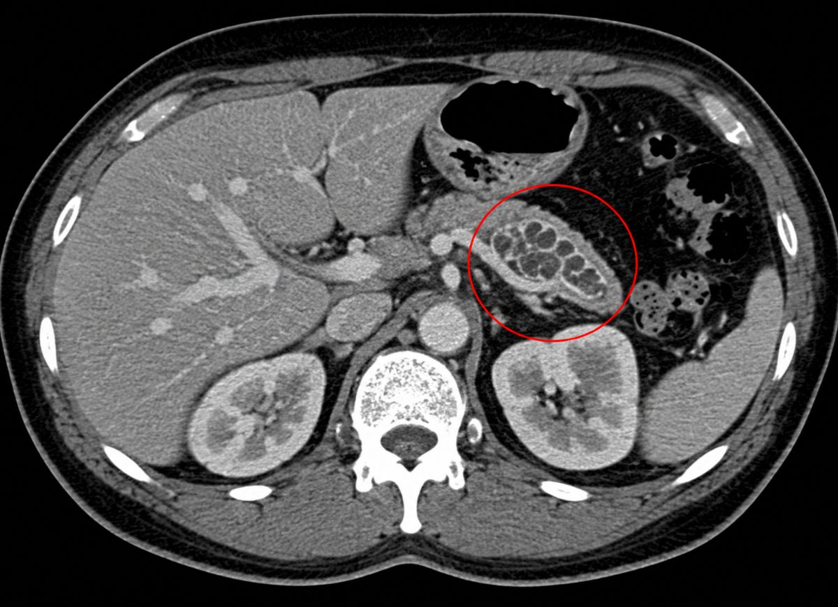
An intraductal papillary mucinous neoplasm IPMN for short is a cyst that forms inside the pancreas’s duct system, the network of small channels that carries digestive juices into the small intestine. As the cyst grows, it produces a thick, gel-like mucus and can cause the duct around it to stretch and widen. Some of these cysts sit quietly for years without changing. Others slowly evolve in ways that raise the chance of pancreatic cancer down the line.
That possibility is exactly why IPMN gets so much medical attention. Hearing the word “precancerous” attached to a cyst on your scan can be alarming but a diagnosis of IPMN is not the same thing as a cancer diagnosis. In fact, most IPMNs are found completely by accident, picked up on a CT or MRI that was ordered for something unrelated, like kidney stones or a bout of abdominal pain.
To figure out how worried to be, doctors look closely at the cyst its size, where it sits, how it’s growing, and whether it shows any “red flag” features such as a solid nodule, a widened main duct, or symptoms like jaundice, pancreatitis, sudden weight loss, or new-onset diabetes. Together, these details determine whether a cyst just needs to be watched over time or investigated further.
This guide walks through what IPMN actually is, why it develops, the symptoms it can cause, how much cancer risk is really involved, and how it’s diagnosed and treated. The intent isn’t to alarm you it’s to make a confusing diagnosis easier to understand, so that with the right monitoring and medical support, most people can manage IPMN safely and catch any trouble early.
What is an Intraductal Papillary Mucinous Neoplasm (IPMN)?
An IPMN is a precancerous growth that develops inside the pancreatic ducts. It’s marked by tiny, finger-like projections and by its signature trait: churning out thick mucin. IPMNs aren’t a single, uniform disease they exist on a spectrum, from mild, low-risk cellular changes all the way to advanced changes that are either right on the edge of becoming invasive cancer or have already crossed that line.
The Term Intraductal Papillary Mucinous Neoplasm
Every word in this name tells you something important about the condition, so it’s worth breaking down.
Intraductal means “inside the duct.” The pancreas is threaded with a system of ducts that ferry digestive enzymes to the intestine, and an IPMN begins growing right on the lining of one of these ducts often pushing the duct to widen as the tumor and its mucus take up space. This location matters clinically, because it gives any future cancer cells a direct route to spread.
Papillary describes what the growth looks like under a microscope: the cells arrange themselves into small, branching, finger-like structures called papillae that project into the duct. This pattern is what sets IPMN apart from other pancreatic cysts, like serous cystadenomas.
Mucinous means the tumor cells churn out large amounts of mucin, a thick, jelly-like fluid. This mucin builds up inside both the cyst and the duct, sometimes enough to cause blockages that trigger pancreatitis. Spotting mucin on imaging is one of the key clues doctors use to diagnose IPMN in the first place.
Neoplasm simply means abnormal tissue growth a tumor. Neoplasms can be harmless, precancerous, or outright malignant. Every IPMN falls into the “has the potential to become cancer” category, which is why none of them are ignored entirely, even the low-risk ones.
Main Types of IPMN
IPMNs are sorted into three categories based on exactly where in the duct system they’re located: main-duct, branch-duct (BD-IPMN), and mixed-type. This isn’t just anatomical trivia location is one of the biggest predictors of how dangerous a given IPMN is, and it drives whether a doctor recommends watchful waiting or surgery.
Main-duct IPMN affects the central channel running through the pancreas. It’s diagnosed when that main duct widens beyond 5 millimeters without any other explanation for the swelling. This is the riskiest category by far studies estimate that well over half of main-duct IPMNs carry either high-grade abnormal cells or invasive cancer. Because the stakes are so high, surgery is almost always recommended for anyone healthy enough to undergo it.
Branch-duct IPMN (BD-IPMN) is the most frequently diagnosed type. It forms in the smaller side channels that feed into the main duct, typically showing up on scans as a cluster of small cysts that resemble a bunch of grapes. The cancer risk here is meaningfully lower than main-duct disease roughly 15–25% over a person’s lifetime, according to most estimates. Because the danger is lower, many BD-IPMNs without worrying features can simply be tracked with periodic imaging rather than removed.
Mixed-type IPMN shows characteristics of both main duct widening alongside branch-duct cysts. Clinically, doctors treat it with the same seriousness as main-duct disease, since involvement of the main duct is what really drives the risk. Surgical removal is usually the recommended path.
Is IPMN a Form of Cancer?
No IPMN itself isn’t cancer. It’s better thought of as an early step on a possible path toward invasive pancreatic cancer, not the destination itself.
The key concept here is dysplasia, which just means the cells have become abnormal in some way. The cells lining an IPMN can gradually shift from normal, to mildly abnormal, to severely abnormal, to cancerous.
Low-grade dysplasia means the cells look slightly off under the microscope but aren’t multiplying out of control or invading nearby tissue. Most small, symptom-free branch-duct IPMNs fall into this bucket, and many stay this way for years without ever progressing.
High-grade dysplasia, sometimes called carcinoma in situ, is a more serious stage. The cells look almost identical to cancer cells and are clearly disorganized but they’re still contained within the duct lining and haven’t broken through into surrounding tissue. This stage is treated as high-risk precisely because it’s the last step before invasive cancer, and the whole point of surgery in high-risk cases is to catch the disease here, before it goes further.
IPMN with associated invasive carcinoma is the point where the disease has fully become cancer. The abnormal cells have pushed through the duct wall into the surrounding pancreatic tissue. From here, it’s managed and staged like any other pancreatic cancer, and the outlook depends heavily on how far it’s spread. The entire purpose of surveillance and early diagnosis is to intervene long before a patient ever reaches this stage.
The Symptoms and Causes of Intraductal Papillary Mucinous Neoplasm
IPMN symptoms are unpredictable many people have none at all while the underlying causes still aren’t fully understood, even though several risk factors are well established. A large share of people with IPMN find out about it purely by chance, during a scan ordered for something else entirely.
When symptoms do show up, they tend to be vague and stem from the cyst physically interfering with the pancreas or nearby organs. Recognizing both the possible warning signs and the risk factors is key to catching the condition early.
Common Signs and Symptoms of IPMN
Not everyone with IPMN experiences symptoms a substantial number of cases are entirely silent. When symptoms do appear, the usual suspects are abdominal pain, acute pancreatitis, jaundice, unexplained weight loss, and new diabetes, and which ones show up often depends on the cyst’s size, location, and whether it’s blocking a duct.
Small branch-duct IPMNs, in particular, are notorious for causing no symptoms whatsoever, which is why incidental discovery on an unrelated scan has become the most common way they’re found.
For people who do have symptoms, abdominal pain is among the most frequent. It’s usually vague, centered in the upper belly, and sometimes radiates toward the back caused either by the cyst stretching pancreatic tissue, pressure from a blocked duct, or an actual episode of pancreatitis.
IPMNs are also a well-known trigger for acute pancreatitis. The thick mucin they produce can plug up the duct, causing digestive enzymes to back up and start damaging the pancreas itself leading to sudden, severe pain, nausea, and vomiting. For some patients, an otherwise unexplained pancreatitis episode is the very first clue that an IPMN is present.
If the IPMN sits in the head of the pancreas, it can grow large enough to press on the common bile duct running through that area, blocking bile flow from the liver. The result is jaundice yellowing skin and eyes, dark urine, and pale stools. Because jaundice caused by a mass is considered a serious warning sign, it typically prompts immediate follow-up.
Unexplained weight loss can occur, either because a blocked duct is interfering with nutrient absorption or as a more general sign that something more serious is going on. Likewise, new-onset diabetes especially in an older adult who isn’t overweight can be an early red flag for a pancreatic problem, since the growing neoplasm may be damaging insulin-producing cells.
Risk Factors for Developing an IPMN
Several factors are linked to a higher chance of developing IPMN: older age, a history of smoking, chronic pancreatitis, and a family history of pancreatic cancer. None of these are proven direct causes, but they show up again and again in people diagnosed with the condition.
Age is the strongest pattern. IPMN is rare before 50 and becomes far more common between ages 60 and 80, suggesting the underlying cellular changes build up gradually over decades.
Smoking is already a well-known risk factor for pancreatic cancer generally, and it’s also tied to IPMN development specifically. The chemicals in tobacco smoke are thought to encourage the kind of genetic mutations that can kick-start these cysts and smokers with an existing IPMN may also face a higher risk of it turning malignant.
Chronic pancreatitis long-running inflammation of the pancreas creates a cellular environment that’s more prone to abnormal growth, since the constant cycle of damage and repair increases the odds of errors creeping into cell division.
A family history of pancreatic cancer particularly in a parent, sibling, or child raises personal risk too, pointing to an inherited genetic component in some families. Certain inherited syndromes, like Peutz-Jeghers syndrome and familial adenomatous polyposis (FAP), are also linked to elevated IPMN risk, though together they account for only a small slice of overall cases. Researchers are still working to pin down the specific genes involved.
Intraductal Papillary Mucinous Neoplasm Diagnosis
IPMN is typically diagnosed using high-resolution imaging CT and MRI/MRCP scans and, when more detail is needed, endoscopic ultrasound (EUS) combined with fine-needle aspiration (FNA). Each of these tools does a different job, and together they build a complete picture of the cyst.
A CT scan with contrast is often the first test used. It shows the cyst’s size, location, and its relationship to nearby organs and blood vessels clearly, and it’s especially good at picking up main-duct widening or larger, more obvious mural nodules. It’s also essential for planning surgery if that becomes necessary.
MRI, and specifically MRCP (magnetic resonance cholangiopancreatography), is extremely sensitive for spotting and characterizing pancreatic cysts and it does this without any injected contrast. MRCP is considered the gold standard for showing exactly how a cyst connects to the duct system, which is critical for telling a branch-duct IPMN apart from other cyst types. It’s more precise than CT at catching subtle duct communication, which makes it a staple of ongoing surveillance.
Endoscopic ultrasound with fine-needle aspiration is more invasive but offers the closest possible look. A thin scope with an ultrasound probe is passed down through the stomach to the duodenum, letting doctors examine the pancreas up close for warning signs small nodules, thickened cyst walls, or duct widening that might not show up on CT or MRI. During the same procedure, a needle can draw fluid from the cyst for lab testing. Elevated levels of a marker called CEA strongly suggest a mucinous cyst like IPMN, and the fluid can also be examined under a microscope for signs of high-grade or cancerous cells.
How do Doctors Decide Between Monitoring (surveillance) and Surgery for IPMN?
The choice between watching an IPMN over time and removing it surgically comes down to a structured risk assessment, guided by international frameworks like the 2017 Fukuoka consensus guidelines. The goal is straightforward: operate on cysts that are genuinely likely to become dangerous, and spare low-risk patients the dangers of major surgery.
Surgery is strongly advised for any suitable candidate whose IPMN shows high-risk stigmata features associated with a greater than 60% chance of malignancy. The two flagship warning signs are jaundice in a patient with a cyst in the pancreatic head, and a solid nodule on the cyst wall that’s 5mm or larger and lights up on contrast imaging.
Surgery is also the default recommendation for every main-duct and mixed-type IPMN, given how consistently risky that category is, regardless of any other features present.
For branch-duct IPMNs without high-risk stigmata, the decision hinges on worrisome features signals of elevated, but not definite, risk. These include a cyst 3cm or larger, thickened or enhancing cyst walls, main duct widening of 5–9.9mm, a mural nodule that doesn’t enhance, an abrupt narrowing of the duct paired with tissue atrophy downstream, or rapid growth.
If any of these worrisome features are present, the usual next step is an endoscopic ultrasound to get a closer look, sometimes with fine-needle sampling. Concerning EUS findings like a clear nodule or abnormal cells can tip the decision toward surgery. If EUS looks reassuring, or if no worrisome features were present to begin with, the patient moves into a surveillance program, with periodic MRI or EUS scans to catch any future changes.
Prognosis and Outlook for Intraductal Papillary Mucinous Neoplasm
Factors Determining the Long-term Prognosis of an IPMN
Long-term outlook after treatment comes down mainly to what a pathologist finds under the microscope once the tissue is removed specifically, the grade of dysplasia present and whether invasive cancer has developed, and if so, how extensive it is.
While imaging and clinical signs guide the treatment decisions along the way, it’s this final tissue analysis that determines the real prognosis. Outcomes are excellent for precancerous disease and get progressively more serious once invasion has occurred.
When surgery removes an IPMN that turns out to contain only low-grade dysplasia, the outlook is essentially as good as it gets close to a 100% five-year survival rate. The surgery is considered curative, since the entire precancerous lesion is gone before it had any chance to progress.
High-grade dysplasia (carcinoma in situ) is a more advanced precancerous stage, but because the abnormal cells remain confined within the duct, surgical removal is still considered curative here too. Five-year survival after complete resection typically exceeds 90% a strong reminder of why catching and removing high-risk IPMNs matters so much before they breach the duct wall.
Once an IPMN has progressed to invasive carcinoma, the picture changes substantially. Outcomes depend on the specific characteristics of the cancer its size, whether it’s reached the lymph nodes, and its subtype (tubular adenocarcinoma tends to behave more aggressively, while colloid carcinoma generally has a gentler course). Even at this stage, early detection and complete removal can still allow for long-term survival, though the odds are meaningfully lower than for non-invasive disease.
Can You Be Cured of IPMN After Treatment?
Yes surgically removing a non-invasive IPMN (whether low-grade or high-grade, as long as no invasive cancer is present) is considered curative.
Once a surgeon removes the portion of pancreas containing the IPMN before it turns malignant, the immediate threat from that lesion is gone. For these patients, survival rates are excellent, and they’re effectively cured of that particular growth.
That said, “cured” comes with an important caveat when it comes to long-term follow-up. The underlying tendency that produced the original IPMN can still be present throughout the rest of the pancreas.
Risk of recurrence or new IPMNs: The remaining pancreatic tissue carries the same susceptibility to forming new cysts, and research shows new IPMNs do develop in the remnant pancreas for a meaningful share of patients, sometimes years after the first surgery. There’s also a small ongoing risk of an entirely separate pancreatic cancer forming later.
Because of this, doctors recommend lifelong surveillance typically annual MRI/MRCP scans for anyone who’s had IPMN surgery, to monitor the remaining pancreas for anything new. The original surgery is curative for the lesion it removed, but staying vigilant afterward remains part of the deal.
Advanced Considerations for Intraductal Papillary Mucinous Neoplasm Patients
IPMN and Other Pancreatic Cysts like MCN or SCN
Telling IPMNs apart from other common pancreatic cysts mucinous cystic neoplasms (MCNs) and serous cystadenomas (SCNs) matters a lot for treatment planning, since their behavior and cancer risk are very different.
The defining trait of IPMN is that it connects directly to the pancreatic duct system something MCNs and SCNs don’t do. SCNs are usually benign, made up of many tiny cysts that give a honeycomb look on imaging, and they contain clear, watery fluid. IPMNs and MCNs, on the other hand, both produce mucin and are both considered precancerous.
MCNs occur almost exclusively in women, usually in their 40s or 50s, and typically sit in the body or tail of the pancreas. IPMNs affect both men and women, usually past age 60, and can appear anywhere along the duct system. SCNs affect both sexes too, generally later in life, and can also appear throughout the gland.
Malignancy potential is really the deciding factor between them. SCNs almost never turn cancerous and are often left alone unless they’re causing symptoms from their size. MCNs do carry a real cancer risk, so removal is frequently recommended. IPMNs sit somewhere in between and vary widely main-duct disease can carry up to a 40–70% cancer risk, while most branch-duct disease is considerably lower, depending on size and features.
Imaging appearance also helps distinguish them: SCNs often show a central scar with calcification, MCNs tend to appear as one or a few large compartments with a thick fibrous wall (sometimes with eggshell-like calcification), and IPMNs especially branch-duct ones often look like a cluster of grapes connected to the main duct.
Surgical Procedures Used to Remove IPMNs
Which operation a surgeon performs depends mainly on where in the pancreas the IPMN sits and how much of the gland is involved, with the goal of removing all diseased tissue while preserving as much healthy pancreas as possible.
Because IPMNs are precancerous, surgery is the only way to cure them, and it’s typically recommended for main-duct disease, mixed-type disease, and branch-duct IPMNs showing worrisome features or high-risk stigmata.
For IPMNs in the head of the pancreas, the standard operation is a Whipple procedure (pancreaticoduodenectomy). It involves removing the pancreatic head, the duodenum, the gallbladder, part of the bile duct, and sometimes a portion of the stomach after which the surgeon reconnects the remaining organs to the small intestine.
For IPMNs in the body or tail, a distal pancreatectomy removes just that section of the gland. Because the spleen sits right next to the pancreatic tail, it’s often removed at the same time (a splenectomy), though spleen-sparing techniques are sometimes an option.
In rare situations where the disease is spread throughout the whole gland, or there are multiple high-risk lesions in different spots, a total pancreatectomy complete removal of the pancreas may be needed. This eliminates future risk within the pancreas entirely, but it also means lifelong dependence on insulin and pancreatic enzyme supplements afterward.
Life After Pancreatic Surgery for IPMN
Recovering from pancreatic surgery usually means permanent adjustments to diet, digestion, and blood sugar control adjustments that scale with how much of the pancreas was removed.
The pancreas has two essential jobs: producing digestive enzymes (exocrine function) and producing hormones like insulin (endocrine function). Removing pancreatic tissue can affect either or both, shaping day-to-day life afterward.
Without enough digestive enzymes, patients can develop malabsorption bloating, gas, cramping, weight loss, and oily or foul-smelling stools. This is managed with pancreatic enzyme replacement therapy (PERT), where patients take enzyme capsules with every meal and snack.
Losing pancreatic tissue can also reduce insulin production enough to cause type 3c (pancreatogenic) diabetes, which requires close blood sugar monitoring, dietary adjustments, and usually insulin therapy.
Many patients shift to smaller, more frequent meals to ease the digestive burden, sometimes alongside a diet lower in complex fats. Ongoing care from a team that includes a surgeon, gastroenterologist, endocrinologist, and dietitian is important for managing these effects, catching nutritional gaps early, and keeping up with cancer surveillance.
Is There a Genetic Link to Developing IPMN?
Yes there’s solid and growing evidence that genetics play a real role in IPMN development for a subset of patients, whether through inherited syndromes or broader familial risk.
Identifying a genetic link matters not just for the patient, but for their relatives too, since it can shape screening decisions across the whole family. A detailed family cancer history is a standard part of evaluating any new IPMN diagnosis.
A few inherited syndromes are clearly tied to higher IPMN risk, including Peutz-Jeghers syndrome (caused by STK11 mutations) and familial adenomatous polyposis (FAP) (linked to APC mutations). People with these syndromes often undergo routine pancreatic screening as part of a broader cancer surveillance plan.
A strong family history of pancreatic cancer two or more first-degree relatives affected, or meeting criteria for familial pancreatic cancer (FPC) also raises risk. Mutations in genes like BRCA1, BRCA2, PALB2, and ATM, more commonly associated with breast and ovarian cancer, are implicated here too.
For patients with a concerning personal or family cancer history, genetic counseling and testing may be recommended. A positive result can confirm an inherited risk, which may influence surgical decisions (such as choosing a more extensive operation in a high-risk individual) and guide screening for relatives who might not otherwise know they’re at risk.
FAQs
1. Is intraductal papillary mucinous neoplasm cancer?
Not always. IPMN is a pancreatic cyst that ranges from benign to precancerous to, in some cases, cancerous. It’s monitored closely because certain types can slowly turn into pancreatic cancer over time.
2. Should IPMN be removed?
Not every case needs surgery. Small, low-risk IPMNs are often just monitored with imaging. Surgery becomes a consideration when the cyst shows high-risk features, grows quickly, causes symptoms, or shows signs of turning cancerous.
3. What percentage of IPMN becomes cancerous?
It depends heavily on the type. Main-duct IPMNs carry a higher risk often cited around 40–60% while most branch-duct IPMNs carry a considerably lower risk.
4. How long can you live with IPMN?
Many people live for years with IPMN, especially when it’s caught early and monitored properly. Outlook depends on the cyst type, its cancer risk, overall health, age, and whether it eventually progresses to invasive cancer.
5. Can IPMNs get smaller?
They typically don’t disappear on their own, though some stay stable for years. Apparent shrinkage on imaging is sometimes just a measurement or fluid-content difference any change should be reviewed by a doctor.
6. What size IPMN needs surgery?
Size isn’t the only factor, but larger cysts generally around 3cm or more usually get a closer look, especially alongside other warning signs like main duct widening, solid nodules, jaundice, pancreatitis, or rapid growth.
7. What’s the most aggressive type of pancreatic cancer?
Pancreatic ductal adenocarcinoma is both the most common and one of the most aggressive forms. It’s often caught late and can spread quickly, which is exactly why monitoring high-risk conditions like IPMN matters.
8. Can IPMN spread to other organs?
A non-invasive IPMN doesn’t spread. But if it progresses into invasive cancer, that cancer can spread to nearby tissue, lymph nodes, the liver, lungs, or elsewhere.
9. How often should I get checked for IPMN?
It depends on the cyst’s size, type, growth pattern, symptoms, and risk features. Some patients need imaging every 6–12 months, while others with small, stable cysts are checked less frequently. Your doctor will tailor a plan to your individual risk.
Conclusion
An IPMN diagnosis can sound frightening, especially with the word “precancerous” attached to it but it doesn’t mean cancer is already present. Many IPMNs remain stable for long stretches of time and are managed safely with regular imaging and medical follow-up.
Understanding your individual risk level is the most important part of this process. Cyst size, location, growth rate, duct involvement, symptoms, and imaging findings all factor into whether monitoring or surgery makes more sense. Main-duct IPMNs generally warrant closer attention, given their higher risk compared with most branch-duct disease.
With the right care plan in place, IPMN can often be tracked carefully long before serious complications arise. Staying consistent with follow-up appointments, flagging new symptoms promptly, and discussing results openly with a pancreatic specialist all help protect long-term health and ensure timely treatment if it’s ever needed.