Nemaline Myopathy in Children: Symptoms, Causes, and Treatment
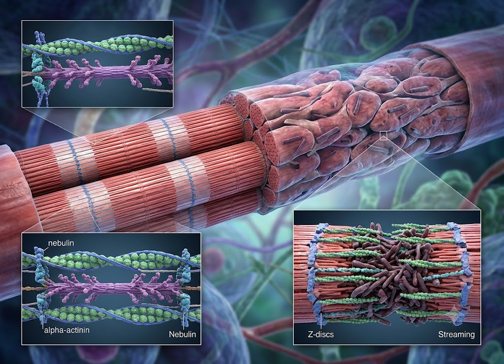
Nemaline myopathy is a rare, inherited disorder of the muscles that affects strength and movement. It results from changes in genes responsible for normal muscle development, which leads to abnormal structures called nemaline bodies forming inside muscle cells.
The condition can first appear in infancy, childhood, or adulthood, and its severity ranges widely, from mild muscle weakness to serious problems with breathing, feeding, and movement.
Understanding nemaline myopathy helps families recognize early warning signs and get the right medical support in place. In children, it can affect motor development, posture, muscle tone, and everyday tasks like walking or eating.
There is no cure, but physical therapy, respiratory care, and nutritional support can significantly improve quality of life and reduce complications. Below is an overview covering symptoms, causes, diagnosis, treatment, and long-term outlook.
What Is Nemaline Myopathy, and What Are Its Common Symptoms?
Nemaline myopathy is a congenital neuromuscular disorder (present from birth) marked by widespread muscle weakness and the presence of distinctive, thread-shaped protein structures known as “nemaline rods” inside muscle fibers. The name comes from the Greek word nema, meaning “thread,” describing exactly how these microscopic rods look under a microscope.
It belongs to a broader family of genetic muscle diseases called congenital myopathies, which affect how muscle tissue forms and works from a child’s earliest days.
Defining a Congenital Myopathy
What separates a congenital myopathy from other muscle disorders is structural disorganization. The genes responsible for building key structural proteins in muscle cells carry mutations, so the muscle cells are assembled incorrectly from the start, leaving them unable to generate normal force.
An important distinction: nemaline myopathy is generally non-progressive, or only very slowly progressive. That sets it apart from muscular dystrophies, where muscle tissue actively degenerates and wastes away over time.
With a congenital myopathy, the muscle fibers are abnormal from day one but don’t actively deteriorate as years pass. The weakness a child shows in infancy is often close to what they’ll carry for life. That said, secondary complications, such as joints locking in place (contractures) or curvature of the spine (scoliosis), can still develop if the weakness isn’t actively managed.
Key Signs in Infants and Children
Symptoms range from mild weakness to severe, life-threatening challenges, and severity varies significantly from child to child. Still, a distinct cluster of signs is typical.
Generalized Muscle Weakness (Hypotonia)
Usually the earliest and most noticeable sign, often called “floppy baby” syndrome. An affected infant feels notably limp when picked up, has poor head control, and struggles to lift their arms or legs against gravity. Their cry may sound weak, and they show less spontaneous movement than expected. As they grow, delays are common in reaching motor milestones: rolling over, sitting independently, crawling, walking. Weakness is usually most pronounced closest to the center of the body: neck, trunk, shoulders, and hips.
Feeding and Swallowing Difficulties (Dysphagia)
When the muscles of the face, jaw, and throat are weakened, basic feeding becomes a real struggle. An infant may have trouble generating enough suction to nurse or bottle-feed, leading to long feeding sessions, frequent coughing or choking, and poor weight gain. The most dangerous risk is aspiration: food, formula, or saliva slipping past the vocal cords into the lungs, which can trigger serious respiratory infections like pneumonia.
Respiratory Insufficiency
This is the most critical, potentially life-threatening element of the condition. Weakness in the diaphragm and the muscles between the ribs leads to shallow breathing, which worsens during sleep as muscle tone naturally drops further. A weak cough also makes it hard to clear mucus and secretions, so an ordinary cold can turn into a severe respiratory event. In moderate to severe cases, mechanical breathing support becomes necessary.
Distinctive Facial Features
Ongoing structural weakness shapes how facial bones develop over time. Many children share a recognizable profile: a long, narrow face, a tented or triangular upper lip, and a highly arched palate. The lower jaw may also be small or set back, adding complexity to speech and swallowing development.
What Causes Nemaline Myopathy, and How Is It Diagnosed?
The diagnostic path moves from clinical observation down to the molecular level. Since the root cause lies entirely in a child’s DNA, a clear diagnosis requires confirming the genetic variant and understanding how it disrupts muscle architecture.
The Genetic Foundations of Nemaline Myopathy
Nemaline myopathy is strictly an inherited or spontaneous genetic disorder; it is never caused by environmental factors or anything occurring during a normal pregnancy.
It stems from mutations in genes that provide the blueprints for proteins inside the sarcomere, the basic unit that allows skeletal muscle to contract. When these genetic instructions are flawed, the resulting proteins are missing, unstable, or misshapen, leaving muscle cells weak and disorganized.
Researchers have mapped mutations across more than ten genes linked to the condition. The two most common are:
- The NEB gene codes for nebulin, a large structural molecule that acts as a molecular ruler stabilizing the thin filaments in muscle.
- The ACTA1 gene codes for alpha-actin, a key contractile building block that works with myosin to generate movement.
Depending on the gene involved, inheritance can follow different patterns:
- Autosomal recessive inheritance: the most common path, especially with NEB mutations. Both parents are healthy, symptom-free carriers, each holding one normal and one mutated copy. A child must inherit the mutated copy from both parents to be affected: a 25% chance per pregnancy, plus a 50% chance of being a carrier and a 25% chance of being unaffected.
- Autosomal dominant inheritance: more common with ACTA1 mutations. Only one mutated copy is needed to cause the disease. An affected parent has a 50% chance of passing it to each child.
- De novo (spontaneous) mutations: in many dominant cases, the mutation arises by chance in the sperm or egg at conception, or very early in development. Neither parent carries it, so the chance of recurrence in future pregnancies is very low.
Confirming the Diagnosis: The Diagnostic Toolset
Specialists use a systematic combination of tests to distinguish nemaline myopathy from similar neuromuscular conditions.
Clinical Evaluation A pediatric neurologist or geneticist performs a focused exam, checking reflexes, muscle tone, and motor milestones, and looking for the characteristic long, narrow facial structure and high-arched palate.
Creatine Kinase (CK) Blood Tests When muscles break down rapidly, as in muscular dystrophies, the enzyme creatine kinase leaks into the bloodstream in large amounts. In nemaline myopathy, the cell membrane stays intact, so CK levels are typically normal or only slightly elevated, a useful marker for ruling out dystrophic disease.
Electromyography (EMG) Thin needle electrodes placed in muscle groups record electrical activity. In a primary myopathy, nerve signals are normal, but the muscle’s response is weak, a pattern that confirms the problem lies within the muscle itself, not the nerves.
Muscle Biopsy For decades, this was the gold standard. A small piece of muscle tissue (often from the thigh) is removed and examined under a microscope. A special stain reveals the hallmark: dense, dark, thread-like structures clustered inside the muscle fibers, the nemaline rods, essentially misassembled clumps of structural protein.
Genetic Sequencing Today, high-throughput genetic sequencing has largely replaced biopsy as the preferred first step. A blood or saliva sample lets geneticists pinpoint the exact mutation, sparing the child a surgical procedure and giving families valuable information about prognosis, long-term care, and family planning.
Management and Treatment Strategies for Nemaline Myopathy
Because there’s no outright cure, modern care focuses on a proactive, lifelong, multidisciplinary approach. Rather than reversing the underlying weakness, a team of specialists works to optimize daily function, conserve energy, and prevent complications.
The Core Rehabilitative Therapies
Physical Therapy (PT) Aims to protect joint mobility and maximize gross motor function. Because weak muscles can’t move joints through their full range, children risk developing contractures, a painful, permanent tightening of muscles and tendons. PT uses gentle stretching, low-impact conditioning, and positional exercises, and therapists help source equipment like ankle braces, standing frames, walkers, or power wheelchairs.
Occupational Therapy (OT) Focuses on fine motor coordination and daily living tasks: introducing built-up utensils for easier self-feeding, low-resistance clothing fasteners, or adaptive keyboards and voice-to-text tools for schoolwork.
Speech-Language Pathology (SLP) Addresses speaking and safe swallowing. SLPs run clinical swallowing evaluations, guide food texture modifications, and recommend positioning techniques like the chin-tuck. When speech is limited, they introduce Augmentative and Alternative Communication (AAC) tools, ranging from picture boards to eye-gaze devices.
Vital Medical and Systemic Support
Pulmonology and Respiratory Care The single most critical element of care. Because the diaphragm and rib muscles lack normal strength, children experience shallow breathing (hypoventilation) that worsens during deep sleep. A pulmonologist monitors lung health with pulmonary function tests and sleep studies, and care may include:
- Non-Invasive Ventilation (NIV): a nighttime BiPAP machine with a nasal mask to assist breathing.
- Mechanical Cough-Assist Devices: machines that apply positive pressure followed by suction to clear mucus from the airway.
- Tracheostomy: in severe cases, a surgical airway connected to a portable ventilator for continuous support.
Gastroenterology and Nutritional Management When swallowing is compromised, eating can burn more calories than it provides, leading to poor weight gain and malnutrition. A dietitian or gastroenterologist may recommend a high-calorie meal plan or, when eating by mouth is unsafe, a feeding tube, typically a gastrostomy tube (G-tube) placed directly into the stomach for secure, long-term nutrition.
Orthopedic and Cardiac Surveillance An orthopedic surgeon monitors for scoliosis, which can compress the chest and worsen lung function if severe; treatment ranges from bracing to spinal fusion surgery. While the heart is typically unaffected, certain rare variants (particularly some ACTA1 or LMOD3 mutations) can cause structural heart issues, so periodic echocardiograms with a pediatric cardiologist are recommended.
Long-Term Outlook for a Child with Nemaline Myopathy
The outlook spans a wide clinical spectrum, from severe infancy-onset forms to mild childhood-onset presentations, so there’s no single, uniform path.
Separating Physical Milestones from Cognitive Potential
Nemaline myopathy is strictly a disease of skeletal muscle; it has no impact on brain development, intellect, or cognitive ability. Children with the condition are intellectually normal and share the same emotional and creative potential as their peers. Physical milestones may be delayed (some children walk independently or with adaptive equipment, while others rely on a power wheelchair), but their minds remain entirely unaffected.
Life Expectancy and the Clinical Spectrum
Life expectancy is closely tied to the specific genetic variant and the degree of respiratory muscle weakness. Presentations generally fall into three categories:
Severe Congenital (Neonatal) Form: the most critical presentation, with profound weakness and respiratory distress at birth. These infants typically need round-the-clock ventilation, and even with intensive care, life expectancy is often limited to the first months or years.
Intermediate Congenital Form: clear weakness in infancy but not immediately life-threatening. With proactive respiratory monitoring (such as nightly BiPAP) and nutritional support via G-tube, many individuals live well into adulthood, with respiratory infections as the main ongoing risk.
Typical Mild Congenital / Childhood-Onset Form: the most common presentation. Motor milestones are delayed, but most individuals achieve independent walking, and weakness stays stable or progresses very slowly over decades. With periodic breathing checkups, life expectancy is normal or near-normal.
Navigating a Full and Meaningful Life
A physical disability doesn’t limit a child’s capacity for a rich, accomplished life. With the right accommodations, individuals with nemaline myopathy can:
- Excel academically and pursue careers in intellectually driven fields, aided by accessible desks, aides, or voice-activated software.
- Build lasting friendships and enjoy adaptive hobbies such as power wheelchair soccer, digital art, music production, or competitive gaming.
- Achieve autonomy and self-advocacy, managing their own care teams and living independently in their communities.
Navigating Advanced Topics and Finding Support for Nemaline Myopathy
The Clinical Classifications of Nemaline Myopathy
Doctors separate the condition into five clinical forms based on onset and severity:
- Typical Congenital Form: the most common, accounting for roughly half of cases. Weakness appears in early infancy, targeting the neck, face, and proximal limb muscles, and is typically static; most children walk independently.
- Severe Congenital (Neonatal) Form: the most critical variant, presenting at birth with total-body weakness and life-threatening respiratory failure, requiring immediate intensive intervention.
- Intermediate Congenital Form: clear weakness at birth or in early infancy; children often reach sitting or early walking milestones but face a high risk of losing ambulatory ability and developing progressive respiratory or spinal issues.
- Childhood-Onset Form: appears between ages 2 and 10, after apparently normal early development, with slowly progressive weakness starting in the feet and lower legs.
- Adult-Onset Form: the rarest form, typically surfacing between ages 20 and 50, sometimes with faster progression involving respiratory and cardiac muscles.
Cellular Distinction: Nemaline Myopathy vs. Muscular Dystrophy
Both are inherited disorders causing significant weakness, but they operate on opposite cellular mechanisms.
Nemaline Myopathy is a structural problem present from birth: muscle cells are miswoven and contain nemaline rods, but the cells themselves remain stable and don’t break down at an accelerated rate.
Muscular Dystrophy is a degenerative process: a missing or flawed protein (such as dystrophin) causes the cell membrane to tear during normal activity, triggering ongoing cell death, inflammation, and replacement of muscle with scar tissue and fat.
| Clinical Marker | Nemaline Myopathy (NM) | Muscular Dystrophy (MD) |
|---|---|---|
| Disease Trajectory | Static or very slowly progressive; baseline strength holds steady | Relentlessly progressive; weakness worsens over time |
| Creatine Kinase (CK) Levels | Normal to mildly elevated | Extremely high |
| Muscle Biopsy Presentation | Structurally intact fibers with dense nemaline rods | Necrosis, scarring, active regeneration, fatty infiltration |
Financial, Medical, and Community Support Channels
Patient Advocacy Foundations
- A Foundation Building Strength (AFBS): the only nonprofit dedicated exclusively to nemaline myopathy, funding research and offering family grants, webinars, and conferences.
- Muscular Dystrophy Association (MDA): supports congenital myopathies broadly, with specialized care centers, equipment assistance, and summer camps.
Navigating Financial and Everyday Assistance
- Government and Social Safety Nets: many children qualify for programs like Supplemental Security Income (SSI) or Medicaid waivers based on disability status; hospital social workers help navigate the process.
- Peer-Led Online Communities: parent-led groups offer practical, day-to-day advice, from traveling with a ventilator to managing skin issues caused by dietary changes.
On the Horizon: The Frontier of Targeted Research
Gene Therapy and Adeno-Associated Viruses (AAV) Because nemaline myopathy is typically caused by a single faulty gene, it’s a strong candidate for gene therapy. Researchers use modified viruses (AAVs) to deliver a working copy of the affected gene (such as TNNT1 in specific severe variants) directly into muscle cells. Preclinical animal studies have shown restored protein expression and improved survival.
Antisense Oligonucleotides (ASOs) and Exon Skipping Rather than replacing a whole gene, ASOs are synthetic genetic strips that bind to the cell’s RNA message, allowing it to “skip” a broken piece of code and produce a shortened but functional protein, such as nebulin.
Muscle Growth and Contractility Modulators Other approaches focus on maximizing existing muscle tissue: myostatin inhibitors to boost muscle mass, and small-molecule drugs targeting calcium handling or filament contractility to help weak sarcomeres generate more force.
The Role of Natural History Studies Groups like the Nemaline Myopathy Clinical Research Network (NM-CTRN) run multi-year Natural History Studies, tracking children over time to establish baseline data that regulators like the FDA require before approving gene therapy trials. Families can follow developments through AFBS or registries like ClinicalTrials.gov.
Conclusion
Nemaline myopathy is a rare genetic muscle condition that affects children differently depending on type and severity. Symptoms may include muscle weakness, delayed motor skills, feeding difficulties, breathing problems, and reduced muscle tone. While there’s no cure, early diagnosis and a coordinated care team of neurologists, physical therapists, respiratory specialists, and others can help children reach their fullest potential. Families who understand the condition and available support can make informed decisions and provide the best possible long-term care.
Frequently Asked Questions
1. What is nemaline myopathy?
Nemaline myopathy is a group of inherited muscle disorders that cause weakness due to abnormalities inside muscle cells. It’s named for nemaline bodies, rod-shaped structures visible under a microscope, caused by genetic changes affecting proteins needed for normal muscle function. Severity ranges from mild weakness to serious complications with breathing and swallowing.
2. What are the symptoms of nemaline myopathy in children?
Children may show low muscle tone, weakness, delayed motor development, and difficulty sitting, standing, or walking. Some also experience feeding difficulties, weak facial muscles, trouble swallowing, or breathing problems. Symptoms often depend on the genetic cause and age of onset, and early signs can sometimes be mistaken for other developmental delays.
3. What causes nemaline myopathy?
It’s caused by genetic mutations affecting proteins involved in muscle fiber structure and function, most commonly involving the ACTA1 and NEB genes. Most cases are inherited, though the exact pattern varies by gene. Genetic testing helps identify the cause and informs family planning.
4. How is nemaline myopathy diagnosed?
Diagnosis combines physical examination, medical history, genetic testing, and specialized muscle evaluations. A muscle biopsy may be performed to look for nemaline bodies, alongside blood tests, breathing assessments, and nerve or muscle studies to gauge severity.
5. How is nemaline myopathy treated in children?
There’s no cure, so treatment focuses on managing symptoms and supporting development: physical therapy for mobility and strength, occupational therapy for daily activities, and respiratory or nutritional support as needed. Care is typically delivered by a coordinated team of specialists.
6. Can children with nemaline myopathy live normal lives?
Outlook varies by type and severity. Many children have mild weakness and participate fully in daily activities, while others need significant medical support. Advances in supportive care, along with regular monitoring and early intervention, have improved outcomes and quality of life for many patients.