10 Things to Know About Peroneal Nerve Injury and Walking Problems
The peroneal (fibular) nerve is responsible for lifting the foot and toes, and it also carries feeling from parts of the lower leg and the top of the foot. When this nerve gets compressed, stretched, or damaged, walking can suddenly become difficult the clearest warning sign being foot drop, where the front of the foot drags or slaps against the floor with each step.
This can happen for many reasons: a knee injury, sitting with legs crossed too often, long periods of squatting, a cast or brace that’s too tight, surgery, diabetes, sudden weight loss, a tumor, or direct trauma near the outer knee. Depending on how severe the damage is, the problem may clear up on its own or linger for much longer. Below are 10 key things to understand about this condition from causes and symptoms to diagnosis, treatment, and recovery.
Primary Causes of Damage to the Peroneal Nerve
Common fibular neuropathy is a notable neurological problem affecting the lower leg. As a major offshoot of the sciatic nerve, the peroneal nerve handles both movement and sensation along the front and outer side of the leg.
It’s especially prone to injury because of where it sits wrapping around the fibular head at the outer knee, just beneath the skin with almost no cushioning. This makes it the leg nerve most frequently injured. Damage generally falls into three categories:
                         [Peroneal Nerve Injury Channels]
                                        │
     ┌──────────────────────────────────┼──────────────────────────────────┐
     ▼                                  ▼                                  ▼
[Direct Physical Trauma]         [External Compression]           [Medical & Surgical Causes]
 ├── Fibular head fractures       ├── Habitual leg crossing        ├── Total knee replacement (TBA)
 ├── Extreme knee dislocations    ├── Occupational squatting       ├── Tight surgical casts/braces
 └── Severe ankle traction sprains└── Bedridden mattress pressure  └── Stirrup positioning ischemia
Direct trauma: A hard blow to the outer knee can crush, stretch, or tear the nerve outright. Fibular head fractures are a common culprit, since broken bone can cut into nearby tissue. Severe knee dislocations can overstretch the nerve, while deep cuts or sports impacts cause direct bruising. Even a bad ankle sprain can yank on the nerve from below, injuring it through traction.
Compression from outside pressure: Constant pressure on the nerve pinches it against the bone underneath what’s known as nerve entrapment. Crossing your legs regularly squeezes one knee against the other and restricts blood flow. Jobs that involve a lot of squatting (carpentry, farming sometimes nicknamed “strawberry picker’s palsy”) press on the nerve over time. A too-tight cast, brace, or even bedsheets pressing on a bedridden patient can do the same.
Medical and surgical causes: Nerve damage can also happen during surgery. Knee replacements, tibial osteotomies, and ligament repairs all carry some risk, whether from how the patient was positioned, tourniquet pressure, or handling of tissue during the procedure. Similarly, a patient positioned in stirrups or lying on their side under anesthesia for too long without proper padding can develop nerve compression.
The Mechanics of Foot Drop
Foot drop is the hallmark symptom of peroneal nerve injury a sudden inability to lift the front of the foot upward (a motion called dorsiflexion).
Why it happens: The nerve can no longer send signals to the tibialis anterior and other muscles at the front of the shin, so those muscles weaken or stop working entirely.
How the body compensates: Since the toes would otherwise catch on the ground, people naturally start lifting their thigh higher than normal with each step almost like climbing stairs just to clear the foot off the floor. This walking pattern is called a steppage (or equine) gait.
The toll it takes: Because the foot can’t be controlled on its way down, it lands flat or slaps audibly onto the ground instead of rolling smoothly from heel to toe. Over time, this abnormal gait is tiring and puts extra strain on the ankle, knee, hip, and lower back.
Beyond Foot Drop: Sensory Alterations and Additional Motor Deficits
Foot drop isn’t the only clue doctors look for. A distinct pattern of numbness and additional weakness helps separate a peroneal nerve problem from a pinched nerve in the spine or a wider sciatic nerve issue.
[Deep Peroneal Branch Injury]     ──► Toe Drop (Loss of toe extension)
[Superficial Peroneal Branch]    ──► Weak Eversion (Ankle turns inward/unstable)
[Dermatomal Sensory Blockade]    ──► Pins & Needles, Numbness on the top of the foot
Sensory changes: Many patients notice tingling, “pins and needles,” or a buzzing feeling along the outer shin and top of the foot. In some cases, the area goes fully numb, which raises the risk of unnoticed cuts or injuries. Pain can range from a mild ache near the outer knee to sharp, burning sensations running down the shin.
Weak ankle eversion: The nerve also controls the muscles that turn the sole of the foot outward. When these weaken, the ankle tends to roll inward, making uneven ground especially risky to walk on.
Toe drop: Because a deep branch of the nerve controls the toe-lifting muscles, some patients lose the ability to raise their toes altogether.
Deep vs. Superficial Symptom Breakdown
The exact symptoms depend on which part of the nerve is affected:
Deep branch damage affects the front of the leg, causing both foot drop and toe drop, along with numbness limited to the space between the big toe and second toe.
Superficial branch damage affects the outer leg muscles, leading to loss of ankle eversion (so the foot turns inward and becomes unstable) plus widespread numbness across the top of the foot and outer shin.
Damage at the common trunk (where both branches are still together, typically at the fibular head) causes the full combination foot drop, toe drop, ankle instability, and numbness across the lower leg often with sharp, radiating pain from the knee downward.
Because the underlying cause and the specific symptoms are so closely linked, any sudden foot drop or ongoing numbness on top of the foot calls for prompt evaluation by a neurologist or orthopedic specialist left unchecked, nerve compression can eventually cause permanent muscle loss.
How is peroneal nerve injury diagnosed?
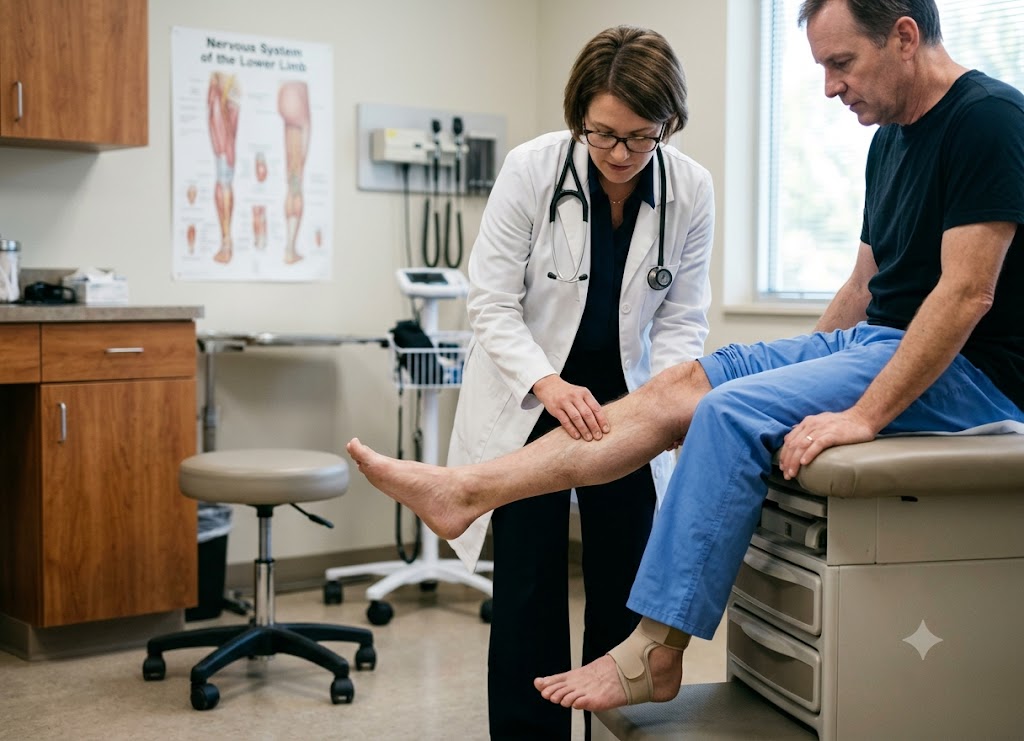
Diagnosis starts with a hands-on physical exam checking gait, strength, and sensation, then moves to electrical tests nerve conduction studies and electromyography to confirm what’s going on.
Initial Clinical Evaluation and Physical Exam
Because the symptoms can resemble other conditions, like a pinched nerve in the lower back, doctors rely on a structured exam built around three checks:
Watching how the patient walks: The clinician looks for the telltale steppage gait an exaggerated knee lift and the sound of the foot slapping the floor after each step.
                     [Clinical Examination Steps]
                                    │
     ┌──────────────────────────────┼──────────────────────────────┐
     ▼                              ▼                              ▼
[Visual Gait Observation]       [Manual Muscle Testing]        [Sensory Mapping Checks]
 ├── Exaggerated knee height     ├── Ankle dorsiflexion pull    ├── Check outer lower shin
 ├── Controlled foot clearance   ├── Foot eversion rotation     ├── Check top surface of foot
 └── Listen for ground slap      └── Grade strength on 0-5 scale└── Trace exact dermatome borders
Testing muscle strength by hand: Pushing against the patient’s foot as they try to flex it upward and turn it outward, then scoring the strength from 0 (no movement) to 5 (fully normal). Anything below a 5 in these movements points toward peroneal nerve weakness.
Mapping sensation: Using a light touch or pinprick, the doctor traces exactly where feeling is reduced or absent on the outer shin and top of the foot. If the numb area lines up with the nerve’s known path (rather than a spinal pattern), that supports the diagnosis.
Definitive Electrodiagnostic Testing
While the physical exam narrows things down, electrical testing confirms it.
[Electrical Pulse Stimulus] ──► Travels Down Peroneal Nerve ──► [Sensor Detects Delay/Weakness]Nerve Conduction Studies (NCS)
Electrodes placed on the skin measure how fast and how strongly a signal travels along the nerve after a small electrical pulse. A slower or weaker signal points to nerve damage, and testing at several points can pinpoint exactly where the problem lies for instance, a sudden drop in speed right at the outer knee suggests entrapment there.
Needle Electromyography (EMG)
A thin needle electrode inserted into a muscle like the tibialis anterior records its electrical activity, both resting and during movement. A healthy resting muscle shows no activity; one that’s lost its nerve connection shows abnormal spontaneous twitching. How the muscle behaves during contraction also tells the doctor whether the damage is recent or long-standing.
Classifying Damage Severity and Prognosis
Combining the NCS and EMG results lets a neurologist classify exactly how bad the damage is which shapes the treatment plan going forward.
                      [Nerve Fiber Damage Classification]
                                     │
     ┌───────────────────────────────┴───────────────────────────────┐
     ▼                                                               ▼
[Demyelination (Mild Block)]                    [Axonal Loss (Severe Damage)]
 ├── Outer protective myelin coating is bruised  ├── Internal structural nerve fibers are broken
 ├── Electrical signals are slowed down          ├── Muscle connection is severed
 └── Excellent outlook for full recovery         └── Guarded outlook; may require surgery
Mild injury (demyelination): The nerve’s inner fibers are intact, but the protective coating around them is bruised. Signals travel slower, but the muscle itself is still healthy. Recovery, once pressure is relieved, is usually excellent.
Severe injury (axonal loss): The nerve’s internal fibers themselves are damaged or severed. The EMG shows signs the muscle has lost its nerve connection, and recovery is slower and less certain the fibers have to regrow the whole distance from knee to foot, and surgery may be needed.
Supplementary Advanced Structural Imaging
Electrical tests show how the nerve is functioning, but not what’s physically pressing on it. When a structural cause is suspected, doctors may add imaging:
Neuromuscular ultrasound lets clinicians watch the nerve move in real time around the fibular head, spotting swelling or compression from a cast, brace, or nearby tissue.
MRI (specifically nerve-focused imaging of the lower leg) can reveal deeper causes like a cyst, soft-tissue growth, or bone fragments left over from an old fracture.
The treatment and recovery options for peroneal nerve injury
Treatment depends heavily on the cause and severity ranging from simple, non-invasive care to surgery in more serious cases.
Spontaneous Healing and the Nerve Regeneration Timeline
Many mild cases, especially those from temporary compression, heal completely without any surgery.
[Bruised Myelin Insulation] ──► Remove Compression Source ──► Myelin Regenerates ──► Normal Signal RestoredThis is most likely with neurapraxia the mildest form of nerve injury, where the fibers themselves are undamaged but the protective coating is bruised or flattened. Once the pressure is removed (loosening a tight cast, cutting back on leg-crossing, or letting swelling go down), that coating rebuilds itself naturally.
The Recovery Pattern
Healing generally takes a few weeks to a few months and tends to follow a predictable order muscles closer to the knee regain strength before those further down the leg. So typically, ankle-turning strength returns before the ability to lift the toes does. Doctors track progress with repeat testing over time and adjust the plan if things stall.
Non-Surgical Treatments for Foot Drop
For most patients waiting on natural healing, treatment focuses on two things: keeping the limb functional and preventing complications.
                   [Conservative Treatment Duo]
                                   │
     ┌─────────────────────────────┴─────────────────────────────┐
     ▼                                                           ▼
[Targeted Physical Therapy]                     [Ankle-Foot Orthosis (AFO)]
 ├── Muscle strengthening drills                 ├── Biomechanical foot support
 ├── Neuromuscular electrical stimulation (NMES) ├── Holds the ankle at a 90-degree angle
 └── Achilles tendon flexibility stretches       └── Eliminates high-steppage gait mechanics
Targeted Physical Therapy
Therapists work on strengthening whatever muscle function remains, often using neuromuscular electrical stimulation (NMES) small painless pulses that make the shin muscles contract to slow down muscle wasting while the nerve heals. Regular stretching of the calf and Achilles tendon is also important, since the tendon can tighten and shorten permanently if the foot can’t move on its own.
Ankle-Foot Orthosis (AFO) Bracing
For patients without a fixable structural cause, an AFO a lightweight L-shaped brace worn inside a shoe holds the foot at a 90-degree angle. This stops the foot from dropping during each step, cutting out the exhausting steppage gait and protecting the knee and back from extra strain.
Surgical Options for Severe Nerve Injuries
Surgery becomes an option when the nerve has been completely torn, something is physically compressing it, or months of monitoring show no improvement.
[Nerve Laceration/Cut]         ──► Direct Microsurgical Suture Repair
[Fibrous Band/Tunnel Pressure] ──► Surgical Decompression (Pressure Release)
[Permanent Axonal Death]       ──► Reconstructive Posterior Tibial Tendon Transfer
Nerve decompression:
If the nerve is being pinched by scar tissue, a cyst, or tight surrounding tissue, a surgeon can release that pressure directly, restoring normal blood flow and letting the nerve heal.
Direct repair or grafting:
If the nerve has actually been cut, surgeons can stitch the ends back together. When there’s a gap too wide to close directly, a graft often a small sensory nerve from elsewhere in the body bridges it so the nerve can regrow across.
Tendon transfer:
When nerve damage is permanent and there’s no sign of recovery, surgeons can reroute a healthy tendon (usually the posterior tibial tendon) from the back of the leg to the top of the foot. This effectively retrains the muscle to lift the foot when the patient pushes down, permanently correcting foot drop.
Broader considerations and related conditions for peroneal nerve injury
Understanding this condition fully means also knowing how it differs from similar nerve problems, how support devices like AFOs work, who’s most at risk, and what long-term recovery generally looks like.
Localized vs. Systemic Pathways: Peroneal vs. Sciatic Nerve Injuries
Peroneal nerve injury and sciatic nerve injury are often confused, since both can cause foot drop but they’re quite different.
The sciatic nerve is the largest nerve in the body, running from the lower spine down the back of the leg. The peroneal nerve is simply a smaller branch that splits off from it just above the knee.
Symptom Location and Distribution
Sciatica typically causes pain or numbness that travels from the lower back or buttock, down through the hamstring, and into the calf and foot. Peroneal nerve injury, by contrast, stays localized symptoms are confined to the outer shin and top of the foot, since the damage happens at the knee.
Root causes:
Sciatica usually stems from spinal issues like a herniated disc or spinal stenosis. Peroneal nerve problems come from direct trauma or compression at the knee leg crossing, prolonged squatting, or a poorly fitted cast.
The Toe-Standing Diagnostic Clue
Checking whether a patient can stand on their toes helps tell the two apart. That movement depends on the tibial nerve, which a peroneal nerve injury doesn’t touch so these patients can usually still stand on their toes. A sciatic nerve injury, however, affects the nerve trunk before it splits, causing weakness in both toe-standing and foot-lifting.
Biomechanical Support: The Multifaceted Role of an AFO
An AFO does more than just hold the foot in place it actively corrects how a person walks.
Normalizing Gait Mechanics
Without a brace, someone with foot drop has to compensate with an exaggerated knee lift or hip swing to keep their toes off the ground. An AFO holds the ankle at 90 degrees, letting the toes clear the floor naturally during each step and restoring a smoother stride.
Conserving Energy and Reducing Fatigue
Walking with untreated foot drop takes noticeably more effort. By normalizing the gait, an AFO reduces that extra energy cost, helping people walk farther with less fatigue.
Joint Protection and Side-to-Side Stability
Many modern carbon-fiber AFOs also stabilize the ankle side-to-side, which matters when eversion weakness makes the ankle prone to rolling. This protects not just the ankle but the knee, hip, and lower back from long-term strain.
Profiling High-Risk Individuals
Some people are more likely than others to injure this nerve, largely because of how exposed it is at the outer knee, where only a thin layer of skin and fat separates it from the surface.
The Impact of Low Body Mass Index (BMI)
A low BMI or rapid weight loss reduces the natural fat padding around the knee, leaving the nerve more exposed to everyday pressure.
Underlying Medical Conditions
Diabetes can weaken peripheral nerves over time, making them more vulnerable to compression. Inherited conditions like Charcot-Marie-Tooth disease can have a similar effect.
Behavioral Positions and Medical Immobility
Jobs involving frequent squatting or kneeling flooring, tiling, farming put ongoing pressure on the outer knee. Patients who are bedridden, in a coma, or under anesthesia for long surgeries are also at risk if their legs aren’t properly padded, since sustained pressure against a mattress or surgical stirrups can cut off blood flow to the nerve.
Long-Term Prognosis and Regeneration Kinetics
Recovery outcomes vary a lot depending on how severe the original damage was.
The Biological Rate of Nerve Regeneration
When the internal fibers are damaged but the outer coating is intact, the long-term outlook is generally good but slow. Nerve fibers regrow at roughly 1 mm a day, or about an inch a month. Since the distance from the knee to the foot can be 12–18 inches, it can take a year or more before function noticeably returns.
The Vital Role of Ongoing Physical Therapy
Without regular movement and stretching, the disconnected muscles can waste away and the joint can stiffen permanently. Ongoing physical therapy keeps everything flexible and ready to work again once the nerve finally reconnects.
Outcomes for Incomplete Regeneration
If the nerve was completely severed, or months of testing show no improvement, foot drop can become permanent. In these cases, long-term management usually means a custom AFO for daily mobility, or tendon transfer surgery as a more permanent fix.
Conclusion
Peroneal nerve injury interferes with walking by weakening the muscles that lift the foot and toes. It can show up as foot drop, tripping, ankle weakness, numbness, tingling, burning pain, or a changed walking pattern. Some cases resolve on their own once pressure on the nerve is relieved; others need bracing, physical therapy, medication, nerve testing, or surgery. If foot drop appears suddenly, gets worse quickly, follows an injury, or comes with severe pain, back symptoms, or weakness elsewhere, it’s important to get it checked out promptly.
Frequently Asked Questions
1. What is peroneal nerve injury?
It’s damage to the nerve responsible for movement and feeling in the lower leg, ankle, and foot particularly the muscles that lift the foot and toes. Injury here can cause weakness, numbness, tingling, or foot drop, and can result from compression, trauma, stretching, surgery, or certain health conditions.
2. How does peroneal nerve injury affect walking?
It makes it hard to lift the front of the foot, so the toes may drag, the foot may slap the ground, or the person may need to lift their knee higher than usual to avoid tripping. This raises fall risk, and bracing or physical therapy can help make walking safer while healing takes place.
3. What causes peroneal nerve injury?
Typical causes include knee injuries, fibula fractures, habitual leg crossing, prolonged squatting, tight casts, positional compression, and surgery near the knee. Rapid weight loss can also increase risk by reducing cushioning around the knee, and conditions like diabetes can make nerves more vulnerable. Imaging or nerve testing is often needed to pin down the exact cause.
4. Can peroneal nerve injury heal?
Yes, especially when the compression isn’t severe though recovery can take weeks to months since nerves heal slowly. More serious injuries, like a tear or major trauma, may lead to longer-lasting weakness or require surgery. Outcome depends on the cause, severity, how quickly treatment started, and the patient’s overall health.
5. How is peroneal nerve injury treated?
Treatment varies with the cause and severity, and may involve relieving pressure on the nerve, using an ankle-foot brace, physical therapy, pain management, and treating any underlying condition. Nerve conduction studies or EMG can help gauge the extent of damage and guide care, while surgery may be considered for severe compression, a physical mass, traumatic damage, or poor recovery over time.