8 Early Warning Signs of Retinoblastoma in Children
For parents, noticing something unusual about a child’s eyes can be easy to dismiss. A strange glow in a photo may seem like a camera effect. A wandering eye might look like a temporary phase, while redness, swelling, or poor vision could be blamed on irritation or tiredness. However, in rare cases, these changes may be linked to retinoblastoma, a childhood cancer that begins in the retina, the light-sensitive tissue at the back of the eye.
Retinoblastoma is rare, but it is the most common cancer that develops inside the eye in children. Estimates suggest that it affects about 1 in 15,000 to 1 in 20,000 live births worldwide, with approximately 8,000 to 9,000 new cases each year. Most cases develop during early childhood, and around 95% occur before the age of 5.
The encouraging news is that early recognition can make an important difference. Retinoblastoma can often be treated successfully when it is detected before it spreads outside the eye. However, the first signs may be subtle. A white reflection in the pupil, crossed eyes, changes in vision, redness, swelling, or difficulty seeing objects clearly may be among the first things parents notice.
This article explains 8 early warning signs of retinoblastoma in children. The purpose is not to make parents fearful of every eye change. Instead, it is to help families recognize unusual patterns that deserve medical attention, especially when a symptom keeps returning, appears in photographs, affects one eye more than the other, or seems to change how a child sees or moves.
What Is Retinoblastoma?
Retinoblastoma is a rare eye cancer that starts in the retina, the specialized light-sensitive tissue lining the back of the eye. It occurs mainly in infants and young children and is the most common primary cancer that develops inside the eye during childhood.
The retina contains specialized nerve cells called photoreceptors, including rods and cones. These cells detect light and color and convert them into signals that travel through the optic nerve to the brain, where they are interpreted as images.
Retinoblastoma develops when immature retinal cells, known as retinoblasts, do not mature normally and instead begin multiplying without control. This abnormal growth is associated with changes in a gene that normally helps regulate cell division. As the tumor grows, it can fill part or all of the inside of the eye. If untreated, it may invade nearby structures, including the optic nerve, and can spread to the brain or other parts of the body.
The Effect of Retinoblastoma on Child’s Vision and Eye Health
Retinoblastoma can affect a child’s vision and overall eye health by interfering with the normal function of the retina. As the tumor grows, it may contribute to complications such as retinal detachment, increased pressure inside the eye, inflammation, and vision loss.
A growing tumor takes up space inside the eye and can damage or block the retinal cells responsible for detecting light. If it develops near the macula, the central part of the retina responsible for detailed vision, the effect on sight may be especially significant.
The tumor can also develop beneath the retina or spread into the vitreous, the gel-like material that fills the eye. This may place pressure on the retina and cause it to separate from the back wall of the eye. A detached retina cannot function normally and may result in major vision loss.
As the tumor becomes larger, it may also interfere with the normal drainage of fluid from the eye. This can increase pressure inside the eye, a condition known as secondary glaucoma. Increased eye pressure can damage the optic nerve and may also cause eye discomfort or pain.
Inflammation can occur as tumor cells break down or as the body’s immune system reacts to the abnormal tissue. This may cause persistent redness, swelling, or irritation that can initially look like an ordinary eye infection.
In advanced cases, continued tumor growth may cause the eye to bulge outward, a condition known as proptosis. Severe disease can damage the internal structures of the eye and may require removal of the affected eye as part of treatment.
Is Retinoblastoma a Common Childhood Cancer?
Retinoblastoma is rare compared with childhood cancers such as leukemia or brain tumors. However, it is the most common primary cancer of the eye in children. It accounts for approximately 2% to 3% of cancers diagnosed in children younger than 15.
In the United States, around 250 to 300 children are diagnosed with retinoblastoma each year. Worldwide, the condition occurs in approximately 1 in every 15,000 to 20,000 live births.
Although the number of cases is relatively small, doctors emphasize awareness because the disease primarily affects infants and young children who may not be able to explain changes in their vision.
When retinoblastoma remains confined to the eye, treatment outcomes are generally very good, with cure rates in developed countries exceeding 95%. Once the cancer spreads beyond the eye, treatment becomes more complicated and aggressive.
Treatment aims first to protect the child’s life, followed by efforts to preserve the eye and as much useful vision as possible. Smaller tumors may sometimes be treated with focused therapies such as laser treatment or cryotherapy. Larger or more advanced tumors may require chemotherapy or surgery.
Approximately 40% of retinoblastoma cases are associated with a heritable change involving the RB1 gene. Identifying this form of the disease can be important for the child’s long-term care and may also have implications for genetic counseling within the family.
8 Key Symptoms of Retinoblastoma
The eight key symptoms associated with retinoblastoma include leukocoria, or a white pupil reflex; strabismus, or crossed or wandering eyes; worsening vision; persistent redness or swelling; a change in iris color; an enlarged pupil; eye pain; and proptosis, or a bulging eye.
Recognizing these changes can be particularly important because retinoblastoma often affects babies and young children who may not be able to explain that their vision has changed.
Even one unusual symptom deserves a thorough eye examination by a qualified medical professional. These signs do not automatically mean a child has retinoblastoma, but they can indicate a serious eye condition that requires evaluation.
Leukocoria (White Pupil or “Cat’s Eye Reflex”)
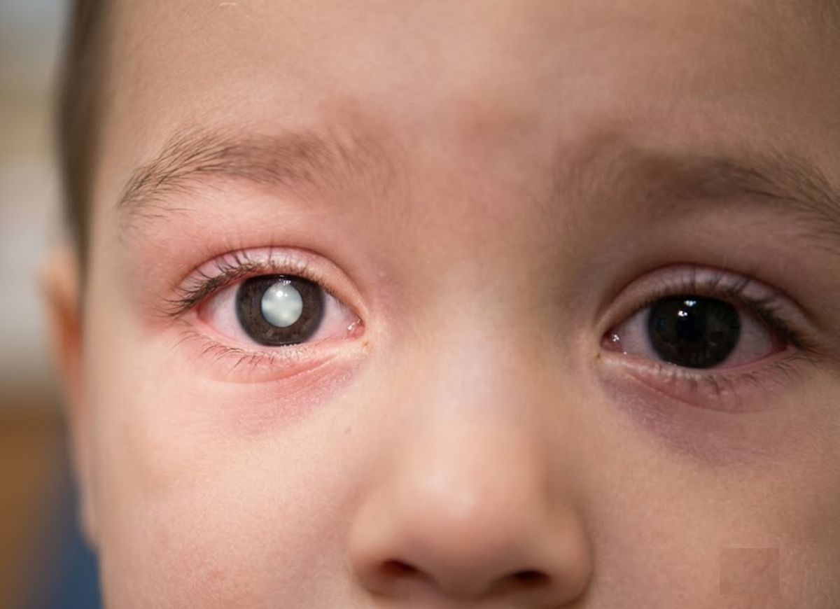
Leukocoria, or a white pupil, is the classic warning sign of retinoblastoma and is reported in about 60% of cases. Parents may notice a white or yellowish reflection in the center of the pupil, particularly in photographs taken with a flash.
Normally, a flash photograph may produce a red reflection because light bounces back from the blood-rich retina. When retinoblastoma is present, the tumor can reflect the light instead, creating a white or yellow appearance.
This is sometimes called the “cat’s eye reflex.” It may be easiest to notice in photographs, although some parents may also see an unusual reflection under certain lighting conditions.
A repeated white reflection in one pupil should never be ignored. It may have causes other than retinoblastoma, but it warrants prompt medical evaluation.
Strabismus (Crossed or Wandering Eyes)
Strabismus occurs when the eyes do not point in the same direction. One eye may turn inward, outward, upward, or downward while the other remains focused forward.
Retinoblastoma can cause strabismus when a tumor interferes with vision in one eye. If the brain receives a poor image from that eye, it may begin relying more heavily on the other eye, which can contribute to misalignment.
Some temporary eye crossing can occur in young infants, but persistent or newly developing strabismus should be evaluated by a healthcare professional. Because crossed or wandering eyes can have many causes, an eye specialist can determine what is behind the change.
Worsening Vision
Retinoblastoma can affect vision in the eye where the tumor develops. Because young children may not be able to describe blurred or reduced vision, parents may notice changes in behavior instead.
A child may appear clumsier than usual, have trouble following objects, avoid making eye contact, bump into things, or hold their head at an unusual angle to see more clearly.
When both eyes are affected, these changes may become more noticeable. Any unexplained change in a child’s ability to see should be evaluated.
Persistent Eye Redness and Swelling
An eye affected by retinoblastoma may become persistently red, irritated, or swollen. These changes may occur because of inflammation or increased pressure within the eye.
If redness or swelling does not improve as expected with treatment for a common condition such as conjunctivitis, further examination may be needed. Persistent symptoms should not automatically be assumed to be an ordinary eye infection.
Change in Iris Color (Heterochromia)
In rare situations, the color of the iris, the colored part of the eye, may change. This is known as heterochromia.
A difference in eye color can have many causes, but a new and unexplained change affecting only one eye deserves medical attention. In some cases involving retinoblastoma, tumor cells may spread toward the front portion of the eye and affect the iris.
An Enlarged Pupil (Mydriasis)
The pupil in the affected eye may appear larger than the other pupil or may not respond normally to bright light.
This can happen when the tumor affects the retina or the nerves involved in controlling the pupil’s response to light. A noticeable difference in pupil size, especially when it develops unexpectedly, should be checked by a medical professional.
Eye Pain
Retinoblastoma is not usually painful during its earliest stages. However, pain can develop as the tumor becomes larger or increases pressure inside the eye.
A young child may show discomfort by becoming unusually irritable, rubbing the affected eye, or avoiding bright light. Older children may be able to describe an eye ache or headache.
Unexplained eye pain, particularly when it occurs along with other eye changes, should be medically evaluated.
Bulging of the Eye (Proptosis)
Proptosis means that the eye appears to bulge or move forward from its normal position. With retinoblastoma, this is generally associated with more advanced disease.
Because a bulging eye can indicate significant disease or another serious condition, it requires immediate medical attention.
What Causes Retinoblastoma in Children?
Retinoblastoma develops when both copies of the RB1 gene in a retinal cell become inactive or altered. The RB1 gene normally acts as a tumor suppressor, helping regulate cell growth and division.
When both copies stop functioning properly in a developing retinal cell, the cell can begin multiplying uncontrollably. Over time, this abnormal growth can form a tumor.
Retinoblastoma can be either heritable or non-heritable. About 40% of cases are associated with a heritable RB1 mutation, while approximately 60% are non-heritable and develop from changes that occur in individual retinal cells.
The distinction between these forms is important because it can affect the age at which the disease develops, whether one or both eyes are affected, and the child’s future health monitoring.
Is Retinoblastoma Always Inherited From a Parent?
No. Retinoblastoma is not always inherited from a parent. Approximately 60% of cases are considered non-heritable and develop from genetic changes that occur within retinal cells.
In non-heritable retinoblastoma, a child generally begins life with normally functioning RB1 genes. Two separate changes can then occur within the same retinal cell, eventually allowing that cell to grow abnormally.
This form often affects one eye and is known as unilateral retinoblastoma. Because the genetic changes are limited to tumor cells, they are generally not present throughout the child’s body.
Heritable retinoblastoma accounts for about 40% of cases. In this form, a child has an RB1 mutation in the body’s cells because the mutation was inherited from a parent or developed around the time of conception.
Because one altered copy of the gene is already present, only another change is needed in a retinal cell for a tumor to develop. This makes multiple tumors and disease affecting both eyes more likely.
Children with heritable retinoblastoma are often diagnosed at a younger age. They also have an increased risk of developing certain other cancers later in life and may have a 50% chance of passing the RB1 mutation to each child.
The Role of The RB1 Gene in Retinoblastoma
The RB1 gene acts as a tumor suppressor. It produces a protein called pRB that helps control the cell cycle and prevents cells from growing and dividing when they should not.
When RB1 is working normally, it helps keep cell growth under control. When both copies of the gene become inactive, this important control mechanism is lost, allowing abnormal cells to multiply.
Retinoblastoma is commonly explained through the “two-hit hypothesis,” a model associated with Dr. Alfred Knudson’s work. The basic idea is that both copies of the RB1 gene must be inactivated for a retinal cell to become cancerous.
In non-heritable cases, a child begins with two functioning copies of RB1. Two separate mutations must occur in the same retinal cell for a tumor to form. Because this is less common, these cases often involve a single tumor in one eye.
In heritable cases, one altered RB1 copy is already present in the child’s cells. A second mutation in a retinal cell can then remove the remaining functional copy. Because many retinal cells are already carrying the first mutation, multiple tumors and disease in both eyes are more likely.
The loss of normal RB1 function removes an important brake on cell division, allowing retinal cells to grow uncontrollably and form a tumor.
When to Seek Medical Help?
Parents should seek medical attention promptly if they notice possible signs of retinoblastoma, especially a white reflection in the pupil or newly developed crossed or wandering eyes.
Early evaluation matters because retinoblastoma can progress, and delayed diagnosis may affect the ability to preserve vision, save the affected eye, or prevent the disease from spreading.
If something about your child’s eyes looks unusual, it is reasonable to have it checked rather than waiting for it to disappear. Contact a pediatrician, family doctor, or eye specialist and explain exactly what you noticed and when it began.
For example, tell the doctor if you noticed a white reflection in one eye in a flash photograph, a new change in eye alignment, or persistent redness that has not improved.
Are These Symptoms Always a Sign of Retinoblastoma?
No. These symptoms do not always mean a child has retinoblastoma. Several other eye conditions can cause similar changes.
However, symptoms such as leukocoria and strabismus should still be evaluated because some of the conditions that cause them can also threaten vision.
For example, Coats’ disease is a rare condition involving abnormal retinal blood vessels. These vessels can leak fluid and contribute to retinal detachment, producing a white or yellow reflection.
Congenital cataracts can also block the normal red reflex and create the appearance of a white pupil. These cataracts may need treatment to support normal visual development.
Persistent Fetal Vasculature (PFV) is another congenital condition that can cause leukocoria and vision problems when blood vessels inside the developing eye do not regress normally.
Retinal detachment can also occur for reasons other than retinoblastoma, including trauma.
Strabismus is often caused by conditions that are not cancerous and may be related to differences in eye muscles or vision. However, new or persistent eye misalignment should still be evaluated, particularly when it occurs alongside other unusual eye symptoms.
The important point is that these signs do not confirm retinoblastoma. Only a proper medical examination can determine the cause.
What to Expect During a Doctor’s Visit for Eye Symptoms?
During an initial visit, the doctor will usually ask about the eye changes you have noticed and perform an examination that may include vision testing and a red reflex test. If anything appears abnormal, your child may be referred to a pediatric ophthalmologist.
The doctor may ask when the symptom began, whether it affects one or both eyes, whether the child has pain or redness, and whether there is a family history of retinoblastoma or other relevant conditions.
Vision testing depends on the child’s age. Older children may be able to use a standard eye chart, while infants and toddlers may be evaluated by observing how well they focus on and follow objects.
The red reflex test is an important screening examination. Using an ophthalmoscope in a dim room, the doctor checks the reflection coming from the back of each eye. A normal reflex should be similar in both eyes.
A white, yellow, dull, or absent reflex, or a significant difference between the two eyes, can indicate a problem and may require urgent referral.
A pediatric ophthalmologist can perform a more detailed examination after dilating the child’s pupils. This allows the specialist to see the retina and optic nerve more clearly.
For very young children who cannot remain still, an examination under anesthesia may sometimes be necessary. If a tumor is suspected or identified, additional tests such as ultrasound or MRI may be used to evaluate the eye and determine the extent of the disease.
Retinoblastoma Diagnosis
The red reflex test and ophthalmoscopy serve different purposes. The red reflex test is a simple screening method, while a detailed dilated eye examination is used to evaluate the retina more thoroughly.
During a red reflex test, a doctor shines a light toward the child’s eyes and looks for the reflection from the retina. A healthy eye generally produces a bright, symmetrical reddish-orange reflection.
An abnormal reflection, including a white, yellow, dull, or uneven appearance, can indicate an obstruction or other eye abnormality. This finding generally requires further evaluation.
Ophthalmoscopy, particularly a dilated fundus examination, provides a much closer view of the back of the eye. The child’s pupils are dilated with special drops, allowing the ophthalmologist to examine the retina, optic nerve, and blood vessels.
If retinoblastoma is suspected, this examination can help the specialist identify and assess an abnormal mass within the eye. Imaging studies may also be used to provide additional information about the tumor.
The Difference Between Unilateral and Bilateral Retinoblastoma
The main difference between unilateral and bilateral retinoblastoma is the number of eyes affected and the genetic pattern commonly associated with the disease.
Unilateral retinoblastoma affects one eye and represents about 60% of cases. Most unilateral cases are non-heritable and result from genetic changes that develop within retinal cells after conception.
Because the mutation is usually limited to the tumor cells in non-heritable disease, the child generally does not have the same increased risk of other cancers associated with a germline RB1 mutation.
Children with unilateral disease are often diagnosed later than children with bilateral disease because symptoms may take longer to become noticeable.
Bilateral retinoblastoma affects both eyes and is associated with heritable disease. The child has an RB1 mutation in cells throughout the body, either because it was inherited from a parent or because a new mutation occurred around conception.
Because retinal cells already carry one altered copy of RB1, tumors can develop in both eyes. These children are often diagnosed during infancy.
A germline RB1 mutation also increases the lifetime risk of certain other cancers. It can also be passed to future children, with a 50% chance of transmission for each pregnancy when a parent carries the mutation.
The Main Treatment Options for Retinoblastoma
Treatment for retinoblastoma depends on factors such as tumor size, location, whether one or both eyes are affected, and whether the disease has spread. Treatment is usually planned by a specialized medical team.
For smaller tumors that remain localized, focal treatments may be used. Laser photocoagulation uses laser energy to treat the tumor and its blood supply, while cryotherapy uses freezing to destroy tumor cells.
Larger or more widespread tumors may require chemotherapy. Systemic chemotherapy delivers medication through the bloodstream to treat tumor cells throughout the body.
Intra-arterial chemotherapy is a more targeted approach in which medication is delivered directly to the blood vessel supplying the affected eye. This can provide a concentrated treatment to the tumor while limiting exposure to other parts of the body.
Intravitreal chemotherapy may be used when tumor cells have spread into the vitreous, the gel-like substance inside the eye. In this treatment, medication is delivered directly into the eye.
When a tumor is very large, the eye has severe damage, or there is a significant risk of the cancer spreading, enucleation, or surgical removal of the eye, may be recommended. An orbital implant and prosthetic eye can then be used to restore the appearance of the eye.
Retinoblastoma and Other Childhood Eye Conditions like Coats’ Disease
Retinoblastoma and Coats’ disease can produce similar signs, including leukocoria, but they are different conditions.
Retinoblastoma is a cancer that develops from retinal cells and is associated with changes in the RB1 gene. It can form a tumor inside the eye and may spread beyond the eye if it is not treated.
Coats’ disease is not a cancer. It involves abnormal blood vessels in the retina that can leak fluid and fatty material into and beneath the retina. Over time, this leakage can contribute to retinal detachment and vision loss.
Retinoblastoma may show characteristic findings on imaging, including calcium deposits within the tumor. Treatment may involve chemotherapy, focal treatments, radiation in selected situations, or surgery depending on the disease.
Treatment for Coats’ disease focuses on controlling the abnormal blood vessels and reducing leakage. Laser therapy or cryotherapy may be used, while advanced cases may require surgery.
Although Coats’ disease is not life-threatening in the same way as retinoblastoma, it can still cause serious and permanent vision problems if left untreated. Because the two conditions can share similar symptoms, professional eye evaluation is important.
FAQs
1. What age do people get retinoblastoma?
Retinoblastoma mainly affects babies and young children. Most cases are diagnosed before the age of 5, and many are identified before age 2.
The disease can affect one eye or both eyes. When both eyes are involved, diagnosis often occurs at a younger age. Retinoblastoma is uncommon in older children and adults, but unusual eye changes should still be evaluated.
2. Is retinoblastoma a cancer?
Yes. Retinoblastoma is a cancer that begins in the retina, the light-sensitive tissue at the back of the eye.
Because the retina plays an important role in vision, a tumor in this area can affect how a child sees and can also cause visible changes in the eye.
Treatment can be highly successful when retinoblastoma is detected early, but untreated disease can grow and spread.
3. Which organ is affected by retinoblastoma?
Retinoblastoma affects the eye, specifically the retina. The retina is located at the back of the eye and helps convert light into signals that the brain uses to create vision.
The disease may involve one eye or both. Early signs can include a white pupil reflection, crossed eyes, reduced vision, redness, swelling, or changes in the way a child looks at objects.
4. What can be mistaken for retinoblastoma?
Several eye conditions can cause symptoms that resemble retinoblastoma. These may include congenital cataracts, Coats’ disease, eye inflammation, injuries, retinal problems, or some common causes of strabismus.
A white pupil reflection or unexplained change in vision should not be diagnosed at home. A professional eye examination is needed to determine the underlying cause.
5. What is stage 4 retinoblastoma?
Stage 4 retinoblastoma generally refers to disease that has spread beyond the eye to distant areas of the body. Depending on the staging system, this may include areas such as the brain, spinal fluid, bones, bone marrow, or other organs.
This represents advanced disease and requires urgent specialist treatment. Staging systems can differ, so families should discuss the exact stage and what it means for their child’s condition with the treating medical team.
Conclusion
Retinoblastoma is rare, but knowing its early warning signs can help parents recognize changes that deserve attention. A white glow in a photograph, crossed or wandering eyes, changes in vision, persistent redness, swelling, pain, or a child struggling to see can sometimes be important clues.
Because retinoblastoma starts in the retina, it can affect both the appearance and function of the eye. When detected early, treatment can often control the disease and may help preserve the child’s eye and vision. Delayed diagnosis can make treatment more difficult, which is why unusual symptoms should be evaluated promptly.
Parents do not need to assume that every eye symptom means cancer. Many eye conditions can cause similar changes. However, if a child’s eye looks different, the change persists, or there is a white pupil reflection or unusual eye movement, arranging a professional eye examination is an important next step.
Recognizing possible warning signs early can help children receive appropriate evaluation and treatment as soon as possible.