10 Warning Signs of Pulmonary Embolism
Picture an ordinary day. You feel fine, and then you suddenly can’t catch your breath, or a sharp pain in your chest gets worse with every breath. For many people, these are the first warning signs of pulmonary embolism (PE), a potentially life-threatening condition in which a blood clot blocks an artery in the lungs. Every year, thousands of people experience these silent dangers without realizing how serious they are.
A pulmonary embolism can strike fast and without warning, leaving people frightened and confused about what is happening to their body. The signs vary widely, from sudden breathlessness to leg swelling, but they all point to the same thing: an urgent need for medical care. What makes PE so dangerous is that it can look subtle or severe, and its symptoms often mimic less dangerous problems.
This article covers the 10 warning signs of pulmonary embolism you should never ignore. Recognizing them early can be the difference between life and death. If you have ever felt sudden, unexplained chest pain or unusual shortness of breath, read on and pay attention.
10 Warning Signs of Pulmonary Embolism
1. Sudden Shortness of Breath (Dyspnea)
Sudden shortness of breath (dyspnea) is one of the most common and immediate signs of PE. It typically appears without warning, often while you are resting and not doing anything physical. The feeling of not being able to catch your breath, even sitting still or sleeping, is alarming. It happens because the clot blocks a pulmonary artery and keeps blood from reaching lung tissue for proper oxygen exchange.
When part of the lung loses its blood flow, oxygen levels drop, and the body tries to compensate by breathing faster and deeper. The result is a distressing sense of air hunger, where every breath feels inadequate. It can range from mild to severe depending on the clot’s size and location, but it should never be ignored, as it can signal a life-threatening emergency.
2. Rapid Breathing (Tachypnea)
Rapid breathing (tachypnea) often accompanies shortness of breath in PE. It is the body’s compensatory response to the low oxygen caused by the clot. To restore oxygen levels, the respiratory system works harder, producing fast, shallow breaths. This higher rate, often with a feeling of breathlessness, is the body’s attempt to take in more oxygen and expel more carbon dioxide.
Healthcare providers can often see this as a key clinical sign. Low blood oxygen (hypoxemia) stimulates the drive to breathe, which speeds up the rate. Rapid breathing can have other causes, such as anxiety or fever, but together with other PE symptoms it becomes a critical indicator that needs immediate medical attention.
3. Sharp, Pleuritic Chest Pain
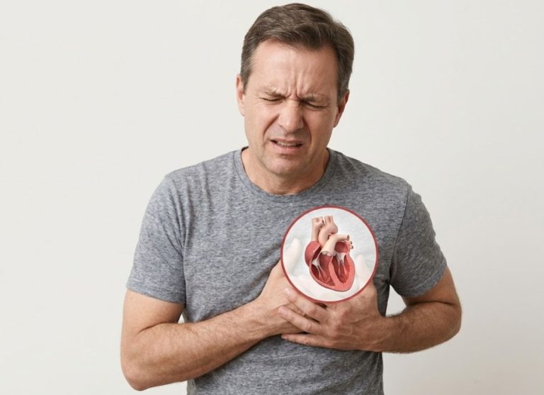
PE chest pain is often sharp and stabbing and usually worse with deep breaths, coughing or sneezing. This is called pleuritic chest pain. It occurs when the pleura, the two-layered membrane around the lungs, becomes inflamed. The inflammation results from lung tissue damage when the clot blocks blood flow. When lung tissue becomes ischemic (starved of oxygen) or necrotic (dead), it irritates the pleura.
The pain can feel localized to one area of the chest and intensify with movement or deep inhalation as the pleura rubs against itself. Unlike the crushing pain typical of a heart attack, pleuritic pain is more acute and localized, often described as a stitch or needle-like sensation. It is important to tell it apart from other kinds of chest pain, because it can indicate a blocked pulmonary artery and needs immediate medical evaluation.
4. Rapid Heartbeat (Tachycardia)
A rapid or irregular heartbeat (tachycardia) is a common circulatory symptom. When a clot obstructs a pulmonary artery, the right side of the heart has to pump harder to push blood past the blockage. The extra workload raises the heart rate in an effort to keep circulation adequate. Tachycardia means a rate above 100 beats per minute and may feel like palpitations, fluttering or a pounding in the chest.
Sometimes an irregular rhythm (arrhythmia) occurs as well, further disrupting the heart’s function. The strain can lead to complications, and these rhythm changes reflect the body’s effort to compensate for reduced blood flow to the lungs. Tachycardia, especially combined with shortness of breath or chest pain, is a critical warning sign that needs immediate medical attention.
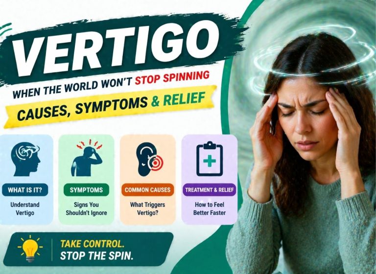
5. Lightheadedness or Dizziness
Lightheadedness or dizziness can result from a reduced oxygen supply to the brain. A clot in a pulmonary artery reduces blood flow through the lungs and therefore the amount of oxygenated blood returning to the left side of the heart. As the heart struggles to pump efficiently, less oxygen reaches the brain. This can cause dizziness, wooziness or a feeling that you are about to faint. In more severe cases, low oxygen can lead to confusion or trouble concentrating.
Lightheadedness is particularly dangerous because it makes falls and accidents more likely. If it comes with shortness of breath, chest pain or a rapid heartbeat, get medical help immediately, as it may mean your body is struggling to maintain normal circulation because of a PE.
6. Fainting (Syncope)
Fainting (syncope) is one of the more alarming signs, especially when a large clot significantly obstructs blood flow to the lungs. The right side of the heart must work harder to pump blood past the blockage, and this strain can cause a sudden drop in blood pressure and cardiac output. The abrupt, severe reduction in blood flow to the brain makes the person lose consciousness.
Syncope often points to a massive PE, which is a medical emergency needing immediate intervention. Fainting alongside other PE symptoms, such as sudden breathlessness, chest pain or a fast heartbeat, is a red flag. A sudden collapse suggests the body cannot compensate for the blockage, and emergency care is essential.
7. Coughing (Sometimes with Blood)
A persistent cough is a common PE symptom and may come with hemoptysis, or coughing up blood. It occurs because the blockage affects lung tissue and airway function. When a clot cuts off blood flow to part of the lung, that tissue can begin to die, irritating and inflaming the airways and causing a persistent dry cough. In more severe cases, the lack of blood flow leads to pulmonary infarction (death of lung tissue), which can cause bleeding in the lungs.
This bleeding can show up as blood-streaked sputum or even frank blood. Hemoptysis is concerning because it indicates significant lung tissue damage. Not everyone with PE coughs up blood, but when it happens it is a clear sign of lung injury and requires immediate medical attention.
8. Excessive Sweating (Diaphoresis)
Excessive sweating (diaphoresis) is another common response, part of the body’s fight-or-flight reaction to the stress and pain caused by the clot. As the clot blocks blood flow to the lungs, oxygen levels fall and the heart works harder. This physiological stress triggers stress hormones like adrenaline, which raise heart rate, blood pressure and sweating.
The sweating is often profuse and clammy and unrelated to exercise or heat. The skin may feel cool and moist even though the person is not exerting themselves. It usually comes with other symptoms like breathlessness and dizziness and signals severe distress. Sweating, especially alongside other PE symptoms, is a warning sign that needs urgent medical evaluation.
9. Symptoms of Deep Vein Thrombosis (DVT)
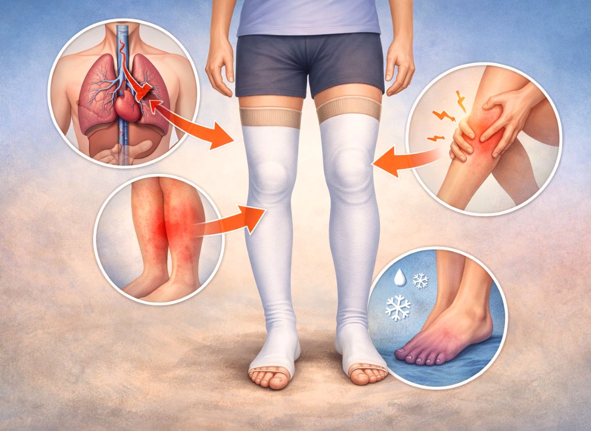
Deep vein thrombosis (DVT) is a blood clot in a deep vein, typically in the legs. The clot can travel to the lungs and cause a PE, so recognizing DVT symptoms can be crucial for preventing one. Common symptoms include leg pain or tenderness, swelling (edema) and skin discoloration. The pain is often a cramping or soreness in the calf or thigh that may worsen with standing or walking.
Swelling occurs because the clot obstructs blood flow, so blood pools in the veins and fluid leaks into the tissues. The skin over the clot may look red, blue or purple and feel warm. If these symptoms appear in one leg, especially with shortness of breath or chest pain, seek medical attention immediately, since untreated DVT can lead to a pulmonary embolism.
10. Leg or Arm Swelling and Tenderness
Swelling in a leg or arm is a classic sign of DVT and can be an early warning that a PE may follow. The clot blocks normal blood flow back to the heart, so blood pools in the veins and the limb visibly swells. The swelling is usually confined to one limb, which may look noticeably larger than the other, and the area may be tender or painful to the touch.
The skin may feel warm from inflammation around the clot and, in severe cases, look red, blue or purple. If you notice these symptoms in one leg or arm, especially with breathing difficulty or chest pain, immediate medical attention is needed to keep the clot from causing a pulmonary embolism.
Causes of a Pulmonary Embolism
Deep Vein Thrombosis (DVT)
DVT causes a pulmonary embolism through a step-by-step process: a clot forms in a deep vein, a piece breaks free, travels through the bloodstream and lodges in a pulmonary artery. This journey from the limbs to the lungs is what makes the disease thromboembolic.
The initial clot is often triggered by sluggish blood flow, blood that clots too readily, or damage to the vein lining. For example, long periods of immobility during a long flight or after surgery cause venous stasis, where blood pools in the leg veins and clots become more likely.
The mechanism unfolds in these steps:
- Thrombus formation: A clot (thrombus) develops in a large vein, most often in the calf, thigh or pelvis. This is the DVT. It is a meshwork of platelets, fibrin and red blood cells stuck to the vein wall.
- Embolization: A sudden movement, muscle contraction or change in blood pressure can break off a piece of the thrombus. This free-floating piece is now an embolus.
- Travel to the heart: The embolus is carried with venous blood up the inferior vena cava, the body’s largest vein, and directly into the right atrium of the heart.
- Passage through the heart: From the right atrium it moves into the right ventricle, which contracts and pumps it into the main pulmonary artery.
- Lodging in the lungs: The pulmonary artery branches into ever smaller vessels. The embolus travels until it reaches a point too narrow to pass and becomes wedged, fully or partly blocking blood flow to the lung tissue beyond it. That blockage is the pulmonary embolism. Size determines how far it travels: a large embolus can block the main pulmonary artery (a saddle embolus), while smaller ones travel further into the lung periphery.
Other Causes of Pulmonary Embolism
Blood clots are overwhelmingly the main cause, but other, much rarer substances can enter the bloodstream and travel to the lungs to create a blockage. These are non-thrombotic pulmonary embolisms, and they arise from very different conditions than DVT.
They account for a small fraction of all PEs but can be equally life-threatening, and they matter in specific contexts such as major trauma or certain medical procedures. These emboli are made of materials other than the usual fibrin and platelet mesh of a clot. The best-known non-thrombotic causes are:
- Fat embolism: Typically follows a fracture of a large long bone such as the femur (thigh bone) or major orthopedic surgery like hip or knee replacement. The trauma can disrupt fat cells in the bone marrow and release microscopic fat globules into torn veins. They travel to the lungs, where they cause a mechanical blockage and a severe inflammatory reaction.
- Air embolism: Air bubbles enter a vein or artery and block it. It can be a rare complication of procedures such as inserting or removing central venous catheters, neurosurgery or scuba diving accidents (decompression sickness). Enough air can create an air lock in the heart or pulmonary artery, obstructing blood flow.
- Tumor embolism: In some people with cancer, a piece of tumor breaks off, enters the bloodstream and travels to the lungs. This is more common with tumors such as renal cell carcinoma, which can invade large veins. The fragments can cause blockages similar to blood clots.
- Amniotic fluid embolism: An extremely rare but catastrophic complication of pregnancy and childbirth. Amniotic fluid, fetal cells or other debris enter the mother’s bloodstream during labor or delivery, triggering an intense allergic-like reaction and obstructing the pulmonary arteries.
- Septic emboli: In severe infection, particularly endocarditis (infection of the heart valves), clumps of bacteria and infected tissue can break off and travel to the lungs, causing blockages and seeding new infections in the lung tissue.
Medical Conditions and Procedures
Many medical conditions and surgical procedures significantly raise PE risk by creating a pro-thrombotic state in the body. Major surgery, especially on the lower body, is among the biggest risk factors.
Vessel wall injury during surgery, the effects of anesthesia and prolonged immobility during recovery combine into a perfect storm for DVT. Chronic conditions affecting circulation or blood composition can also raise risk persistently. The key medical and procedural risk factors are:
- Major surgery: Operations on the pelvis, hip or leg, such as hip or knee replacement, carry a very high risk. Manipulating large veins can injure them, and the bedridden recovery period severely slows blood flow in the legs.
- Prolonged immobility: Anything that keeps you immobile for long periods lets blood pool in the veins and promotes clotting. This includes long hospital stays, bed rest for illness and paralysis from spinal cord injury.
- Cancer and cancer treatment: Many cancers, particularly of the pancreas, lung, ovary and brain, release substances that increase clotting. Many chemotherapy agents also damage blood vessels and raise the risk of thrombosis.
- Heart and lung disease: Conditions like heart failure reduce the heart’s pumping ability, causing blood pooling and congestion. Atrial fibrillation, an irregular heart rhythm, can form clots in the heart’s upper chambers that travel to the lungs. Chronic obstructive pulmonary disease (COPD) also raises PE risk.
- Inherited clotting disorders (thrombophilia): Some people inherit gene mutations that make blood clot more easily, most commonly Factor V Leiden and the prothrombin gene mutation. A family history of blood clots or PE is a significant red flag.
- Previous DVT or PE: Anyone who has had one clot is at much higher risk of another.
- Inflammatory bowel disease: Crohn’s disease and ulcerative colitis involve chronic inflammation that can increase clot risk.
- Trauma: Severe injuries such as leg or pelvic fractures can damage veins and cause prolonged immobility, both strong DVT risk factors.
Lifestyle and Personal Factors
Various lifestyle choices and personal characteristics can substantially raise PE risk, mainly by creating conditions favorable to clotting. These factors are often modifiable, so changes in habits can lower your risk.
Long periods of sitting, body weight, smoking and hormonal factors all influence blood thickness and circulation. One factor may add only a little risk, but several together can have a significant cumulative effect. The most prominent are:
- Prolonged inactivity (travel): Sitting for long stretches on a plane, in a car or on a train lets blood pool in the leg veins. This “economy class syndrome” is a well-known risk factor. On long journeys the legs are often bent and still, which compresses veins and slows circulation, an ideal setting for DVT.
- Obesity: A body mass index (BMI) of 30 or higher is a significant risk factor. Extra weight increases pressure on the veins in the pelvis and legs and can impede blood flow. Fat tissue is also metabolically active and can produce substances that promote inflammation and clotting.
- Smoking: Tobacco damages the lining (endothelium) of blood vessels, which can start the clotting process. It also makes platelets stickier and raises levels of certain clotting factors, making clots more likely.
- Supplemental estrogen: Estrogen in birth control pills or menopausal hormone replacement therapy (HRT) increases the concentration of clotting factors in the blood, raising DVT and PE risk, particularly in women who also smoke or have other risk factors.
- Pregnancy and the postpartum period: Risk is elevated during pregnancy and for up to six weeks after giving birth. Higher estrogen raises blood clotting tendency, the growing uterus presses on pelvic veins and slows blood return from the legs, and blood vessels can be injured during delivery.
- Age: PE risk rises with age, particularly after 60, likely because of decreased mobility and more medical risk factors such as heart disease and cancer.
Diagnose of Pulmonary Embolism
Diagnosing PE is a multi-step process combining clinical assessment with specific imaging and blood tests to confirm a clot in the lungs. A physician first evaluates your symptoms, reviews your history for risk factors such as recent surgery or previous clots, and performs a physical exam.
If PE is suspected, several tools come into play. The most common initial blood test is the D-dimer, which measures a substance released when a clot breaks down. A negative result in a low-risk patient can often rule out PE, but a positive result isn’t definitive, since levels can rise from infection or recent surgery, and it calls for further imaging.
The gold standard for confirming PE is CT pulmonary angiography (CTPA), an advanced X-ray using contrast dye to create detailed images of the pulmonary arteries and clearly show any blockage. For patients who can’t receive contrast dye, a ventilation/perfusion (V/Q) scan is a valuable alternative, comparing airflow and blood flow in the lungs to spot mismatches that indicate a clot.
Since most PEs originate from leg clots, a Doppler ultrasound is often performed to look for DVT. To identify and confirm PE accurately, providers follow a structured pathway:
- Initial clinical evaluation: Scoring systems like the Wells score or Geneva score estimate the pre-test probability of PE from symptoms, signs (such as tachycardia or signs of DVT) and risk factors.
- Definitive imaging studies: CTPA is preferred for its high sensitivity and specificity in visualizing clots. A V/Q scan is the main alternative, while a traditional pulmonary angiogram, though highly accurate, is more invasive and rarely used today.
- Supporting diagnostic tests: Besides D-dimer, an electrocardiogram (ECG) may show heart strain, and chest X-rays can help rule out conditions like pneumonia or a collapsed lung, although they are often normal in PE.
Prevention Strategies for Pulmonary Embolism
The most effective prevention targets its primary cause, deep vein thrombosis. Proactive measures are essential, particularly for people with risk factors such as prolonged immobility, recent major surgery, a personal or family history of clotting disorders, or certain chronic illnesses.
One of the simplest yet most powerful steps is regular movement. During long periods of sitting, such as on a flight or road trip, get up, walk around and stretch every one to two hours. If you can’t walk, do in-seat exercises like ankle pumps, foot circles and knee lifts to stimulate calf blood flow and prevent pooling.
For higher-risk people, physicians often recommend graduated compression stockings, which apply gentle pressure to the legs to keep blood moving back toward the heart. Staying well hydrated also matters, since dehydration can thicken the blood and make it more prone to clotting.
In high-risk situations, such as after orthopedic surgery or during hospitalization for a serious illness, doctors may prescribe anticoagulants (blood thinners) as a preventive measure. A multi-faceted approach is key to significantly reducing the risk of DVT and a subsequent PE.
- Maintaining an active lifestyle: Regular physical activity improves circulation, strengthens the cardiovascular system and helps maintain a healthy weight, all of which lower clot risk.
- Medical interventions for high-risk patients: Beyond anticoagulants, some hospitalized patients benefit from intermittent pneumatic compression (IPC) devices, inflatable leg sleeves that regularly squeeze the muscles to promote blood flow.
- Awareness and management of risk factors: Managing chronic conditions like heart failure or cancer, quitting smoking and discussing estrogen-containing medications with a doctor are important steps in controlling modifiable risk factors.
Long-Term Recovery After a Pulmonary Embolism
Long-term recovery focuses on preventing new clots and managing any lingering health effects. The cornerstone of treatment is anticoagulation therapy, commonly called blood thinners. Most patients take them for at least three to six months, though some people with ongoing risk factors or an unprovoked PE may need lifelong therapy.
The goal isn’t to dissolve the existing clot, which the body does over time, but to prevent new clots and stop the current one from growing. During this period, regular follow-ups and blood tests confirm the medication is working effectively and safely. Alongside medication, patients are encouraged to adopt a healthier lifestyle: regular gentle exercise to improve circulation and lung function, a healthy weight and avoiding prolonged immobility.
Most people recover fully with no lasting consequences, but a small percentage develop long-term complications. The most serious is chronic thromboembolic pulmonary hypertension (CTEPH), where old, organized clots permanently block pulmonary arteries, causing high blood pressure in the lungs, progressive shortness of breath and heart strain.
Another possible issue is post-thrombotic syndrome, which can affect the leg where a DVT began, causing chronic pain, swelling and skin changes. Recovery is highly individual and requires diligent management and monitoring.
- Duration and type of anticoagulation: The choice of drug (for example, warfarin versus newer direct oral anticoagulants) and how long to take it are tailored to the individual, balancing the risk of a new clot against the risk of bleeding.
- Monitoring for complications: Patients learn to watch for signs of CTEPH, such as returning or worsening shortness of breath, and to report persistent leg symptoms that could indicate post-thrombotic syndrome.
- Psychological and emotional recovery: Surviving a life-threatening event like PE can cause significant anxiety or depression. Emotional support through counseling or support groups is an important, if sometimes overlooked, part of a full recovery plan.
FAQs
1. How is a pulmonary embolism different from a panic attack?
There are critical differences in the nature and context of the symptoms. PE chest pain is typically sharp, stabbing and pleuritic, meaning it intensifies with deep breathing, coughing or movement. Chest discomfort in a panic attack is more often a crushing tightness, pressure or dull ache in the center of the chest.
A PE is also a physical event, often linked to risk factors like recent immobility or surgery, and may come with signs of DVT such as swelling, tenderness or redness in one leg. A panic attack is a psychological event, an abrupt surge of intense fear or discomfort that often has no clear physical trigger and frequently brings trembling, sweating, a choking feeling, numbness or tingling in the extremities, and an overwhelming sense of doom or losing control.
PE onset can be sudden but the symptoms persist, whereas the acute symptoms of a panic attack typically peak within minutes and then gradually fade.
- Symptom triggers and context: PE symptoms may worsen with exertion, while panic attack symptoms are rooted in psychological distress and can occur even at rest. A key question is whether physical signs, like a swollen leg, are present, which strongly points toward a clot.
- Accompanying symptoms: PE may bring a low-grade fever or a cough with bloody sputum, which panic attacks do not. A panic attack instead involves neurological and psychological symptoms such as derealization (feeling detached from reality) or a fear of dying.
- Pain characteristics: The pleuritic nature of PE chest pain is a major differentiator. Asking whether the pain worsens with a deep breath is a crucial diagnostic question, as panic attack chest pain doesn’t typically change with breathing.
2. What are the red flags for a pulmonary embolism?
Red flags include:
- Sudden shortness of breath without physical exertion
- Sharp, pleuritic chest pain that worsens with deep breathing or coughing
- Rapid heart rate (tachycardia) or palpitations
- Lightheadedness, dizziness or fainting
- Excessive sweating and clammy skin
- Coughing up blood (hemoptysis)
- Swelling, pain or tenderness in one leg (signs of DVT)
- Bluish skin or lips (cyanosis) from lack of oxygen
If you have any combination of these, especially if they come on suddenly, seek immediate medical help, because a pulmonary embolism is a medical emergency.
3. Can a pulmonary embolism resolve on its own?
A PE is a serious medical emergency. Some very small clots may break down on their own, but it is extremely rare for a PE to resolve without medical intervention. The body has natural mechanisms, such as the fibrinolytic system, that help dissolve clots, but these are usually not enough for larger or more dangerous ones. Untreated, a PE can lead to severe complications such as heart failure, lung damage or even death.
4. How long can you have a pulmonary embolism without knowing?
A PE can go unnoticed for hours, days or even weeks, especially if it is small or causes subtle symptoms. Some people have only mild or occasional shortness of breath or chest discomfort that they dismiss as something minor. But symptoms can worsen suddenly, depending on the clot’s size and how much of the pulmonary arteries it blocks.
Left undiagnosed and untreated, a PE can cause severe complications, including long-term damage to the lungs and heart, or even death. Pay attention to any unexplained breathing trouble, chest pain or leg swelling, and seek medical attention if you notice them.
Conclusion
A pulmonary embolism is a severe, life-threatening condition that requires immediate medical attention. Recognizing the warning signs, such as sudden shortness of breath, chest pain, a rapid heartbeat and symptoms of deep vein thrombosis, can help you get prompt care. Some signs may appear gradually, but the rapid onset of multiple symptoms should be taken seriously.
If you have any combination of these symptoms, especially if they come on suddenly, get emergency medical help immediately. Early intervention can save lives and prevent further complications.
Is post mein bhi structure, intent aur H2 headings same rakhe hain, FAQs ke questions H3 mein hain, aur 10 warning signs numbered H3 mein hain. Note: original mein date “March 26, 2026” thi (baaki posts mein March 27), maine wahi rakhi hai.