What Size Of Kidney Cyst Is Dangerous? Signs You Should Be Concerned
If you have recently been diagnosed with a kidney cyst, one of the first questions you may have is, “At what point should I be concerned?” Kidney cysts are quite common, and many people have them without experiencing any symptoms or even knowing they exist. While most simple cysts are harmless, certain cysts can become problematic and may lead to complications.
The size of a kidney cyst can play an important role in determining whether it is likely to cause symptoms. Smaller cysts often remain unnoticed and generally do not interfere with kidney function. As a cyst becomes larger, however, it may put pressure on surrounding tissues, cause discomfort, affect normal kidney function, or contribute to other health problems. This raises an important question: how large does a kidney cyst need to be before it becomes concerning?
Knowing the warning signs of a growing or complicated cyst can help you understand when medical evaluation is necessary. At the same time, discovering a kidney cyst does not automatically mean something is wrong. Understanding the cyst’s size, structure, symptoms, and imaging characteristics is much more useful than focusing on size alone.
In this article, we’ll look at the sizes of kidney cysts that may become concerning, symptoms that should not be overlooked, different types of kidney cysts, possible complications, and situations in which you should speak with a healthcare professional. With the right information, you can better understand your kidney health and take appropriate steps when necessary.
What Size Of Kidney Cyst Is Dangerous?
The Typical Sizes for Simple Kidney Cysts
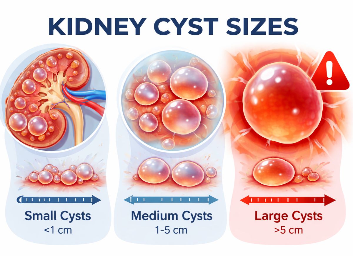
Simple kidney cysts are commonly grouped by size into small cysts measuring less than 1 cm, medium cysts measuring about 1 to 5 cm, and large cysts measuring more than 5 cm. Most simple kidney cysts fall into the small-to-medium range and do not produce noticeable symptoms. These fluid-filled sacs become increasingly common with age and are frequently discovered accidentally during an ultrasound, CT scan, or other imaging test performed for an unrelated reason.
Cysts smaller than 1 cm are generally considered clinically insignificant when they have the typical appearance of a simple cyst. They usually have a thin, smooth wall and contain clear fluid. Such cysts rarely cause symptoms, interfere with kidney function, or raise concern for cancer. In most cases, they do not require follow-up unless the individual has another condition, such as polycystic kidney disease.
Cysts measuring between 1 and 5 cm represent a common size range for incidentally discovered kidney cysts. These cysts are usually benign and do not cause symptoms. However, a cyst approaching the upper end of this range, such as one measuring 4 or 5 cm, may sometimes be monitored through follow-up imaging. A healthcare provider may suggest another ultrasound within several months to determine whether the cyst remains stable or develops characteristics that make it more complex.
When a cyst grows beyond 5 cm, it is generally considered large, and the possibility of symptoms becomes greater. Cysts measuring between 5 and 10 cm may produce an aching sensation in the flank, side, or upper abdomen. Very large cysts, sometimes reaching 10 cm or more, can place pressure on surrounding kidney tissue or nearby structures. In certain cases, this pressure may affect kidney function or contribute to elevated blood pressure. Symptomatic or very large cysts are more likely to require consideration of treatment.
Size of Kidney Cyst Requires Monitoring or Intervention
A kidney cyst may receive closer monitoring once it reaches several centimeters in size, particularly if it is growing or has other concerning characteristics. Treatment is generally considered when a cyst becomes sufficiently large to produce significant symptoms or complications. However, there is no single size that automatically means every cyst requires treatment. The cyst’s structure, location, growth, symptoms, and the patient’s overall health all matter.
For an uncomplicated 3-to-5 cm simple cyst that does not cause symptoms, doctors commonly take a watch-and-monitor approach. Follow-up imaging, often with ultrasound, may be recommended to observe changes in size and appearance. The purpose of monitoring is not only to measure growth but also to check for changes such as thicker walls, internal septations, or solid components.
If the cyst remains stable and continues to have a simple appearance over time, additional monitoring may eventually become less frequent or may no longer be considered necessary.
When a simple cyst becomes larger than approximately 5 to 10 cm and begins producing symptoms, treatment may become an option. Persistent flank or abdominal pain, difficult-to-control high blood pressure, or obstruction of urine flow may be reasons for intervention. One treatment approach is percutaneous aspiration with sclerotherapy. During this procedure, fluid is removed from the cyst with a needle, and a solution may be introduced to help prevent the cyst from filling again. Very large or recurring cysts may sometimes be treated surgically, including with laparoscopic procedures.
Size is much less important when dealing with a complex kidney cyst. A relatively small cyst can still require close evaluation if it contains suspicious characteristics. Cysts classified as Bosniak III or IV, for example, may have a significant risk of malignancy. In these situations, the internal appearance and behavior of the cyst are more important than its diameter.
The Relationship Between Size of Kidney Cyst and The Risk of Rupture
As kidney cysts become larger, the possibility of rupture may increase. A larger cyst has a more expanded wall and may be more vulnerable to trauma or changes in pressure. Although spontaneous rupture of a simple kidney cyst is uncommon, larger cysts may be more susceptible than smaller ones.
A cyst smaller than approximately 5 cm generally has a relatively small volume and is surrounded by protective tissues within the body. Rupture is uncommon and would generally be associated with significant trauma rather than occurring spontaneously.
As a cyst grows beyond 5 to 10 cm, its wall may become increasingly stretched. A very large cyst can also extend beyond the normal contour of the kidney, potentially making it more vulnerable to injury. A fall, direct impact, or contact-related injury may therefore pose a greater concern for a very large cyst.
If a cyst ruptures, the sudden release of fluid can produce severe flank or abdominal pain. In some situations, rupture may also result in bleeding or infection. Sudden severe pain following an injury or other symptoms suggesting internal bleeding or infection should receive urgent medical evaluation.
Kidney Cyst Types
Kidney cysts are not classified by size alone. Their internal appearance and structural characteristics are also important. The two broad categories are simple cysts, which are usually benign, and complex cysts, which may contain features that require additional investigation.
Radiologists commonly use the Bosniak classification system to evaluate complex kidney cysts. This system categorizes cysts from Bosniak I, representing a simple benign cyst, through Bosniak IV, which has characteristics strongly associated with malignancy. Features such as wall thickness, internal septations, calcifications, and enhancement after contrast administration help determine the category.
The Difference Between A Simple and A Complex Kidney Cyst
Simple kidney cysts are the most common type. On ultrasound or CT imaging, they typically appear round or oval, have a thin and smooth wall, and contain clear fluid. They do not contain solid tissue and generally do not show enhancement after contrast administration.
A typical simple cyst falls into the Bosniak I category and is considered benign. If it remains small and does not cause symptoms, treatment is usually unnecessary.
Complex kidney cysts differ from simple cysts because they contain one or more unusual structural features. These characteristics can sometimes be associated with a cystic kidney tumor and therefore require more careful assessment.
Important features may include:
- Thick or Irregular Walls: Thickening, irregularity, or nodules along the cyst wall can increase concern.
- Septations: These are internal partitions that divide the cyst into separate sections. A small number of very thin septa may have little clinical significance, while numerous, thick, or irregular septa may be more concerning.
- Calcifications: Calcium deposits can occur in cyst walls or septa. Their significance depends on their size, thickness, and pattern.
- Solid Components & Enhancement: Solid tissue that becomes enhanced after contrast administration is an important concerning feature because it may indicate tissue with its own blood supply.
Cysts with more suspicious characteristics may fall into Bosniak III or IV categories and may require specialist evaluation and, depending on the circumstances, surgical treatment.
A Kidney Cyst Is Septated or Has Calcifications
When an imaging report describes a kidney cyst as septated or calcified, it means the cyst does not have the completely simple appearance of an uncomplicated fluid-filled cyst. These findings do not automatically mean cancer is present, but they may require closer evaluation.
Septations are thin internal walls that divide a cyst into two or more compartments. Their significance depends on their number, thickness, smoothness, and whether they enhance with contrast.
A cyst containing only one or a few extremely thin and smooth septa may remain in a relatively low-risk category. However, numerous septations, thickened partitions, irregular surfaces, or enhancement can increase concern. Depending on the imaging findings, such cysts may be categorized as Bosniak IIF or III and may require ongoing imaging or further treatment.
Calcifications are deposits of calcium that can develop within the cyst wall or internal partitions. Small, thin, or peripheral calcifications may occur in benign cysts and are not necessarily dangerous.
On the other hand, thick, irregular, or nodular calcifications may be considered more concerning, particularly when they occur alongside other suspicious findings. The overall appearance of the cyst, rather than the presence of calcium alone, helps doctors determine its risk category.
Warning Signs of Kidney Cyst
Most kidney cysts cannot be felt from outside the body because the kidneys are positioned deep inside the abdomen and protected by muscles, fat, and the lower ribs. A cyst generally has to become extremely large before it can create a noticeable lump or bulge.
The kidneys are located in the retroperitoneal space, behind the main abdominal cavity. Their deep position makes it difficult to physically detect an ordinary kidney cyst during an examination. Even a cyst several centimeters in diameter will usually not create a visible or palpable abnormality.
In rare cases, a very large renal cyst can displace nearby organs and push against the abdominal wall. A person with a giant cyst may notice abdominal fullness, bloating, an unusual bulge, or an asymmetry on one side of the body. A physician may also be able to feel the mass during an examination.
For most people, however, kidney cysts are discovered through imaging rather than by touch.
Dull Ache in the Flank, Back, or Side
A persistent dull ache around the flank, back, or side is one of the more common symptoms associated with a large kidney cyst. As the cyst expands, it can place pressure on the kidney capsule or nearby tissues, producing discomfort.
The pain is often described as a deep, persistent ache rather than a sudden stabbing sensation. Pressure on surrounding organs or nerves can also contribute to discomfort.
Fever, Chills, and Tenderness
Fever and chills may indicate that a cyst has become infected. In some cases, bacteria can enter a cyst and cause an accumulation of infected fluid or pus.
An infected cyst may produce fever, chills, localized tenderness, and worsening pain around the affected kidney. Because an infection can become serious, these symptoms require prompt medical assessment and may require antibiotics and drainage of the infected fluid.
Hematuria (Blood in the Urine)
Blood in the urine can occasionally occur when a kidney cyst affects or ruptures into the urinary collecting system. The urine may become visibly pink, red, or cola-colored, or blood may only be detected through laboratory testing.
Although there are several possible causes of hematuria, blood in the urine should not simply be attributed to a kidney cyst without proper evaluation. A healthcare professional may recommend additional testing to determine the underlying cause.
High Blood Pressure (Hypertension)
A large kidney cyst may sometimes place pressure on blood vessels supplying the kidney. In certain situations, this can affect the kidney’s regulation of blood pressure and contribute to hypertension.
When reduced blood flow to the kidney triggers hormonal responses that increase blood pressure, the condition may be difficult to control with medication. In selected cases, treating the cyst may help address the underlying pressure problem.
Can a Benign Kidney Cyst Turn Into Cancer?
A typical simple kidney cyst is considered benign and does not generally transform into kidney cancer. The main concern is not that an ordinary simple cyst will suddenly become cancerous, but that a lesion initially thought to be a cyst may actually have complex or cancerous characteristics.
Simple cysts are fluid-filled structures lined by cells and generally grow by expanding rather than invading surrounding tissue. A cyst confidently identified as Bosniak I is considered benign and usually does not require long-term surveillance.
The situation is different with complex cystic lesions. Bosniak IIF, III, and IV cysts may contain features such as thick walls, enhancing septa, nodules, or solid components. These findings can be associated with renal cell carcinoma.
In other words, a simple cyst does not typically “turn into” cancer. Instead, a cystic kidney cancer may have cancerous tissue from the beginning and can appear partly fluid-filled on imaging. This is why the Bosniak classification is useful: it helps doctors distinguish harmless simple cysts from lesions that may require closer observation or treatment.
The Potential Risk of Kidney Cyst
A ruptured kidney cyst can cause sudden and intense pain in the flank or abdomen as fluid escapes into the surrounding tissues. Although rupture is uncommon, especially with smaller simple cysts, it can sometimes result in bleeding or infection.
The effects of rupture depend on the size and contents of the cyst and whether nearby blood vessels are affected. Severe pain is often the most immediate symptom. Irritation caused by leaked fluid can trigger inflammation in the surrounding tissues and may resemble other causes of acute abdominal or flank pain.
In some cases, rupture may damage a small blood vessel and cause bleeding around the kidney. Significant bleeding can result in symptoms such as dizziness, weakness, low blood pressure, or a rapid heartbeat. Severe bleeding may require medical intervention.
An infected cyst that ruptures can present another serious concern. Infected fluid may spread into the surrounding area and contribute to an abscess or more widespread infection. Fever, chills, severe pain, and marked illness in this situation require urgent treatment.
Doctors may use imaging, such as CT scanning, to determine whether bleeding, fluid leakage, or infection has occurred. Minor ruptures may sometimes be treated conservatively with rest and pain management, while significant bleeding or infection may require more intensive treatment.
Advanced Kidney Cyst Diagnosis
Imaging plays a central role in identifying and monitoring kidney cysts. Ultrasound is often used first because it is non-invasive and can quickly determine whether a kidney lesion appears fluid-filled.
A typical simple cyst on ultrasound has a smooth, thin wall and clear fluid. When these characteristics are present and the person has no symptoms, additional imaging may not be necessary.
If the cyst has unusual features or the ultrasound does not provide enough information, a CT scan may be recommended. CT imaging provides detailed cross-sectional views of the kidneys and can help doctors evaluate the cyst wall, internal septations, calcifications, and contrast enhancement.
MRI can provide additional information when a lesion remains uncertain or when more detailed soft-tissue imaging is needed. It may be particularly useful for further characterizing complex or indeterminate cystic lesions.
When ongoing surveillance is recommended, imaging may be repeated periodically to determine whether the cyst has changed in size or appearance.
The Bosniak Classification System for Kidney Cysts
The Bosniak system is a standardized method used by radiologists to categorize cystic kidney lesions according to their imaging characteristics and potential risk of malignancy. The classification helps guide decisions about surveillance, additional testing, or treatment.
- Bosniak I: These are classic simple cysts with thin walls and no concerning internal structures. They are considered benign and generally require no follow-up.
- Bosniak II: These cysts may contain a few thin septa or small areas of calcification but are still considered overwhelmingly benign. Routine follow-up is generally unnecessary.
- Bosniak IIF: The “F” stands for follow-up. These cysts have more complex features than Bosniak II lesions and are usually monitored with repeat imaging to look for changes.
- Bosniak III: These lesions have more concerning features, such as thickened or irregular walls or septa that enhance with contrast. They have a meaningful risk of malignancy and may require specialist assessment and treatment.
- Bosniak IV: These cystic lesions contain clearly suspicious enhancing solid components and have a high likelihood of malignancy. Surgical treatment is commonly considered for these lesions.
FAQs
1. Is a 4 cm kidney cyst considered large?
A 4 cm kidney cyst is generally not considered extremely large. It falls within the medium-sized range described for simple kidney cysts. However, size alone does not determine whether a cyst is dangerous.
A 4 cm cyst that appears simple on ultrasound or CT, with a thin smooth wall and clear fluid, is generally considered very likely to be benign. If it causes no symptoms, doctors may choose observation and, depending on the circumstances, repeat imaging to confirm that it remains stable.
The situation is different if the cyst causes flank pain, blood in the urine, high blood pressure, or other symptoms. Likewise, a 4 cm cyst with complex characteristics such as thick walls, septations, calcifications, or solid components may need additional evaluation.
2. Can an ultrasound tell if a kidney cyst is cancerous?
Ultrasound can provide useful information about a kidney cyst, including its size, shape, location, and whether it appears simple or complex. However, ultrasound alone cannot always determine with certainty whether a cyst is cancerous.
If the lesion has concerning characteristics, a doctor may recommend additional imaging, such as a contrast-enhanced CT scan or MRI. In selected situations, additional testing may be necessary to establish the diagnosis.
3. What is the root cause of kidney cysts?
Kidney cysts can develop for several different reasons. Some occur as part of inherited conditions such as polycystic kidney disease, in which numerous cysts develop in the kidneys.
Simple kidney cysts, however, are often associated with aging and become more common as people get older. Other cysts may be associated with previous kidney injury, infection, or chronic kidney conditions.
In many cases, the precise reason a simple cyst develops is not known. They generally arise when fluid accumulates within a small abnormal area of kidney tissue.
4. Should I worry about cysts on my kidneys?
Having a kidney cyst does not automatically mean that you have a serious health problem. Many simple cysts cause no symptoms and remain harmless throughout a person’s life.
Concern is greater when a cyst becomes very large, produces symptoms, changes in appearance, becomes infected, bleeds, or interferes with normal urinary or kidney function. Complex cysts also require more careful evaluation because some may have a risk of malignancy.
Regular medical follow-up and appropriate imaging can help determine whether a cyst needs treatment.
5. How do doctors treat a kidney cyst?
Treatment depends on the cyst’s size, appearance, location, symptoms, and underlying cause. Small simple cysts that do not cause symptoms generally do not need treatment.
When a cyst becomes painful or causes another complication, a doctor may consider draining it or using another procedure to reduce the chance of it returning. An infected cyst may require antibiotics and drainage.
Cysts with highly suspicious imaging characteristics may require surgical treatment. For people with polycystic kidney disease, management focuses on controlling symptoms, protecting kidney function, and addressing complications associated with the condition.
6. What are 5 signs your kidneys are not working properly?
Several symptoms may occur when kidney function is impaired. Five possible warning signs include:
- Swelling: Fluid retention can cause swelling in the face, legs, ankles, or feet.
- Changes in urination: Urine may become unusually dark or foamy, or you may notice changes in how frequently you urinate.
- Fatigue or weakness: Reduced kidney function can contribute to persistent tiredness and weakness.
- Pain: Ongoing pain in the back or side can occur with certain kidney conditions, although pain has many possible causes.
- High blood pressure: Kidney problems can affect the body’s regulation of fluid and salt, contributing to elevated blood pressure.
Conclusion
The size of a kidney cyst can provide useful information, but it is not the only factor that determines whether a cyst is concerning. Small simple cysts are often harmless, while larger cysts may sometimes cause pressure, pain, bleeding, infection, or other complications.
Pay attention to symptoms such as persistent flank or abdominal pain, changes in urination, blood in the urine, fever, swelling, or unexplained high blood pressure. Complex features seen on imaging also deserve careful medical evaluation.
Regular monitoring and timely medical care can help identify changes early and determine whether treatment is necessary. If you have been diagnosed with a kidney cyst, discussing its size, appearance, and follow-up plan with your healthcare provider can help you understand your individual situation and protect your kidney health.