5 Symptoms of a Macular Hole That Affect Central Vision
Your eyesight shapes how you read, drive, recognize the people you love, and handle small daily tasks. So when your central vision starts to blur or warp, it’s worth paying attention it could point to a retinal problem called a macular hole.
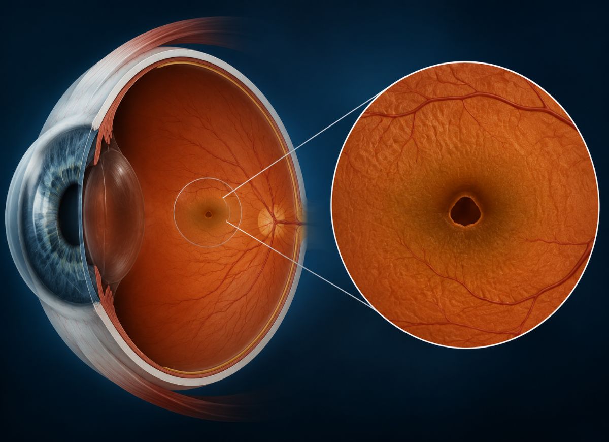
A macular hole is a small break that develops in the macula, the part of the retina that gives you sharp, straight-ahead sight. Unlike vision loss at the edges of your visual field, which can go unnoticed for a while, a macular hole gets in the way of fine detail almost immediately, which is why it tends to disrupt daily life even in its earliest stages.
Studies suggest macular holes mostly show up in people past 60, affecting roughly 7 to 8 out of every 100,000 people each year, with women slightly more likely to develop one than men. The main driver is age: the vitreous, a gel-like substance inside the eye, naturally shrinks over time. As it pulls away from the retina, it can tug hard enough on the macula to tear it.
What makes a macular hole tricky is how quietly it can start. Many people notice a little blurring or a slight kink in their vision and chalk it up to getting older or tired eyes. Left alone, though, this can progress into wavy lines, trouble with small print, or a dark patch sitting right in the middle of their view.
Because those early signs are so easy to dismiss, plenty of people wait too long before seeing a doctor but catching a macular hole early usually means better odds with treatment, including surgery that can bring vision back.
Below, we break down five central-vision symptoms worth watching for, so you know when it’s time to get your eyes checked.
What Is a Macular Hole and How Does It Affect Vision?
A macular hole is a full-thickness tear in the macula the small, central patch of the retina that handles detailed, sharp vision. Because this area does the heavy lifting for tasks like reading and recognizing faces, even a small hole here can have an outsized effect on how you see.
What Part of the Eye Does a Macular Hole Affect?
The macula sits at the back of the eye, right in the center of the retina the light-sensitive layer that works much like film in a camera or the sensor in a digital one. Light enters the eye, gets focused by the lens, and lands on the retina, which turns it into signals the optic nerve carries to the brain.
While the whole retina plays a part in vision, the macula is built for the hardest jobs. It’s crammed with cone cells, and those cones give you two things: crisp detail and rich color. The dense cluster of cones is what lets you make out fine patterns, read small text, and reach 20/20 sharpness and because cones work best in good light, this is also why your central vision looks sharpest during the day.
Because so much visual processing depends on this one small area, a hole here isn’t a minor glitch. It physically removes photoreceptors from that spot, so light landing there simply doesn’t register. The result is a blurry patch or blind spot exactly where your eyesight should be at its best.
The Role of Central Vision
Central vision handles the tasks that need precision: reading, driving, recognizing faces, and any close, detailed work. Side (peripheral) vision is good at picking up movement and giving you a general sense of your surroundings, but it’s your central vision powered by the macula that lets you zoom in on detail.
Reading depends entirely on this sharp central view. When a macular hole disrupts it, letters can blur, vanish into a blind spot, or twist out of shape, turning something as simple as reading a text message into a struggle.
Driving safely also leans heavily on central vision reading signs, spotting traffic lights, staying in your lane, and checking your speedometer all need a clear, focused gaze. Losing that sharpness in the middle of your vision can make it genuinely unsafe to drive.
The same goes for recognizing people. Picking out the details of a face eyes, smile, expression takes fine-detail vision. A hole in the macula can blur those features or blot them out entirely, taking away something as basic as knowing who you’re looking at.
Hobbies and everyday chores suffer too. Cooking, sewing, working on a laptop, or simply seeing what’s on your plate all rely on the same sharp focus that a macular hole disrupts.
In short, central vision is your window into detail and when a macular hole damages it, the ripple effects touch independence, safety, and daily life.
5 Key Central Vision Symptoms of a Macular Hole
Blurry or Distorted Central Vision
One of the earliest signs of a macular hole is a condition called metamorphopsia distorted central vision where straight lines look bent, wavy, or crooked. A door frame or a telephone pole might appear to curve; a grid pattern might look rippled, almost as if you’re viewing it through textured glass or shimmering heat.
This happens because the pulling force of the shrinking vitreous disturbs the neat, orderly layout of photoreceptor cells in the macula. Once those cells shift out of position, the signals they send to the brain get scrambled, and the brain interprets that jumbled input as a warped image.
In the early stages, the distortion can be so mild it’s barely noticeable maybe a slight bend in a straight line that glasses can’t fix. As the hole grows, though, the distortion usually becomes more obvious, making it harder to read or recognize shapes and structures.
Eye doctors often use a simple tool called an Amsler grid a pattern of straight lines with a dot in the center to catch this kind of distortion early, sometimes before a patient notices it on their own. Because metamorphopsia is such a specific red flag for macular problems, it’s one symptom that should always prompt a trip to the eye doctor.
Blind Spot In Central Vision
A macular hole can also cause a central scotoma a dark, gray, or blank patch sitting right in the middle of your vision. This tends to appear as the hole develops from an early-stage defect into a full-thickness break, and the size of the blind spot generally tracks with the size of the hole.
The reason is simple: where the hole exists, retinal tissue is missing. Without photoreceptors in the fovea the very center of the macula there’s nothing there to register light, so the brain gets no signal from that spot and reads it as blank or dark.
Early on, this might just look like a hazy or grayed-out patch. As the hole grows (typically at stage 3 or 4), the blind spot becomes denser and more absolute a true gap in vision.
This can be especially disruptive. Looking at someone’s face, the blind spot might block out their nose and mouth. While reading, it can sit directly over the word you’re trying to focus on. Unlike floaters, which drift around, a central scotoma stays fixed in the middle of your gaze no matter where you look which is part of what makes it such a clear sign of macular trouble.
Difficulty Reading Small Print
Struggling to read small text is one of the most noticeable, real-world symptoms of a macular hole, since reading depends so heavily on the sharp central vision the macula provides. This difficulty is really a combination of the other symptoms blur, distortion, and the blind spot all working against you at once.
Many people find that even their best reading glasses don’t help. Bent or wavy letters (from metamorphopsia) can make lines of text look like they’re curving, and words may blur together. Meanwhile, the blind spot can literally erase letters or whole words as you scan a sentence, and your brain may try to “fill in” the missing pieces leading to misreading or confusion.
In the beginning, brighter lighting or a magnifying glass might help. But as the hole progresses, these workarounds stop being enough, since they can’t fix the underlying distortion or cover for a genuine blind spot.
This kind of struggle isn’t limited to books it shows up in threading a needle, checking a wristwatch, scrolling through photos, or signing a document. Often, it’s this everyday frustration that finally pushes someone to see a doctor.
Objects Appearing Smaller With The Affected Eye
A less common but telling symptom is micropsia when objects viewed through the affected eye look smaller, and sometimes farther away, than they do through the healthy eye. It’s a distinctive marker of macular trouble, caused by the retina physically stretching as the hole forms.
The leading explanation involves the photoreceptors getting pulled apart. Normally, the brain judges an object’s size by how many photoreceptors its image activates. When retinal stretching spreads those cells further apart, the same object now lights up fewer cells than before so the brain reads it as smaller or more distant.
You can sometimes spot this yourself by covering one eye at a time while looking at something fixed, like a TV or door frame, and switching between eyes. If the object seems to shrink when viewed through the affected eye, that’s micropsia and it can also throw off depth perception.
While distorted shape (metamorphopsia) is reported more often, distorted size (micropsia) is just as strong a signal that something’s wrong with the macula, and it’s worth a full eye exam either way.
Sudden Decrease In Vision Acuity
A drop in sharpness sudden or gradual is one of the clearest indicators of a macular hole, since the fovea (the macula’s center) is entirely responsible for your best possible visual acuity. Any damage there shows up directly on a standard eye chart.
In the earliest stage, the drop might be small say, from 20/20 down to 20/30 or 20/40 subtle enough that you only notice it by covering your good eye. But as a full hole forms and grows, acuity can fall much further, sometimes to 20/80, 20/200, or worse with 20/200 marking the threshold for legal blindness in the U.S.
Importantly, this loss stays confined to central vision. Peripheral sight remains intact, so someone with a macular hole can still move around a room and notice motion to the sides they just can’t make out detail in whatever they look at directly.
Any unexplained drop in sharp central vision whether over days or weeks is a serious sign that glasses alone won’t fix, and it calls for a prompt, dilated eye exam.
What Causes a Macular Hole to Develop?
Aging
Age is by far the most common cause, largely because of a natural process called posterior vitreous detachment (PVD). The vitreous a clear gel filling about 80% of the eyeball is firmly attached to the retina in youth, but it liquefies and shrinks with age, gradually pulling away.
For most people, this separation happens cleanly, with maybe some new floaters or flashes of light. But in some, the vitreous stays stubbornly stuck to the center of the macula. As it keeps shrinking, it drags on that tissue a condition called vitreomacular traction stretching the macula and often triggering the first signs of blur and distortion.
If that pulling continues, it can eventually tear the tissue, starting as a partial hole (stage 1) and progressing to a full-thickness hole (stage 2) and beyond, as fluid seeps in and the hole widens further. This whole sequence is simply a byproduct of aging it isn’t linked to diet or lifestyle, but to how the vitreous and macula happen to be attached in certain eyes.
Eye Injury or Trauma
A hard, direct blow to the eye from a ball, an accident, or similar impact can also cause what’s called a traumatic macular hole, though this is far less common than the age-related kind.
The mechanism here is different: the force of impact rapidly compresses and then re-expands the eyeball, creating intense stress on the retina. That sudden deformation can yank on the macula hard enough to tear it instantly.
Traumatic macular holes can strike at any age, including younger, otherwise healthy people involved in sports injuries or accidents. Interestingly, some of these especially in younger patients have been known to close up on their own without surgery. That said, this isn’t the norm, and anyone with a serious eye injury followed by vision changes should get checked immediately, since other issues like retinal detachment can also be involved.
Certain Medical Conditions
A handful of medical and eye conditions can raise the odds of developing a macular hole by weakening the retina or adding extra tractional stress.
People with high myopia (severe nearsightedness) have eyes that are longer than normal front to back, which stretches the retina thin and makes it more prone to tears and holes.
Macular pucker, or epiretinal membrane, involves scar tissue forming over the macula. As this membrane tightens over time, it can pull hard enough to trigger a hole, similarly to vitreous traction.
Advanced diabetic eye disease can cause swelling in the macula (diabetic macular edema), and that chronic stress sometimes combined with abnormal vitreous changes linked to diabetes can contribute to hole formation.
A previous retinal detachment in the same eye also raises risk, since surgical repair and the healing process that follows can leave scar tissue capable of pulling on the macula later on.
Finally, vitreomacular traction (VMT) syndrome is essentially an early warning stage active pulling on the macula that hasn’t yet torn all the way through. People diagnosed with VMT are at high risk of it progressing into a full macular hole if the traction isn’t resolved.
Macular Hole Diagnosis
Diagnosing a macular hole starts with a full, dilated eye exam paired with imaging technology. The doctor first checks visual acuity to measure how much central vision has already been affected, then often uses an Amsler grid a simple chart of straight lines to screen for distortion or blind spots.
If lines on the grid look bent, broken, or missing to the patient, that’s a strong clue something’s wrong with the macula. From there, the gold-standard test is Optical Coherence Tomography (OCT) a non-invasive scan that uses light waves to create detailed, cross-sectional images of the retina.
An OCT scan lets the specialist see the macula’s structure up close: confirming whether a hole exists, measuring its size, and determining whether it’s a partial (lamellar) or full-thickness hole. This detail is essential for staging the hole correctly and planning treatment including spotting active vitreomacular traction or any fluid buildup around the hole that’s adding to the blur.
Different Stages of a Macular Hole
Macular holes are classified into four stages under the Gass system, tracking their progression from an early warning sign to a fully developed hole. Staging helps doctors predict outcomes and decide when to step in with treatment.
Stage 1 sometimes called an impending hole starts when the shrinking vitreous begins tugging on the fovea, the very center of the macula, causing it to lift slightly. This may show up as a small yellow spot on exam, along with mild blur or distortion, though no true hole exists yet.
Stage 2 marks the appearance of a genuine full-thickness opening, usually under 400 microns across, with noticeably worse blur and distortion.
Stage 3 is a bigger hole over 400 microns where the vitreous has fully separated from the macula, though a small flap of tissue may still hover nearby. Central vision loss becomes substantial here.
Stage 4 is the most advanced: a large, fully developed hole alongside complete separation of the vitreous from the entire retina.
Spontaneous healing is rare overall but most likely at stage 1. From stage 2 onward, surgery is typically the recommended path, since vision loss at that point usually won’t resolve on its own and the size and duration of the hole strongly influence how well surgery is likely to work.
Surgery for a Macular Hole
The standard treatment is a procedure called pars plana vitrectomy, performed by a retinal specialist to relieve traction on the macula and give it the chance to heal. It generally involves three steps.
First, the surgeon removes the vitreous gel entirely, eliminating any pulling force on the retina. Second, many surgeons also peel away the internal limiting membrane (ILM) the retina’s innermost layer around the hole, which makes the tissue more flexible and encourages the cells needed for healing to move in. Third, the eye is filled with a temporary gas bubble (commonly SF6 or C3F8), which acts like an internal splint, pressing the hole’s edges together while it heals. Over several weeks, the body’s natural fluid gradually replaces this bubble.
Positioning after surgery matters a lot. Patients are usually told to keep their face pointed down for several days to a week, since the lighter-than-fluid gas bubble naturally rises face-down positioning keeps it pressed right over the macula where it’s needed.
Flying or traveling to high altitudes is off-limits until the gas bubble is fully absorbed, since pressure changes can make it expand dangerously and spike eye pressure. Strenuous activity is also restricted for a while to let the eye heal undisturbed.
Macular Hole vs. Macular Degeneration
Macular holes and age-related macular degeneration (AMD) both affect the macula and can cause similar blur or distortion, but they’re fundamentally different conditions. A macular hole is a physical, structural tear usually caused by mechanical pulling from the shrinking vitreous and it’s an acute problem with a defined starting point.
AMD, by contrast, is a chronic, degenerative disease. In dry AMD (the more common form), light-sensitive cells slowly break down and yellow deposits called drusen build up under the retina, causing gradual vision loss. In wet AMD, abnormal blood vessels grow beneath the retina and leak fluid or blood, causing faster, more severe damage.
These differences shape how each is treated. A macular hole is usually addressed with one surgery a vitrectomy that closes the hole successfully more than 90% of the time and often meaningfully improves vision. AMD, on the other hand, is managed over the long term rather than cured: dry AMD is often slowed with AREDS2 supplements, while wet AMD needs ongoing anti-VEGF injections to control abnormal blood vessel growth.
A macular hole also tends to appear suddenly, with vision changing over days or weeks, while AMD especially the dry form progresses much more gradually across years. And while a macular hole is mechanical in origin, AMD stems from biological aging, genetics, and lifestyle factors like smoking.
FAQs
1. How serious is a macular hole?
It’s a serious condition because it directly targets central vision the part you rely on for reading, driving, and recognizing faces. It rarely causes total blindness, but it can significantly reduce clarity in the affected eye, with severity depending largely on how advanced the hole is.
2. Can you fix a macular hole?
Yes surgery, typically a vitrectomy, successfully closes most macular holes. The vitreous gel is removed and a gas bubble takes its place to help seal the hole, often restoring or improving vision, especially when caught early.
3. What happens if a macular hole is left untreated?
It tends to worsen over time, with central vision loss increasing and daily tasks becoming harder. The hole can also enlarge, which can make treatment less effective. Peripheral vision usually stays intact, but central vision damage can become permanent if it goes on too long.
4. How can I stop a macular hole from getting worse?
Not every hole can be prevented from progressing, but early diagnosis makes a real difference. Avoiding eye trauma, keeping up with regular eye exams, and following your ophthalmologist’s guidance including strict post-surgery instructions all help protect your outcome.
5. What shouldn’t I do after macular hole surgery?
Avoid flying, heavy lifting, and strenuous activity for the period your doctor specifies. If a gas bubble was placed in your eye, follow the required positioning instructions closely, and don’t skip follow-up appointments both are key to proper healing.
6. Can stress cause a macular hole?
Not directly. The condition is mainly tied to age-related changes in the vitreous gel, not stress. That said, staying generally healthy and attentive to your body can help you notice and act on symptoms sooner.
7. Can I fly if I have a macular hole?
Flying itself isn’t usually a problem before surgery, but it’s strongly discouraged right after a vitrectomy if a gas bubble is in place, since pressure changes at altitude can be dangerous. Always check with your ophthalmologist for guidance specific to your case.
8. What vitamins support macular health?
Vitamin C, vitamin E, lutein, zeaxanthin, and zinc are commonly recommended for overall eye health, found naturally in leafy greens and colorful produce. They won’t treat an existing hole, but they can support general retinal health.
9. Can a macular hole heal without surgery?
Occasionally, small early-stage holes close on their own, but this is the exception rather than the rule. Most full-thickness holes need surgical repair to restore vision and prevent further damage, so regular monitoring by a specialist is important.
Conclusion
A macular hole targets the part of your eye responsible for your sharpest, most detailed vision, which is why even a small one can make everyday tasks noticeably harder. It rarely leads to complete blindness, but left unaddressed, it can meaningfully affect your quality of life.
Recognizing the five symptoms covered here blur, wavy or distorted lines, blind spots, trouble reading, and shrinking objects in one eye gives you a head start on catching the problem early, when treatment tends to work best.
Vitrectomy surgery has a strong track record, but like most eye conditions, earlier detection generally means better outcomes. If you notice any lasting change in your central vision, don’t wait it out get it checked. Regular eye exams, especially after 60, remain one of the best tools for catching a macular hole before it does lasting damage.