10 Things to Know About Microscopic Colitis and Chronic Diarrhea
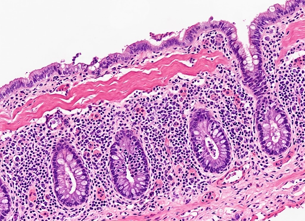
Microscopic colitis is an inflammatory condition of the large intestine that commonly causes persistent, watery diarrhea. Unlike other forms of colitis, the colon typically looks normal during a colonoscopy; the inflammation only becomes visible under a microscope, after small tissue samples (biopsies) are examined. Because of this, the condition can go undiagnosed for a long time, even when digestive symptoms have lasted for weeks or months.
Microscopic colitis isn’t the same as Crohn’s disease or ulcerative colitis, but it can still take a real toll on daily life. Frequent diarrhea, cramping, urgency, bloating, fatigue, and unintended weight loss can interfere with work, travel, sleep, and social plans.
It’s more common in older adults, particularly women, and certain medications, smoking, autoimmune conditions, and genetic factors appear to raise the risk. Here are 10 important things to understand about microscopic colitis: its symptoms, causes, diagnosis, treatment, and long-term outlook.
1. The Core Paradox: Inflammation That Doesn’t Show Up on a Colonoscopy
Microscopic colitis is a chronic inflammatory bowel condition marked by persistent, watery, non-bloody diarrhea. During a colonoscopy, the lining of the large intestine looks entirely normal to the naked eye, with none of the ulcers, bleeding, or redness seen in Crohn’s disease or ulcerative colitis.
Because of this, the condition can’t be diagnosed through visual inspection alone. A gastroenterologist has to collect multiple small tissue biopsies from different sections of the colon, which a pathologist then examines under a microscope to find the hidden inflammation, distinguishing the condition from functional disorders like irritable bowel syndrome (IBS).
2. Two Microscopic Subtypes: Collagenous and Lymphocytic
Pathologists divide the disease into two subtypes based on the specific cellular changes seen in colon tissue. Although they look different under a microscope, both cause nearly identical symptoms and respond to the same treatments.
Collagenous colitis involves an abnormally thick band of collagen, the main structural protein in connective tissue, sitting just beneath the surface cells of the colon lining.
Lymphocytic colitis involves a sharp rise in lymphocytes (a type of white blood cell) within the colon’s surface lining. A small number of these immune cells are normally present, but in lymphocytic colitis their numbers climb past 20 per 100 surface cells, a sign that the immune system is actively attacking the body’s own tissue and disrupting the colon’s normal fluid balance.
3. The Clinical Hallmark: Secretory Chronic Diarrhea
The main symptom is chronic, watery, non-bloody diarrhea, not just occasional loose stool, but frequent, large-volume bowel movements that can start gradually or hit suddenly. A few patterns define this diarrhea:
It’s secretory in nature. The inflamed intestinal lining actively pumps salt and water into the bowel instead of absorbing it, so the diarrhea typically continues even during fasting.
It’s frequent, and dehydration is a real risk. Patients commonly have four to ten watery bowel movements a day; in severe cases that can exceed ten a day, leading to weight loss, dehydration, and electrolyte imbalances.
It often strikes at night. Waking up with an urgent need to use the bathroom is a strong sign of an organic, physical disease rather than a functional issue like IBS, which rarely disrupts sleep.
There’s no visible blood. Since the lining doesn’t develop deep, bleeding ulcers, the stool stays non-bloody, a key distinction from ulcerative colitis, where rectal bleeding is common.
4. Related Symptoms Beyond Diarrhea
Widespread inflammation in the large intestine disrupts the bowel’s normal contraction patterns, leading to a cluster of related symptoms:
Cramping and pain. The colon walls spasm intermittently while trying to manage the excess fluid, causing mild to moderate cramping across the mid to lower abdomen.
Bloating and gas. Fluid shifts and altered cell function can slow digestion, causing uncomfortable pressure and visible abdominal swelling.
Urgency and incontinence. A sudden rush of watery fluid into the rectum can create an intense, hard-to-control need to go, which can cause real anxiety around travel, work, and social life.
5. Causes and Key Risk Factors
The exact cause isn’t known, but microscopic colitis is thought to result from an abnormal immune response in genetically susceptible people, triggered by a combination of factors, including certain medications, autoimmune disease, and demographic patterns favoring older women.
Medications are a leading suspect. For many patients, reviewing their full prescription and over-the-counter history is the most important step in tracking down a cause. Several drug classes have been strongly linked to the condition:
- NSAIDs (common pain relievers): long-term use can weaken the tight junctions in the colon wall, letting antigens leak into tissue and trigger inflammation.
- Proton pump inhibitors (PPIs), often taken for acid reflux, can shift the gut microbiome and prompt inflammation.
- SSRIs (certain antidepressants) are linked to altered gut motility and immune cell activation in the intestinal lining.
- Other agents include cholesterol-lowering statins, certain blood pressure beta-blockers, and the diabetes medication acarbose.
Because medication is such a common trigger, doctors often recommend a supervised “drug holiday,” pausing a suspected medication under medical guidance. If diarrhea resolves during this trial, it confirms a drug-induced cause and can eliminate the need for long-term immunosuppressive treatment.
Autoimmune connections are common. Microscopic colitis frequently occurs alongside other autoimmune conditions rather than as an isolated issue:
- Celiac disease has the strongest known link; the risk of developing microscopic colitis is up to 70 times higher in celiac patients.
- Thyroid disorders, like Hashimoto’s thyroiditis and Graves’ disease, frequently appear alongside colon inflammation.
- Rheumatoid arthritis and type 1 diabetes share similar genetic pathways of immune dysregulation.
Tracking these overlapping conditions helps doctors screen for issues like celiac disease early in the care plan.
Age and gender patterns are distinct. The condition is most often diagnosed in people in their 60s and 70s and is relatively rare under 45, suggesting that years of cumulative exposure to triggers and medications eventually bring the disease out in susceptible people. Women face a much higher risk too, with female-to-male diagnosis ratios ranging from 3:1 up to 7:1, a pattern that mirrors other autoimmune diseases and may be tied to hormonal factors like estrogen’s effects on the immune system. A typical profile is a postmenopausal woman over 50 with unexplained, severe, watery diarrhea. Smoking has also been linked to a higher risk, particularly for the collagenous subtype.
6. How Diagnosis Works, and How It’s Told Apart From Other Conditions
Because the bowel looks healthy on standard imaging, diagnosis relies entirely on a gastroenterologist collecting multiple tissue biopsies during a colonoscopy or flexible sigmoidoscopy.
Why a visual exam alone isn’t enough. During a standard colonoscopy, the colon lining typically looks smooth, pink, and healthy. At most, a doctor might notice minor changes like slight redness, but nothing that clearly points to disease. A proper diagnosis requires:
- Broad tissue sampling, usually 8 to 10 biopsies from several sections of the colon (ascending, transverse, descending, and sigmoid), since the inflammation can be patchy and easy to miss with limited sampling.
- Microscopic confirmation, where a pathologist looks for a thickened collagen band (collagenous colitis) or a high concentration of lymphocytes in the lining (lymphocytic colitis). Without this step, the true cause is often missed, sometimes for years.
Distinguishing it from IBS. Microscopic colitis can closely resemble diarrhea-predominant IBS, since both cause chronic loose stools, cramping, and urgency. But IBS is a functional disorder, meaning the bowel is overly sensitive and irregular but shows no structural damage; both the colonoscopy and biopsy come back normal in IBS. Signs pointing toward microscopic colitis instead include sudden onset after age 50, large volumes of watery stool, and nighttime diarrhea.
Distinguishing it from IBD. Microscopic colitis is much milder than classic inflammatory bowel disease (Crohn’s disease and ulcerative colitis). Classic IBD causes visible, destructive inflammation, deep ulcers, and tissue that bleeds easily, along with bloody stools, severe pain, fever, and significant weight loss. Microscopic colitis, by contrast, produces non-bloody diarrhea with inflammation confined to the microscopic level. Importantly, while long-term IBD raises colorectal cancer risk substantially, microscopic colitis does not increase that risk beyond the general population.
| Marker | IBS | Microscopic Colitis | IBD |
|---|---|---|---|
| Visual colonoscopy | Normal | Normal or near-normal | Abnormal (ulcers, bleeding) |
| Microscopic biopsy | Normal | Abnormal (thickened collagen or excess white cells) | Abnormal (severe structural damage) |
| Stool | Loose, watery, or pasty | Profuse, watery, non-bloody | Frequent, loose, often bloody |
| Nighttime symptoms | Rare | Common | Common and severe |
| Typical onset | Under 45 | Over 50 | Teens to young adulthood |
| Colorectal cancer risk | Not increased | Not increased | Significantly increased |
7. How Chronic Diarrhea Is Managed and Treated
Treatment follows a stepwise approach, starting with conservative measures and moving toward medication when needed.
First-line steps usually include:
- Reviewing medications for known triggers like NSAIDs, PPIs, or SSRIs, and pausing them under medical supervision when a trigger is suspected; this alone can resolve diarrhea within a few weeks.
- Adjusting diet, since avoiding caffeine, alcohol, dairy, artificial sweeteners, and heavily spiced or high-fat foods often eases symptoms. Because of the celiac disease connection, a doctor may also suggest checking for gluten sensitivity or trying a gluten-free diet temporarily.
- Over-the-counter anti-diarrheals, such as loperamide (which slows gut motility) or bismuth subsalicylate (which offers mild anti-inflammatory effects alongside diarrhea control).
Budesonide is the go-to prescription treatment for moderate to severe cases that don’t respond to lifestyle changes. Clinical trials show it helps roughly 80% of patients reach full symptom remission within 6 to 8 weeks. It belongs to the same family of anti-inflammatory drugs as prednisone, but it’s specifically designed to act on the gut: a special coating lets it pass safely through the stomach and small intestine before releasing into the colon, where the inflammation is. About 90% of the drug is broken down by the liver on its first pass through the bloodstream, which limits the classic systemic side effects of steroids, like mood changes, fluid retention, high blood sugar, or bone density loss.
A typical treatment plan starts with a 9 mg daily dose for 6 to 8 weeks, followed by a gradual taper rather than stopping abruptly. Even so, up to 60% of patients see symptoms return after stopping the drug completely. If that happens, doctors may recommend a repeat course, or for those with frequent flare-ups, a low-dose long-term maintenance plan (often 3 to 6 mg daily or every other day).
8. Long-Term Dietary and Lifestyle Adjustments
There’s no single diet that cures microscopic colitis, but targeted changes can meaningfully help manage it long-term. The goal is to cut out foods and drinks that irritate the colon or act as natural laxatives.
- Caffeine and alcohol speed up gut motility and can trigger flare-ups, so cutting them is often a helpful first step.
- Sugar and artificial sweeteners like sorbitol and mannitol draw extra water into the intestine, worsening watery stools.
- Lactose intolerance often coexists with microscopic colitis, so a temporary dairy-free trial can reduce gas, bloating, and bowel frequency.
Identifying personal triggers matters too. Given the strong link to celiac disease, many gastroenterologists recommend a strict gluten-free trial for patients whose diarrhea persists despite first-line treatment. A food diary can help pinpoint less obvious triggers, and for those with significant cramping and bloating, a temporary low-FODMAP diet (limiting certain fermentable carbohydrates) may also help.
9. Prognosis: Does It Ever Go Away?
The answer depends on how an individual responds to treatment and ongoing triggers. For a small number of patients, the condition resolves completely after one course of treatment and never returns. For most, though, microscopic colitis follows a relapsing-remitting course, meaning symptoms clear during treatment but can come back weeks, months, or years after stopping medication.
Even so, the long-term outlook is reassuring compared to classic IBD: microscopic colitis doesn’t cause destructive scarring, doesn’t require surgery, and doesn’t raise colon cancer risk above that of the general population. For patients with frequent relapses, doctors typically focus on a low-dose maintenance plan or a clear protocol for restarting treatment at the first sign of a flare.
10. Bile Acid Malabsorption: An Often-Overlooked Contributor
If diarrhea persists despite standard treatment, doctors will look for other contributing issues, and bile acid malabsorption (BAM) is a common one that gets overlooked.
Bile acids, produced by the liver to help digest fat, are normally reabsorbed (about 95% of them) in the final section of the small intestine. But tissue changes from microscopic colitis can disrupt this recycling process, letting excess bile acids spill into the large intestine. Once there, they act as a natural laxative, forcing the bowel to secrete large volumes of water and electrolytes, producing diarrhea that looks just like standard microscopic colitis symptoms.
Doctors can confirm BAM using a specialized nuclear medicine test called a SeHCAT scan, or by evaluating how a patient responds to targeted treatment. Since standard anti-inflammatory medication won’t help BAM-related diarrhea, treatment instead relies on bile acid sequestrants, such as cholestyramine or colesevelam, which bind the excess bile acids and let them pass safely without irritating the colon.
A Note on Subtype Treatment
Whether someone is diagnosed with collagenous or lymphocytic colitis, treatment is generally the same and is guided by symptom severity rather than the specific cell changes seen under the microscope. Mild cases in either subtype start with over-the-counter anti-diarrheals and dietary changes, while moderate to severe cases in both use budesonide as the first-line treatment.
Some data suggests lymphocytic colitis may have slightly higher rates of spontaneous remission, while the collagenous subtype can be a bit more persistent. If either subtype doesn’t respond to budesonide, the next steps, such as immunomodulators or biologic medications, are the same regardless of subtype.
Conclusion
Microscopic colitis is a treatable cause of chronic watery diarrhea, but it’s easy to overlook since the colon often looks normal on a routine colonoscopy. Diagnosis usually requires biopsies to reveal inflammation that isn’t visible to the naked eye.
Treatment may involve medication changes, dietary adjustments, anti-diarrheal medications, or prescription anti-inflammatory therapy. If diarrhea persists for several weeks, causes dehydration or weight loss, or comes with blood in the stool, severe abdominal pain, or fever, it’s worth seeking medical evaluation to find the underlying cause and start appropriate treatment.
Frequently Asked Questions
1. What is microscopic colitis?
It’s a type of inflammatory bowel disease that causes chronic, watery diarrhea due to inflammation in the colon lining. Unlike other forms of colitis, the colon usually looks normal during a colonoscopy, and diagnosis relies on examining biopsy samples under a microscope. There are two main types: collagenous colitis and lymphocytic colitis.
2. What are the symptoms of microscopic colitis?
The most common symptom is frequent, non-bloody watery diarrhea that can last for weeks or months. Other symptoms include cramping, bloating, urgency, fecal incontinence, fatigue, and mild weight loss. Symptoms often come and go, with periods of improvement followed by flare-ups, and because they overlap with other digestive disorders, medical evaluation is needed for an accurate diagnosis.
3. What causes microscopic colitis?
The exact cause isn’t fully understood, but it’s believed to involve an abnormal immune response in the colon. Certain medications, smoking, autoimmune diseases, infections, and genetic factors have all been linked to higher risk. Some people develop symptoms after using NSAIDs, proton pump inhibitors, or SSRIs, though in many cases no single cause can be pinpointed.
4. How is microscopic colitis diagnosed?
Doctors typically diagnose it through a colonoscopy with multiple biopsies taken from different parts of the colon. Even when the colon looks healthy during the procedure, examining the tissue under a microscope can reveal the characteristic inflammation. Blood and stool tests may also be used to rule out infections or other causes, since imaging and colonoscopy alone can’t confirm the diagnosis.
5. How is microscopic colitis treated?
Treatment depends on symptom severity and any underlying triggers. A doctor may recommend stopping medications that could be contributing, making dietary changes, or using anti-diarrheal medications. For moderate to severe cases, corticosteroids like budesonide are often effective at reducing inflammation and controlling diarrhea, and some people may need additional medication if symptoms persist or return.
6. Can microscopic colitis go away?
Many people experience periods of remission, especially with appropriate treatment. Some improve after stopping a medication that may have triggered the condition, while others need ongoing therapy to prevent flare-ups. Relapses can occur, but the condition doesn’t typically raise colon cancer risk, and regular follow-up with a healthcare provider can help manage symptoms over time.