Esophagram vs Endoscopy: 6 Differences Patients Should Know
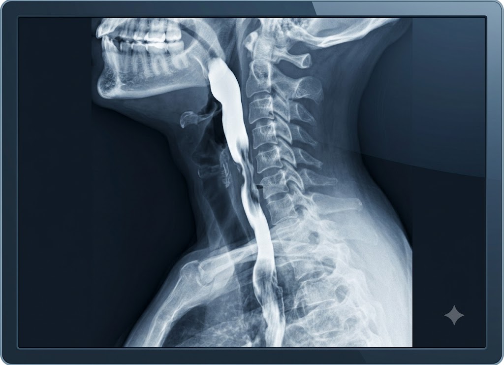
An esophagram is an X-ray test. You swallow a contrast liquid, usually barium, and the images show the shape of your esophagus and how well swallowed material travels toward the stomach. An endoscopy is different. A thin, flexible tube with a camera lets the doctor look directly at the lining of the esophagus, the stomach, and the first part of the small intestine. Both tests can help explain trouble swallowing, reflux, pain, narrowing, or suspected structural problems, but they work in very different ways.
Knowing how they differ can help you feel prepared when your doctor picks one over the other. An esophagram is great for watching movement, shape, strictures, and swallowing in real time. Endoscopy is usually better for seeing inflammation, ulcers, bleeding, tumors, and Barrett’s esophagus up close, and for taking biopsies. This article covers six differences worth knowing before you’re tested.
What is an Esophagram and What is an Upper Endoscopy?
An esophagram is a non-invasive X-ray study that checks how the esophagus is shaped and how it works. An upper endoscopy is a minimally invasive procedure that uses a camera to see the lining of the upper digestive tract directly. Looking at what each test involves makes it easier to see which one suits your situation.
Comparing Functional Silhouettes with Direct Visual Exploration
The biggest difference is how each test sees the upper digestive tract.
                    [Diagnostic Imaging Breakdown]
                                    │
     ┌──────────────────────────────┴──────────────────────────────┐
     ▼                                                             ▼
[Esophagram Radiology Silhouette]                    [Upper Endoscopy Mucosal Feed]
 ├── External outline of the entire tract             ├── Direct, internal, high-def video feed
 ├── Tracks movement and muscle coordination          ├── Evaluates surface texture and tissue health
 └── Highlights blockages and wide hernias            └── Allows for immediate biopsy collection
An esophagram, often called a barium swallow, is a non-invasive X-ray study. It shows an outline of the throat and esophagus and how they move. It is very good at showing muscle coordination, narrowed areas, and larger structural changes such as hiatal hernias. What it cannot show is the surface texture or color of the inner lining.
An upper endoscopy, medically called an esophagogastroduodenoscopy (EGD), is done under sedation. A gastroenterologist guides a thin, flexible tube with a high-definition camera into the digestive tract. Instead of an outline, you get a live, close-up view of the lining of the esophagus, stomach, and duodenum. That lets the doctor spot inflammation, small ulcers, and early tumors that an esophagram would miss.
Real-Time Tracking and Motility Assessment
An esophagram shows swallowing as it happens, not just a still picture.
[Oral Ingestion of Contrast] ──► Real-Time Fluoroscopy ──► Live Video Tracking of PeristalsisYou drink a thick, chalky barium liquid that coats the throat and esophagus. Because barium blocks X-rays, the walls of the esophagus stand out clearly on the radiologist’s screen.
During the test, you stand or lie on a tilting X-ray table while a fluoroscope records continuous images. The radiologist may ask you to swallow different textures, such as thin liquids, thick pastes, or a barium-coated cracker.
This lets the doctor follow peristalsis, the wave-like muscle squeezes that push food down. That makes the test useful for finding motility problems such as diffuse esophageal spasm and achalasia, as well as complex swallowing difficulties.
Direct Internal Visualization and Tissue Sampling
Where an esophagram shows an outline, an endoscopy gets inside. The doctor can look at the tissue itself, take samples, and sometimes treat problems in the same sitting.
                            [Endoscopic Treatment Capabilities]
                                         │
     ┌───────────────────────────────────┼───────────────────────────────────┐
     ▼                                   ▼                                   ▼
[High-Precision Tissue Biopsy]       [Immediate Medical Control]         [Mechanical Dilation]
 ├── Collects surface tissue cell     ├── Seals open bleeding ulcers      ├── Stretches narrowed spaces
 └── Confirms Barrett's esophagus     └── Removes pre-cancerous polyps    └── Resolves food impaction
Because you are sedated, the scope passes through the mouth into the esophagus without triggering a gag reflex. The live color image lets the gastroenterologist look for signs of irritation, such as reflux-related esophagitis, or find the source of bleeding.
The main advantage over a barium test is that treatment can happen right away:
- Targeted biopsies: Small instruments pass through a channel in the scope to collect tissue. A pathologist then checks it for conditions such as celiac disease, H. pylori infection, Barrett’s esophagus, or cancer.
- Direct treatment: If the doctor finds active bleeding, a precancerous polyp, or a badly narrowed section, they can seal the vessel, remove the polyp, or widen the passage during the same procedure.
Operational Comparison: Timeline, Risks, and Preparation
Choosing between the tests means weighing preparation, recovery, and typical uses.
| Feature | Esophagram | Upper Endoscopy (EGD) |
|---|---|---|
| Invasiveness | Non-invasive X-ray imaging from outside the body | Minimally invasive; a camera is inserted |
| Sedation | None; you stay awake and alert | Conscious or deep sedation for comfort |
| Preparation | Fast from food and liquids for 8 hours | Strict fasting; blood thinners may need adjusting |
| Main focus | Swallowing coordination, motility, overall shape | Surface changes, tissue health, biopsies |
| Afterward | Resume normal activities; drink plenty of water | Need a driver; sedation takes hours to wear off |
Preparing for an esophagram is simple, and you can drive yourself home afterward. An endoscopy takes a little more planning because of the sedation.
When doctors want to see both movement and structure, they may order a cine esophagram or a timed barium esophagram first to check muscle function. An endoscopy can then follow to examine the lining and take biopsies.
6 Key Differences Between an Esophagram and an Endoscopy
The six main differences are invasiveness, sedation, imaging technology, diagnostic versus treatment ability, patient experience, and safety risks. Each is explained below so you can see why your doctor might choose one test over the other.
Invasiveness and Clinical Sedation Protocols
The first difference is how invasive each test is and whether sedation is needed.
[Patient Ingests Contrast Awake] ──► Entirely External XR Imaging ──► Immediate Discharge (Self-Drive)
[IV Sedation Administered]        ──► Scope Enters GI Cavity        ──► Recovery Ward Stay (Requires Driver)
An esophagram is entirely non-invasive. Everything happens outside the body, so there is no sedation, no IV line, and no pain medication. You stay awake, drink the contrast, and change positions on the X-ray table as the radiologist asks. There is no recovery time. You can drive home and go back to your normal routine right away.
An upper endoscopy is minimally invasive because the scope is passed through your mouth and down into the digestive tract. To keep you comfortable, calm the gag reflex, and prevent muscle spasms, you receive conscious sedation (often called “twilight sleep”) or general anesthesia.
Sedation means an IV line, close monitoring of your vital signs, and a 30-to-60-minute stay in a recovery room. You can’t drive, operate machinery, or make major decisions for the rest of the day while the medication wears off.
Imaging Technology: Fluoroscopic Silhouettes vs. High-Definition Video
The two tests use very different technology, and each is built to find different kinds of problems.
Fluoroscopy Esophagram Mechanics
An esophagram relies on fluoroscopy, a continuous X-ray beam that records live video of the upper digestive tract. Soft tissue like the esophagus doesn’t show up well on ordinary X-rays, so you drink a contrast material such as barium.
As the liquid coats the walls of the throat and esophagus, it forms a bright white outline on the screen. The doctor isn’t looking at the lining itself. They are tracking the organ’s shape, edges, and movement to judge how well the muscles squeeze food downward.
Endoscopic Optical Technology
An upper endoscopy uses direct viewing. The tip of the flexible scope holds a tiny high-definition camera and a bright light. Together they send a live, full-color, magnified picture from inside the digestive tract to a monitor.
The gastroenterologist can study the real surface of the lining and notice small changes in color, blood vessel patterns, or texture. These would be invisible on an esophagram and include early inflammation, tiny areas of bleeding, and precancerous changes.
Diagnostic Targets: Motility Profiles vs. Mucosal Pathology
Because the technology differs, each test is better suited to different conditions.
                          [Diagnostic Target Selection]
                                         │
     ┌───────────────────────────────────┴───────────────────────────────────┐
     ▼                                                                       ▼
[Esophagram (Structure & Motility)]                         [Endoscopy (Tissue & Mucosal Health)]
 ├── Achalasia (sphincter failure)                           ├── Esophagitis and acute gastritis
 ├── Diffuse esophageal spasms                               ├── Barrett's esophagus tracking
 └── Structural hiatal hernias                               └── Peptic ulcers and mucosal cancers
Conditions Best Diagnosed by an Esophagram
An esophagram is ideal for checking how swallowing works and for spotting structural changes along the upper digestive tract. It is often the first test for dysphagia, the feeling that food is stuck in the chest or throat, or when a muscle coordination problem is suspected.
It is especially good at finding:
- Motility disorders: Achalasia (the lower esophageal sphincter fails to open), diffuse esophageal spasm, and weak or irregular contractions.
- Structural problems: Large hiatal hernias, Schatzki rings, esophageal diverticula (abnormal pouches in the wall), and long-standing strictures.
- Oropharyngeal dysphagia: Coordination trouble in the first phase of swallowing, as food moves from the mouth into the throat.
Conditions Best Diagnosed by an Endoscopy
Endoscopy is considered the gold standard for judging the health of the inner lining. If you have chronic heartburn, severe reflux, unexplained upper abdominal pain, ongoing nausea, or signs of internal bleeding, it is usually the definitive test.
It is essential for finding and confirming:
- Inflammation: Active esophagitis, gastritis, duodenitis, and eosinophilic esophagitis (EoE).
- Reflux complications: Damage from long-term acid reflux and screening for Barrett’s esophagus.
- Ulcers and related disease: Open peptic ulcers in the stomach or small intestine, and signs of celiac disease.
- Cancer screening: Finding, mapping, and sampling suspicious growths, polyps, or tumors.
Treatment Capabilities: Pure Imaging vs. Active Surgical Interventions
An esophagram is purely diagnostic. An endoscopy can be both diagnostic and therapeutic.
[Barium Esophagram] ──► Identifies Stricture or Narrowing ──► Purely Observational ──► Requires Scheduling Second Procedure
[Upper Endoscopy]   ──► Identifies Stricture or Narrowing ──► Active Intervention ──► Balloon Dilation Performed Immediately
With an esophagram, the radiologist watches, records, and reports. No treatment can happen during the scan. If it reveals a serious problem, such as a severe narrowing, you need a separate procedure to fix it.
An upper endoscopy offers “see and treat.” The scope has working channels that let the gastroenterologist pass long, flexible tools into the digestive tract. Treatment can happen during the diagnostic exam itself, which often saves you a second procedure.
Common treatments during an endoscopy include:
- Dilation: A balloon passed through the scope stretches open a severe narrowing or stricture.
- Biopsy and polypectomy: Tissue samples are taken, or precancerous polyps are removed with small wire loops.
- Hemostasis: Active bleeding from an ulcer is stopped with heat, medical clips, or injections.
- Foreign body retrieval: Food or objects stuck in the esophagus are removed.
Patient Experience and Post-Procedure Recovery Timelines
Preparation, the experience itself, and recovery are quite different for each test.
| Esophagram | Upper Endoscopy (EGD) | |
|---|---|---|
| Preparation | Fast from food, drinks, and tobacco for 8 hours | Strict fasting; blood thinners may be paused |
| Duration | Usually 30 to 60 minutes | Procedure itself takes 15 to 30 minutes |
| What it feels like | Drinking chalky liquid and changing positions on a table | Sleeping under sedation, with no memory of the scope going in |
| Immediate recovery | No downtime; you can drive home | 30 to 60 minutes in a recovery area |
| Common after-effects | Temporary white stools; slight risk of constipation | Mild sore throat, bloating, lingering grogginess |
Preparing for an esophagram is simple: fast for eight hours so the esophagus is empty. During the test, your job is to drink the barium and follow the radiologist’s instructions. There is no recovery time afterward, though you should drink plenty of water to flush out the contrast and prevent constipation.
An endoscopy is shaped by the sedation. You fast beforehand and get an IV when you arrive. Most people fall asleep and don’t remember the procedure.
Afterward, you rest in a recovery area while the medication wears off, and you may feel groggy for several hours. Mild side effects include a sore throat or bloating from the air used to gently open the digestive tract for a clearer view.
Risk Profiles and Procedural Safety Considerations
Both tests are very safe in experienced hands, but their risks are different.
               [Procedural Risk Breakdown]
                                 │
     ┌───────────────────────────┴───────────────────────────┐
     ▼                                                       ▼
[Esophagram Safety Profile]                             [Endoscopy Safety Profile]
 ├── Low ionizing radiation exposure                     ├── Cardiovascular/respiratory sedation reactions
 ├── Rare risk of barium aspiration                      ├── Mechanical tissue perforation or tearing
 └── Uses water-soluble agents if tears exist            └── Localized bleeding at tissue biopsy sites
The main concern with an esophagram is exposure to ionizing radiation. The dose from a standard barium test is low, roughly what you’d absorb naturally from your surroundings over a few months. Allergic reactions to the contrast flavorings are rare.
The main clinical worry is aspiration, which is breathing the contrast liquid into the lungs. It is mostly a risk for people with severe neurological swallowing problems. If the doctor suspects a tear or hole in the esophagus, they use a water-soluble contrast such as Gastrografin instead of barium, because the body can safely absorb it if it leaks.
For an upper endoscopy, most risks come from sedation. Modern anesthesia is safe, but it carries a small chance of breathing or heart rhythm problems, especially in older adults or people with other medical conditions.
Procedure-related risks include perforation, a rare but serious tear in the lining of the digestive tract caused by the scope, and localized bleeding, which can happen after a polyp is removed or a biopsy is taken.
What Other Factors Should Patients Consider?
Beyond how the tests differ, you should think about cost, medical reasons a test might not be safe, alternative tests, and preparation. Together, these help you and your doctor choose a path that is medically appropriate and also practical and safe for you.
Financial Planning and Resource Demands
An esophagram generally costs much less than an upper endoscopy. The gap comes from the complexity, setting, and resources each one needs.
                [Financial Cost Multipliers]
                                 │
     ┌───────────────────────────┴───────────────────────────┐
     ▼                                                       ▼
[Standard XR Esophagram Study]               [Upper Endoscopy Hospital Series]
 ├── Uses standard x-ray equipment            ├── Requires specialized surgical suites
 ├── Run by a radiologist & tech              ├── Involves a gastroenterologist & nurses
 └── No sedation or IV drugs used             └── Requires anesthesia team & IV sedation
Sedation is the biggest cost driver for an endoscopy. It requires an anesthesiologist or a Certified Registered Nurse Anesthetist (CRNA), monitoring equipment, and medications, each billed separately.
Endoscopy also takes place in a specialized suite with reusable scopes that need careful sterile processing, followed by a stay in a recovery room staffed by specialized nurses. An esophagram uses ordinary X-ray equipment found at local imaging centers, and since you stay awake, there are no anesthesia fees.
Insurance plans generally cover both tests when they are medically necessary, but your final cost depends on several things:
- Deductibles and coinsurance: On a high-deductible plan, you may pay the full cost of either test until your deductible is met. After that, you typically pay a percentage (coinsurance) of the bill.
- Network status: The cost can vary a lot depending on whether the imaging facility, the gastroenterologist, and the anesthesia provider are all in your insurer’s network.
- Pre-authorization: Insurers often require approval in advance for an endoscopy. Skipping it can lead to a denied claim and leave you with the full bill.
Clinical Contraindications and Patient Safety
Both tests are effective, but neither is right for everyone. Certain medical conditions, called contraindications, mean a test should be avoided.
                      [Procedural Contraindications]
                                   │
     ┌─────────────────────────────┴─────────────────────────────┐
     ▼                                                           ▼
[Esophagram Safety Risks]                                   [Endoscopy Safety Risks]
 ├── Suspected tract perforation (tear)                     ├── Severe or unstable heart/lung disease
 ├── Active bowel blockages or obstructions                 ├── Inability to cooperate while awake
 └── Documented contrast allergies                          └── Very recent stomach or throat surgeries
For a barium esophagram, the concern is the contrast liquid. If an esophageal perforation (a tear or hole in the esophageal wall) is suspected, you should not have a standard barium swallow. Leaking barium can settle in the chest cavity and cause severe, life-threatening inflammation called mediastinitis. In urgent cases, an iodine-based, water-soluble agent like Gastrografin is used instead because the body can absorb it safely.
People with an active bowel obstruction should also avoid barium, since it can dry out and harden in the intestines and make the blockage worse. Known contrast allergies are another concern.
For an upper endoscopy, the concerns are anesthesia and the path of the scope. Factors that make someone a poor candidate include:
- Severe cardiopulmonary disease: People with unstable heart or lung conditions, such as a recent heart attack, severe heart failure, or active breathing distress, face a much higher risk of heart rate or breathing problems under sedation.
- Inability to cooperate: Sudden movement during scope insertion can cause scratches or tears inside, so the procedure can’t safely be done on someone who can’t cooperate unless they receive full general anesthesia.
- Recent gastrointestinal surgery: Passing a scope through an area recently operated on risks disturbing staples or stitches, which can cause serious bleeding or tears.
Alternative Functional Diagnostic Modalities
Sometimes an esophagram or endoscopy looks normal, but symptoms like trouble swallowing or chronic chest pain continue. Doctors then turn to tests that measure how the esophagus works rather than how it looks.
[Esophageal Manometry] ──► Passes pressure-sensitive nasal tube ──► Maps muscle coordination & wave strength
[24-Hour pH Monitor]   ──► Places internal nasal probe         ──► Measures precise acid volume over a full day
Esophageal Manometry
This test measures the pressure and coordination of muscle waves in the esophagus. A thin, flexible, pressure-sensitive tube is passed through the nose and down into the stomach.
You then take small sips of water while computerized sensors record the strength and timing of each contraction. Manometry is the gold standard for complex motility disorders such as achalasia or diffuse esophageal spasm, which can be hard to catch on a standard esophagram.
24-Hour pH Study
This test measures how much stomach acid flows back into the esophagus over a full day. A thin probe is passed through the nose and placed just above the stomach. It stays there for 24 hours and tracks acid levels while you eat, sleep, and go about your routine.
It is useful for confirming gastroesophageal reflux disease (GERD) when symptoms are unusual, or for verifying that acid reflux is the real cause before anti-reflux surgery.
Pre-Appointment Checklists and Logistics
Good preparation keeps the procedure safe and helps the team get clear images. Ignoring the instructions can get your appointment canceled or rescheduled.
                     [Pre-Procedure Action Plan]
                                        │
     ┌──────────────────────────────────┼──────────────────────────────────┐
     ▼                                  ▼                                  ▼
[The Fasting Baseline]               [Medication Modifications]          [Mandatory Ride Home]
 ├── NPO status for 6-8 hours         ├── Discuss all blood thinners      ├── Required for all endoscopies
 ├── No water, gum, or tobacco        ├── Adjust insulin for fasting      ├── Must be a designated driver
 └── Ensures an empty tract           └── Prevents biopsy bleeding        └── Public transit is not allowed
Fasting Guidelines (For Both Procedures)
Don’t eat or drink anything for at least 6 to 8 hours before either test. This is called being NPO (nil per os, Latin for “nothing by mouth”). An empty upper digestive tract gives the doctor a clear view during a barium swallow. It also prevents aspiration, the dangerous inhaling of stomach contents into the lungs while sedated. That means no water, gum, or tobacco either.
Medication Management (Primarily for Endoscopy)
Go over your full medication list with your provider well before the appointment. If you take blood thinners such as warfarin, clopidogrel, or daily aspirin, you may be told to stop them several days before an endoscopy.
This lowers the risk of internal bleeding if the gastroenterologist needs to remove a polyp or take a biopsy. If you take diabetes medication, the dose will need adjusting to match your fasting period.
Arranging Transportation (Endoscopy Only)
Sedation causes lingering grogginess, so you shouldn’t drive for the rest of the day. You need a responsible adult to bring you to the clinic, wait during the procedure, and take you home.
A taxi or public transit on your own is usually not allowed, since medical centers require a designated companion to make sure you are safe as the sedation wears off.
Conclusion
An esophagram and an endoscopy can both help doctors look into esophageal symptoms, but each answers a different question. An esophagram uses X-rays and swallowed contrast to show shape, movement, and possible blockage. An endoscopy uses a camera to inspect the lining and take tissue samples when needed.
Some people need only one test, and others need both for a full diagnosis. If you have trouble swallowing, chest discomfort, unexplained weight loss, vomiting blood, black stools, severe reflux, or symptoms that keep getting worse, a healthcare provider can decide which test fits best.
Frequently Asked Questions
1. What is an esophagram?
An esophagram is an imaging test that checks the esophagus, the tube carrying food and liquid from the mouth to the stomach. You swallow a contrast liquid, often barium, while X-ray images or fluoroscopy show how it moves. Doctors use it to look for narrowing, swallowing problems, reflux, hiatal hernia, or abnormal movement. Many medical settings call it a barium swallow.
2. What is an endoscopy?
Endoscopy uses a thin, flexible tube with a camera to look inside the digestive tract. An upper endoscopy can examine the esophagus, stomach, and duodenum. It can help detect inflammation, ulcers, bleeding, tumors, narrowing, or Barrett’s esophagus. Unlike an esophagram, it can also let the doctor take biopsies or perform certain treatments.
3. How is an esophagram different from endoscopy?
An esophagram uses X-rays and swallowed contrast to show the shape and movement of the esophagus. An endoscopy gives a direct camera view of the lining inside the esophagus and nearby digestive organs. The esophagram is better for watching swallowing motion and some structural changes, while endoscopy is better for seeing tissue detail and taking samples. The best test depends on your symptoms and what the doctor needs to find out.
4. Does an esophagram require sedation?
Usually not. Most patients stay awake and swallow the contrast while images are taken. Endoscopy often uses sedation or anesthesia to keep you comfortable while the scope passes through the mouth. Because sedation can affect driving and alertness, you may need someone to take you home after an endoscopy.
5. Can an esophagram replace endoscopy?
Not always, because the tests give different information. An esophagram may show narrowing, movement problems, or reflux patterns, but it can’t take a biopsy. Endoscopy may be needed when doctors must inspect the lining closely or test tissue for inflammation, infection, precancerous changes, or cancer. Sometimes doctors use both tests together for a clearer diagnosis.