7 Warning Signs of Angle-Closure Glaucoma That Need Immediate Care
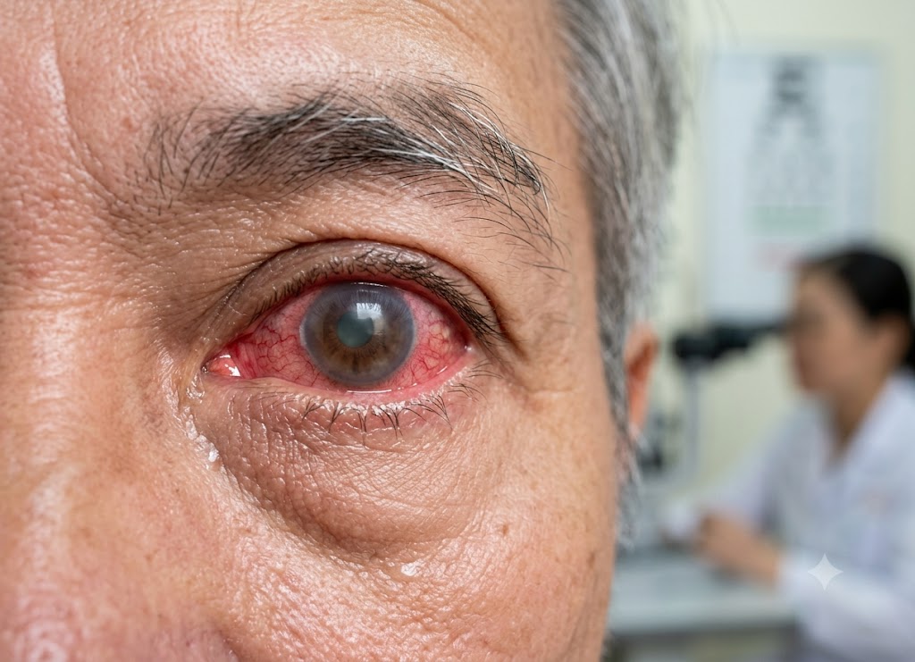
Angle closure glaucoma is a serious eye condition that develops when the drainage angle inside the eye becomes blocked. As a result, aqueous humor, the clear fluid inside the eye, cannot drain normally. This can cause eye pressure to rise quickly and may damage the optic nerve, potentially resulting in permanent vision loss if treatment is delayed.
Unlike open-angle glaucoma, which often develops slowly without noticeable symptoms, an acute angle-closure attack can happen suddenly and requires urgent medical attention.
Recognizing the warning signs is important because prompt treatment can help protect vision. Common symptoms include intense eye pain, headache, blurred vision, halos around lights, nausea, vomiting, and eye redness. This article covers seven important warning signs, along with common causes, risk factors, diagnosis, treatment, and prevention.
What is Acute Angle-Closure Glaucoma and Why Is It an Emergency?
Acute angle-closure glaucoma occurs when the normal drainage pathway inside the eye becomes suddenly blocked. This prevents aqueous humor from leaving the eye and causes intraocular pressure (IOP) to rise rapidly.
The condition is considered an eye emergency because prolonged high pressure can damage the optic nerve and cause permanent vision loss.
The front portion of the eye contains aqueous humor, a clear fluid that helps nourish the cornea and lens while maintaining the eye’s shape. Normally, this fluid flows through the drainage angle between the iris, the colored part of the eye, and the cornea, the clear surface at the front of the eye.
A structure called the trabecular meshwork helps the fluid leave the eye. In some people, this drainage angle is naturally narrow, making it easier for the iris to block the normal flow of fluid.
An acute attack can occur when the pupil becomes enlarged. As the pupil dilates, the iris may move forward and block the drainage angle. The fluid then becomes trapped inside the eye, causing pressure to rise quickly.
The optic nerve is particularly sensitive to increased eye pressure. If the pressure remains elevated, blood flow to the optic nerve may be affected and nerve fibers can become damaged. Because damaged optic nerve cells do not regenerate, untreated acute angle closure can result in permanent vision loss.
7 Key Symptoms of an Angle-Closure Glaucoma Attack
An acute attack of angle closure glaucoma can cause several sudden eye and whole-body symptoms. Because the condition can progress quickly, recognizing these signs and seeking emergency care is extremely important.
1. Unbearable, Radiating Eye Pain
One of the most noticeable symptoms of acute angle closure glaucoma is sudden, severe pain in or around the affected eye.
Skyrocketing Intraocular Pressure ──► Mechanical Strain on Sclera & Cornea
 └──► Trigeminal Nerve Compression ──► Deep, Radiating Facial Pain
The pain may feel deep, intense, or throbbing and can sometimes spread to nearby areas of the face.
- Abrupt and Unilateral: The pain usually begins suddenly and commonly affects one eye.
- Radiating Pathway: Because the eye and parts of the face share sensory nerve pathways, pain may spread toward the forehead, temple, or jaw on the same side.
Severe eye pain combined with other symptoms such as blurred vision or nausea should be treated as an emergency.
2. Sudden, Profoundly Blurry Vision
A sudden decrease in visual clarity is another common sign of closed angle glaucoma. Instead of gradually worsening, vision may become cloudy or hazy over a short period.
One reason for this change is corneal edema, or swelling of the cornea. Increased pressure can interfere with the cornea’s ability to remain clear, causing light to scatter instead of passing normally through the eye.
As a result, a person may feel as though they are looking through fog or a heavily steamed window.
3. Vibrant Halos and Rainbow Rings Around Lights
Seeing halos or rainbow-colored rings around lights can occur during an acute angle-closure attack.
This symptom is associated with swelling of the cornea. When the cornea becomes swollen, changes in the tissue can cause incoming light to scatter and create colored rings around bright objects.
The effect may become especially noticeable in darker surroundings, where bright lights stand out more strongly.
4. Systemic Nausea and Vomiting
Nausea and vomiting can occur along with severe eye pain during an acute glaucoma attack. Although these symptoms may initially seem unrelated to the eyes, they can be part of the body’s response to intense pain.
Intense Ocular Pain ──► Trigeminal Nerve Transmission ──► Brainstem Overstimulation ──► Activation of the Vomiting CenterThis can sometimes make the condition difficult to recognize because severe headache, nausea, and vomiting may resemble other illnesses.
Clinical Warning: Sudden eye pain combined with blurred vision, halos, redness, headache, nausea, or vomiting should not simply be assumed to be a migraine or stomach illness. Immediate medical evaluation is important.
5. An Intense, “Angry” Red Eye
A very red eye can be another visible warning sign of angle-closure glaucoma.
The increased pressure inside the eye can affect normal blood flow and cause the small blood vessels on the surface of the eye to become more prominent. Redness may be particularly noticeable around the edge of the cornea, a pattern sometimes called ciliary flush.
When severe redness appears together with eye pain or sudden vision changes, urgent medical attention is needed.
6. A Severe, One-Sided Headache
A strong headache on the same side as the affected eye is another possible symptom.
The headache may occur around the eyebrow, forehead, or temple. This happens because the eye and surrounding areas share sensory nerve pathways, allowing pain originating in the eye to be felt in other parts of the head.
The headache may become more severe as eye pressure increases and may improve when the pressure is successfully reduced.
7. A Fixed, Mid-Dilated Pupil
During an acute angle-closure attack, the affected pupil may become enlarged and less responsive to light.
Ischemic Blood Flow Restriction ──► Oxygen/Nutrient Starvation ──► Paralysis of the Iris Sphincter Muscle ──► Fixed, Mid-Dilated PupilNormally, the pupil becomes smaller in bright light and larger in darkness. During an angle-closure crisis, increased pressure and changes in the iris can interfere with this normal response.
The pupil may remain partly dilated and may appear slightly irregular. An eye specialist can examine the pupil’s response during an emergency evaluation.
Symptom Overview Matrix
| Symptom | Possible Mechanism | Clinical Appearance / Description | Diagnostic Importance |
|---|---|---|---|
| Severe Eye Pain | Increased eye pressure irritates sensitive structures and nerves. | Deep or intense pain around one eye that may spread to the face. | Important warning sign of an acute eye emergency. |
| Sudden Blurry Vision | Increased pressure can cause corneal swelling. | Rapidly developing cloudy or hazy vision. | Indicates that normal light transmission through the cornea has been affected. |
| Halos Around Lights | Corneal swelling can scatter incoming light. | Colored or rainbow-like rings around bright lights. | A common sign associated with corneal edema. |
| Nausea & Vomiting | Severe eye pain can trigger a systemic response. | Nausea and vomiting accompanying eye symptoms. | May cause confusion with migraine or gastrointestinal illness. |
| Eye Redness | Increased pressure can affect blood vessels around the eye. | Noticeably red eye, sometimes strongest around the cornea. | Supports the presence of significant ocular inflammation or pressure changes. |
| Severe Headache | Referred pain through shared nerve pathways. | One-sided pain around the eyebrow or temple. | May occur alongside an acute glaucoma attack. |
| Fixed Pupil Changes | Pressure and iris dysfunction can interfere with the pupil response. | Partly dilated pupil that responds poorly to light. | An important objective sign during examination. |
What Causes the Sudden Onset of Angle-Closure Glaucoma?
Acute angle-closure glaucoma usually develops when a person has a naturally narrow drainage angle and something causes the pupil to dilate.
A shallow space at the front of the eye can make the iris more likely to block the drainage pathway. This anatomical pattern can be more common in people with farsightedness and tends to become more likely with age as the natural lens becomes thicker.
Other risk factors include being female, having a family history of glaucoma, and belonging to certain populations with a higher prevalence of narrow drainage angles.
Once a narrow angle is present, pupil dilation can trigger an attack. Possible triggers include:
Entering a Dark Environment: Moving into a dark room can naturally cause the pupils to enlarge. In an eye with a narrow drainage angle, this may contribute to blockage.
Certain Medications: Some medications can cause pupil dilation or affect the eye’s drainage system. These may include certain antihistamines, decongestants, antidepressants, anticholinergic medicines, and other drugs. People with known narrow angles should ask their doctor or pharmacist about the safety of new medications.
Emotional Stress or Excitement: Strong emotional responses can affect pupil size. In people with anatomically narrow angles, significant pupil dilation may contribute to an attack.
Some people may have narrow or potentially closable angles without experiencing a major attack. Early eye examinations can identify this risk before an emergency occurs.
Immediate Actions To Take for Suspected Angle Closure Glaucoma
An acute angle closure glaucoma attack requires immediate medical attention. Delaying treatment can increase the risk of permanent optic nerve damage and vision loss.
If sudden severe eye pain occurs along with blurred vision, halos around lights, nausea, vomiting, redness, or a change in the pupil, seek emergency medical care immediately.
1. Step-by-Step Emergency Protocol
If you suspect an active attack, take the following steps:
1. Seek Immediate Medical Care: Do not wait for symptoms to disappear on their own. Contact an eye specialist urgently or go to the nearest emergency department.
2. Arrange Safe Transportation: Do not drive yourself if your vision is blurred or your symptoms are severe. Ask someone to take you or use emergency transportation when necessary.
3. Maintain Proper Posture and Lighting: Stay upright while waiting for medical care. Avoid delaying treatment by trying to rest at home.
4. Advocate at the Emergency Desk: Clearly explain that you have sudden eye pain and vision changes and that you are concerned about an acute glaucoma attack. This can help medical staff understand that an urgent eye examination is needed.
2. Acute Medical Management
Once a patient reaches a medical facility, the first priority is usually to lower the elevated intraocular pressure as quickly and safely as possible.
[Skyrocketing IOP] ──► Medical Combo: Topical Drops + Systemic Agents ──► Opens Drainage Angle & Drops PressureTreatment may involve a combination of eye drops and systemic medications.
Topical Eye Drops
Doctors may use several types of eye drops to reduce eye pressure:
- Beta-Blockers (e.g., Timolol): These reduce the production of aqueous humor.
- Alpha-Agonists (e.g., Apraclonidine): These can reduce fluid production and may improve fluid movement out of the eye.
- Miotic Agents (e.g., Pilocarpine): These medicines can constrict the pupil and may help relieve the blockage in appropriate situations.
Systemic Medications
Additional medicines may be used when a rapid reduction in eye pressure is needed:
- Carbonic Anhydrase Inhibitors (e.g., Acetazolamide / Diamox): These reduce the production of aqueous humor and can be given orally or intravenously.
- Osmotic Diuretics (e.g., Intravenous Mannitol): In severe cases, an osmotic medicine may be used to help lower pressure quickly.
3. Definitive Intervention: Laser Peripheral Iridotomy (LPI)
After the initial pressure is controlled, doctors may recommend Laser Peripheral Iridotomy (LPI) to address the underlying pupillary-block mechanism.
Laser Created Iris Hole ──► New Fluid Bypass Route ──► Equalizes Front/Back Pressure ──► Flattens Iris Away From AngleDuring LPI, an ophthalmologist uses a focused laser to create a very small opening in the peripheral iris. This opening provides an alternative pathway for fluid to move between areas of the eye and can reduce the chance of the iris blocking the drainage angle.
Because the anatomical risk may be present in both eyes, an ophthalmologist may recommend preventive treatment for the other eye as well.
Emergency Action Summary Matrix
| Phase of Care | Specific Action / Medication | Purpose | Critical Goal |
|---|---|---|---|
| Immediate Self-Care | Seek urgent medical attention and avoid driving yourself. | Helps ensure rapid professional evaluation. | Prevent delays in treatment. |
| Topical Therapy | Pressure-lowering eye drops such as timolol or other appropriate medicines. | Reduces aqueous humor production and may help lower pressure. | Begin controlling the pressure quickly. |
| Systemic Therapy | Acetazolamide or other appropriate systemic treatment. | Provides additional pressure reduction when needed. | Lower dangerously elevated IOP. |
| Definitive Treatment | Laser Peripheral Iridotomy (LPI). | Creates an alternative fluid pathway through the iris. | Reduce the risk of future pupillary-block attacks. |
What Else Should You Know About Glaucoma Risk and Management?
Understanding the risk factors, diagnostic tests, and preventive options can help people with narrow drainage angles protect their vision.
Because angle-closure glaucoma is closely linked to the structure of the eye, identifying narrow angles before an acute attack can be especially valuable.
1. High-Risk Demographics and Anatomical Profiles
An attack of closed angle glaucoma is more likely when the eye has a crowded or narrow drainage angle.
Anatomical Crowding (Farsightedness/Aging Lens) ──► Shallower Anterior Chamber ──► High Risk for Physical OcclusionEye specialists may consider several risk factors:
Age and Gender: The risk of angle closure generally increases with age. Changes to the natural lens can gradually make the drainage angle narrower. Women also have a higher risk than men.
High-Risk Ethnicities: Certain populations, including people of East Asian or Inuit ancestry, have a higher prevalence of anatomically narrow drainage angles.
Hyperopia (Farsightedness): Farsighted eyes are often shorter from front to back, which can be associated with a shallower anterior chamber and narrower drainage angle.
2. Advanced Clinical Diagnostics and Treatment
When someone develops closed angle glaucoma symptoms, doctors need to assess eye pressure and the drainage angle quickly.
Diagnostic Testing
- Tonometry: This test measures intraocular pressure.
- Gonioscopy: This examination allows an ophthalmologist to evaluate whether the drainage angle is open, narrow, or closed.
Additional eye examinations and imaging may be used depending on the patient’s condition.
Targeted Medical and Surgical Intervention
After an acute attack is confirmed, treatment is focused on quickly reducing eye pressure and then addressing the underlying cause.
[Emergency Evaluation] ──► Medical Lowering (Drops/IV) ──► Defuse Acute Spike ──► Laser Peripheral Iridotomy (LPI)Immediate Pressure Control: Doctors may use pressure-lowering eye drops and systemic medicines to bring IOP down.
Definitive Laser Surgery: Once the eye is stable, Laser Peripheral Iridotomy (LPI) may be performed to create an alternative pathway for fluid and reduce the risk of another pupillary-block episode.
3. Structural Contrasts: Angle-Closure vs. Open-Angle Glaucoma
It is important to understand that not all types of glaucoma develop in the same way. Angle closure glaucoma and open-angle glaucoma differ in their drainage anatomy, symptoms, and typical progression.
| Feature | Open-Angle Glaucoma | Angle-Closure Glaucoma |
|---|---|---|
| Drainage Angle Status | The drainage angle remains open, but fluid outflow may become less effective. | The drainage angle can become narrowed or blocked by the iris. |
| Onset Profile | Usually develops gradually over time. | Can develop suddenly and rapidly during an acute attack. |
| Symptom Expression | Often has few or no early symptoms. | May cause severe eye pain, headache, blurred vision, halos, nausea, and redness. |
| Clinical Urgency | Usually managed through long-term monitoring and treatment. | Acute attacks require immediate medical evaluation and treatment. |
4. Proactive Attack Prevention Strategies
People with narrow or potentially closable angles may be able to reduce their risk through regular eye care and appropriate preventive treatment.
Routine Gonioscopy Exam ──► Identifies High-Risk "Narrow Angles" ──► Preventative Prophylactic LPIProphylactic Laser Peripheral Iridotomy (LPI)
If an eye examination shows that the drainage angle is dangerously narrow or at risk of closing, an ophthalmologist may recommend preventive LPI.
The procedure creates a tiny opening in the iris that provides another route for fluid movement. This can reduce the chance of an acute attack caused by pupillary block.
Vigilant Medication Screening
People who have been diagnosed with narrow angles should discuss new medications with their healthcare professional. Some medicines can affect pupil size or other aspects of the eye and may increase the risk of angle closure in susceptible individuals.
High-Risk Medications to Discuss With Your Doctor:
- Some over-the-counter allergy medicines and cold decongestants.
- Certain motion sickness medicines and antidepressants.
If you have been told that you have narrow angles, always ask your doctor or eye specialist whether a new medication is appropriate for you.
Conclusion
Angle closure glaucoma is a serious eye emergency that can threaten vision when treatment is delayed. Warning signs can include sudden severe eye pain, blurred vision, halos around lights, nausea, vomiting, headache, eye redness, and changes in the pupil.
If these symptoms develop suddenly, immediate medical evaluation is essential. Treatment may involve medicines to lower eye pressure followed by laser treatment or another procedure to address the underlying drainage problem.
Regular eye examinations can also help identify narrow drainage angles before they lead to an emergency. Early diagnosis, appropriate treatment, and ongoing eye care can help protect vision.
Frequently Asked Questions
1. What is angle closure glaucoma?
Angle closure glaucoma occurs when the drainage angle between the iris and cornea becomes narrowed or blocked. This prevents aqueous humor from draining normally and can cause intraocular pressure to rise. An acute attack can damage the optic nerve and requires urgent medical treatment.
2. What are the warning signs of angle closure glaucoma?
Warning signs may include sudden severe eye pain, headache, blurred or hazy vision, halos around lights, nausea, vomiting, and a red eye. These symptoms can develop quickly and should be evaluated immediately.
3. What causes angle closure glaucoma?
Angle closure glaucoma is often associated with a naturally narrow drainage angle. Risk can increase with factors such as farsightedness, aging-related lens changes, family history, and certain anatomical characteristics. Pupil dilation and some medications can trigger an attack in susceptible eyes.
4. How is angle closure glaucoma diagnosed?
Doctors may measure intraocular pressure with tonometry and examine the drainage angle using gonioscopy. A comprehensive eye examination and additional imaging may also be used when appropriate.
5. How is angle closure glaucoma treated?
Treatment aims to lower eye pressure quickly and protect the optic nerve. Doctors may use pressure-lowering eye drops and systemic medications during an acute attack. Laser Peripheral Iridotomy (LPI) may then be performed to create an alternative fluid pathway and reduce the risk of future attacks.
6. Can angle closure glaucoma cause permanent blindness?
Yes. If an acute attack is not treated promptly, increased eye pressure can damage the optic nerve and cause permanent vision loss. Seeking emergency care as soon as symptoms appear is critical.
7. Who is at risk for angle closure glaucoma?
People with narrow drainage angles, farsightedness, increasing age, or a family history of glaucoma may have a higher risk. Certain ethnic populations, including East Asian and Inuit groups, also have a higher prevalence of narrow angles. Regular eye examinations can help identify people at risk before an acute attack occurs.